北京大学学报(医学版) ›› 2020, Vol. 52 ›› Issue (5): 931-937. doi: 10.19723/j.issn.1671-167X.2020.05.023
生物活性玻璃预处理对牙本质粘接界面耐久性的影响
李秋菊,宫玮玉,董艳梅( )
)
- 北京大学口腔医学院·口腔医院,牙体牙髓科 国家口腔疾病临床医学研究中心 口腔数字化医疗技术和材料国家工程实验室 口腔数字医学北京市重点实验室,北京 100081
Effect of bioactive glass pretreatment on the durability of dentin bonding interface
Qiu-ju LI,Wei-yu GONG,Yan-mei DONG()
- Department of Cariology and Endodontology, Peking University School and Hospital of Stomatology & National Clinical Research Center for Oral Diseases & National Engineering Laboratory for Digital and Material Technology of Stomatology & Beijing Key Laboratory of Digital Stomatology, Beijing 100081, China
摘要:
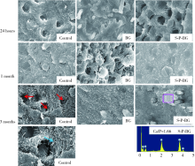
目的:研究生物活性玻璃(bioactive glass, BG)预处理对维持牙本质粘接界面耐久性的作用。方法:选取30颗无龋坏第三磨牙,去除冠部釉质制备牙本质平面,随机均分对照组、BG组、三偏磷酸钠(sodium trimetaphosphate, STMP)-聚丙烯酸(polyacrylic acid, PAA)-BG组(S-P-BG组)。各组均使用35%(质量分数)磷酸酸蚀牙本质样本,BG组再使用0.5 g/L BG涂擦酸蚀后的牙本质样本;S-P-BG组先使用5%(质量分数)STMP、5%(质量分数)PAA浸泡酸蚀后的牙本质样本1 min,再使用0.5 g/L BG涂擦牙本质样本。各组样本使用3M Single Bond 2粘接剂及3M Z350XT复合树脂粘接,并制备微拉伸柱状样本,每颗牙的柱状样本按时间随机分为24 h、1个月、3个月组。各组样本保存在37 ℃人工唾液(artificial saliva, AS)中相应时间后,进行微拉伸粘接强度测试,并使用单因素方差分析及LSD法进行统计学分析,扫描电镜下观察粘接断裂界面形貌。另选取27颗无龋坏第三磨牙制备牙本质平面,随机分为对照组、BG组、S-P-BG组,并按上述分组处理牙本质样本,再使用含0.1%(质量分数)罗丹明B的3M Single Bond 2粘接剂完成粘接。去除样本牙根暴露髓腔,并保存在 37 ℃ AS中24 h、1个月、3个月后,髓腔内放置0.1(质量分数)荧光素钠溶液染色1 h,激光共聚焦显微镜观察粘接界面形态及混合层微渗漏。结果:AS中浸泡24 h、1个月后,3组微拉伸粘接强度间的差异无统计学意义(P>0.05);浸泡3个月后,S-P-BG组微拉伸粘接强度为(36.91±7.07) MPa,高于对照组粘接强度(32.73±8.06) MPa,且差异有统计学意义(P=0.026);对照组、BG组3个月的微拉伸粘接强度较24 h呈下降趋势,且差异有统计学意义(对照组P=0.017,BG组P=0.01);S-P-BG组3个月微拉伸粘接强度较24 h粘接强度[(37.99±7.98) MPa]下降,但差异无统计学意义(P>0.05)。扫描电镜观察24 h粘接断裂面,3组均未见明显矿化;1个月、3个月后,BG组、S-P-BG组的粘接界面可见矿物质形成,S-P-BG组无明显胶原暴露。激光共聚焦显微镜观察对照组、BG组与S-P-BG组树脂突形成的形态及数量无明显差异;3组样本粘接24 h后粘接界面混合层均有渗漏,3个月后对照组微渗漏增加,BG组和S-P-BG组混合层微渗漏减少。结论:BG预处理牙本质粘接界面能够在粘接界面形成矿物质,减少粘接混合层微渗漏;STMP、PAA 与BG共同预处理牙本质粘接界面,可能在一定程度上维持牙本质粘接修复的耐久性。
中图分类号:
- R783.1


| [1] |
Pashley DH, Tay FR, Breschi L, et al. State of the art etch-andrinse adhesives[J]. Dent Mater, 2011,27(1):1-16.
doi: 10.1016/j.dental.2010.10.016 |
| [2] |
Van Meerbeek B, Yoshihara K, Yoshida Y, et al. State of the art of self-etch adhesives[J]. Dent Mater, 2011,27(1):17-28.
doi: 10.1016/j.dental.2010.10.023 |
| [3] |
Ikeda T, De Munck J, Shirai K, et al. Effect of evaporation of primer components on ultimate tensile strengths of primer-adhesive mixture[J]. Dent Mater, 2005,21(11):1051-1058.
doi: 10.1016/j.dental.2005.03.010 |
| [4] | Sabatini C, Pashley DH. Mechanisms regulating the degradation of dentin matrices by endogenous dentin proteases and their role in dental adhesion[J]. Am J Dent, 2014,27(4):203-214. |
| [5] |
Opdam NJM, Van de Sande FH, Bronkhorst E, et al. Longevity of posterior composite restorations[J]. J Dent Res, 2014,93(10):943-949.
doi: 10.1177/0022034514544217 |
| [6] | 李景辉, 刘慧, 乔佳, 等. 2%氯己定对牙本质自酸蚀粘接强度影响的初步研究[J]. 北京口腔医学, 2010,18(3):132-134. |
| [7] |
Imazato S, Ma S, Chen J, et al. Therapeutic polymers for dental adhesives: Loading resins with bio-active components[J]. Dent Mater, 2014,30(1):97-104.
doi: 10.1016/j.dental.2013.06.003 |
| [8] | Liu SQ, Gong WY, Dong YM, et al. The effect of submicron bioactive glass particles on in vitro osteogenesis[J]. Rsc Advances, 2015,5(49):38830-38836. |
| [9] | Sheng X, Gong W, Hu Q, et al. Mineral formation on dentin induced by nano-bioactive glass[J]. Chin Chem Lett, 2016,27(9):1509-1514. |
| [10] |
Hu S, Chang J, Liu M, et al. Study on antibacterial effect of 45S5 Bioglass[J]. J Mater Sci Mater Med, 2009,20(1):281-286.
doi: 10.1007/s10856-008-3564-5 pmid: 18763024 |
| [11] | Osorio R, Yamauti M, Sauro S, et al. Zinc Incorporation improves biological activity of beta-tricalcium silicate resin-based cement[J]. J Endod, 2014,40(11):1840-1845. |
| [12] |
Tezvergil-Mutluay A, Seseogullari-Dirihan R, Feitosa VP, et al. Effects of composites containing bioactive glasses on demineralized dentin[J]. J Dent Res, 2017,96(9):999-1005.
doi: 10.1177/0022034517709464 pmid: 28535357 |
| [13] | 王瑜, 龚玲, 计艳, 等. 生物活性玻璃改性树脂粘接剂对牙本质脱矿及树脂粘接强度的影响[J]. 华西口腔医学杂志, 2016,34(4):350-353. |
| [14] | Wang Z, Shen Y, Haapasalo M, et al. Polycarboxylated microfillers incorporated into light-curable resin-based dental adhesives evoke remineralization at the mineral-depleted dentin[J]. J Biomater Sci Polym Ed, 2014,25(7):679-697. |
| [15] |
Liu Y, Li N, Qi Y, et al. Intrafibrillar collagen mineralization produced by biomimetic hierarchical nanoapatite assembly[J]. Adv Mater, 2011,23(8):975-980.
doi: 10.1002/adma.201003882 pmid: 21341310 |
| [16] |
Abuna G, Feitosa VP, Correr AB, et al. Bonding performance of experimental bioactive/biomimetic self-etch adhesives doped with calcium-phosphate fillers and biomimetic analogs of phosphoproteins[J]. J Dent, 2016,52:79-86.
doi: 10.1016/j.jdent.2016.07.016 pmid: 27472956 |
| [17] |
De Munck J, Van Landuyt K, Peumans M, et al. A critical review of the durability of adhesion to tooth tissue: methods and results[J]. J Dent Res, 2005,84(2):118-132.
doi: 10.1177/154405910508400204 pmid: 15668328 |
| [18] | 闫晶. 龋病内层牙本质和正常牙本质粘接强度及粘接界面密合性的比较研究[D]. 第四军医大学, 2006. |
| [19] |
Okuda M, Pereira PN, Nakajima M, et al. Long-term durability of resin dentin interface: nanoleakage vs. microtensile bond strength[J]. Oper Dent, 2002,27(3):289-296.
pmid: 12022462 |
| [20] | 孙秋榕, 麦穗. 树脂-牙本质粘接界面的渗透性与粘接耐久性研究进展[J]. 国际口腔医学杂志, 2016,43(3):338-342. |
| [21] |
Cerruti MG, Greenspan D, Powers K. An analytical model for the dissolution of different particle size samples of Bioglass in TRIS-buffered solution[J]. Biomaterials, 2005,26(24):4903-4911.
doi: 10.1016/j.biomaterials.2005.01.013 |
| [22] |
Vollenweider M, Brunner TJ, Knecht S, et al. Remineralization of human dentin using ultrafine bioactive glass particles[J]. Acta Biomater, 2007,3(6):936-943.
doi: 10.1016/j.actbio.2007.04.003 pmid: 17560183 |
| [23] |
Sauro S, Osorio R, Fulgencio R, et al. Remineralisation properties of innovative light-curable resin-based Dent Mater containing bioactive micro-fillers[J]. J Mater Chem B, 2013,1(20):2624-2638.
doi: 10.1039/c3tb00205e pmid: 32260950 |
| [24] |
Profeta AC, Mannocci F, Foxton RM, et al. Bioactive effects of a calcium/sodium phosphosilicate on the resin-dentine interface: a microtensile bond strength, scanning electron microscopy, and confocal microscopy study[J]. Eur J Oral Sci, 2012,120(4):353-362.
doi: 10.1111/j.1600-0722.2012.00974.x |
| [25] |
Nudelman F, Lausch AJ, Sommerdijk NAJM, et al. In vitro models of collagen biomineralization[J]. J Struct Biol, 2013,183(2):258-269.
doi: 10.1016/j.jsb.2013.04.003 pmid: 23597833 |
| [26] |
Toroian D, Lim JE, Price PA. The size exclusion characteristics of type Ⅰ collagen[J]. J Biol Chem, 2007,282(31):22437-22447.
doi: 10.1074/jbc.M700591200 pmid: 17562713 |
| [27] |
Wang Z, Jiang T, Sauro S, et al. Dentine remineralization induced by two bioactive glasses developed for air abrasion purposes[J]. J Dent, 2011,39(11):746-756.
doi: 10.1016/j.jdent.2011.08.006 pmid: 21864641 |
| [28] |
Niu L, Zhang W, Pashley DH, et al. Biomimetic remineralization of dentin[J]. Dent Mater, 2014,30(1):77-96.
doi: 10.1016/j.dental.2013.07.013 |
| [29] |
Liu Y, Mai S, Li N, et al. Differences between top-down and bottom-up approaches in mineralizing thick, partially demineralized collagen scaffolds[J]. Acta Biomater, 2011,7(4):1742-1751.
doi: 10.1016/j.actbio.2010.11.028 pmid: 21111071 |
| [1] | 黄丽东,宫玮玉,董艳梅. 生物活性玻璃对人脐静脉血管内皮细胞增殖及成血管的作用[J]. 北京大学学报(医学版), 2021, 53(2): 371-377. |
| [2] | 唐仁韬,李欣海,于江利,冯琳,高学军. 复合树脂与玻璃陶瓷微拉伸粘接强度的体外研究[J]. 北京大学学报(医学版), 2020, 52(4): 755-761. |
| [3] | 朱晓鸣,齐璇,李德利,张玉玮,李和平,谭建国. 不同温度新型大气压冷等离子体处理对牙本质粘接强度的影响[J]. 北京大学学报(医学版), 2019, 51(1): 43-48. |
| [4] | 龙赟子,刘思毅,李稳,董艳梅. 生物活性玻璃盖髓剂的理化性质[J]. 北京大学学报(医学版), 2018, 50(5): 887-891. |
| [5] | 李爽,张清. 玷污层对新型三氧化矿物凝聚体根尖封闭性的影响[J]. 北京大学学报(医学版), 2018, 50(3): 560-563. |
| [6] | 朱林,王聿栋,董艳梅,陈晓峰. 缓释米诺环素的介孔纳米生物玻璃载药系统[J]. 北京大学学报(医学版), 2018, 50(2): 249-255. |
| [7] | 宫玮玉,刘绍清,董艳梅,高学军,陈晓峰. 纳米生物活性玻璃促进兔颅骨临界骨缺损修复[J]. 北京大学学报(医学版), 2018, 50(1): 42-48. |
| [8] | 李皓,刘玉华,罗志强. 生物活性玻璃用于缓解活髓牙全冠预备后敏感的效果评价[J]. 北京大学学报(医学版), 2017, 49(4): 709-713. |
| [9] | 信义,王赛楠,崔彩云,董艳梅. 生物活性玻璃和牙本质浸提蛋白对人牙髓细胞的作用[J]. 北京大学学报(医学版), 2017, 49(2): 331-336. |
| [10] | 刘意,王赛楠,崔彩云,董艳梅. 精氨酸-甘氨酸-天冬氨酸-丝氨酸序列在生物活性玻璃对人牙髓细胞生物作用中的影响[J]. 北京大学学报(医学版), 2017, 49(2): 326-330. |
| [11] | 王月,梁宇红. 次氯酸钠溶液表面处理对牙本质粘接强度的影响[J]. 北京大学学报(医学版), 2017, 49(1): 105-109. |
| [12] | 胡佳,邹晓英,庄姮,高学军. 根管封闭剂对牙周膜细胞生物相容性的影响[J]. 北京大学学报(医学版), 2016, 48(5): 871-877. |
| [13] | 乔迪, 董艳梅, 高学军. 体外评价新型根尖倒充填材料iRoot的生物学性能[J]. 北京大学学报(医学版), 2016, 48(2): 324-329. |
| [14] | 张路, 袁重阳, 田福聪, 王晓燕, 高学军. 自酸蚀粘接剂系统对变形链球菌的抑制作用[J]. 北京大学学报(医学版), 2016, 48(1): 57-62. |
| [15] | 谢窈, 张笋, 葛立宏. 乳牙应用Er:YAG激光备洞充填微渗漏效果的离体研究[J]. 北京大学学报(医学版), 2014, 46(3): 474-477. |
|
