北京大学学报(医学版) ›› 2021, Vol. 53 ›› Issue (1): 69-75. doi: 10.19723/j.issn.1671-167X.2021.01.011
数字化评估CAD/CAM个性化基台与成品基台影响粘接剂残留的体外研究
岳兆国1,张海东1,杨静文2,侯建霞1,Δ( )
)
- 1.北京大学口腔医学院·口腔医院,牙周科 国家口腔疾病临床医学研究中心 口腔数字化医疗技术和材料国家工程实验室 口腔数字医学北京市重点实验室,北京 100081
2.北京大学口腔医学院·口腔医院,修复科 国家口腔疾病临床医学研究中心 口腔数字化医疗技术和材料国家工程实验室 口腔数字医学北京市重点实验室,北京 100081
Comparison of residual cement between CAD/CAM customized abutments and stock abutments via digital measurement in vitro
YUE Zhao-guo1,ZHANG Hai-dong1,YANG Jing-wen2,HOU Jian-xia1,Δ()
- 1. Department of Periodontology, Peking University School and Hospital of Stomatology & National Clinical Research Center for Oral Diseases & National Engineering Laboratory for Digital and Material Technology of Stomatology & Beijing Key Laboratory of Digital Stomatology, Beijing 100081, China
2. Department of Prosthetics, Peking University School and Hospital of Stomatology & National Clinical Research Center for Oral Diseases & National Engineering Laboratory for Digital and Material Technology of Stomatology & Beijing Key Laboratory of Digital Stomatology, Beijing 100081, China
摘要:
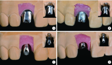
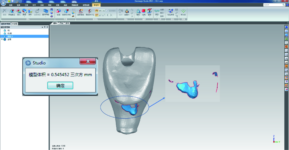
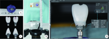
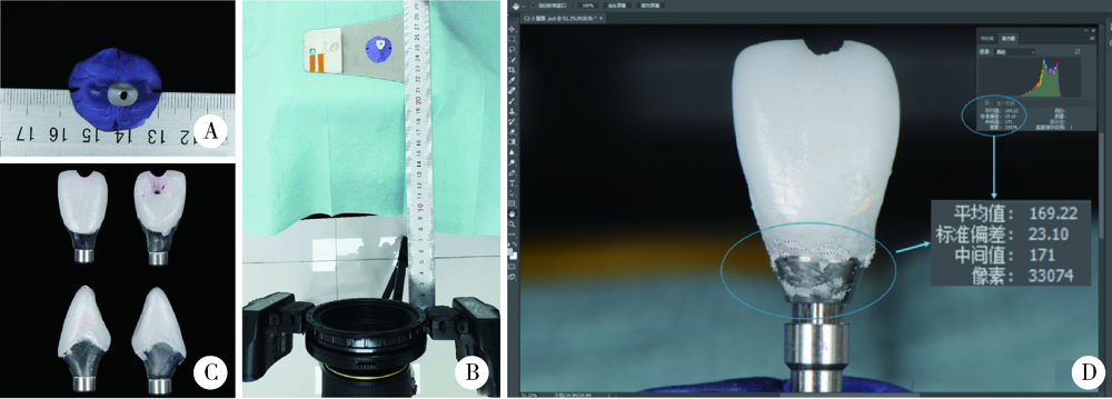
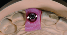
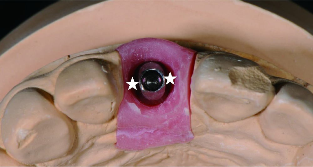
目的: 通过体外模型比较计算机辅助设计/计算机辅助制造个性化基台(computer aided design/computer aided manufacturing customized abutments,CCA)与成品基台(stock abutments,SA)对粘接剂残留的影响,同时初步评价数字化三维扫描技术定量评估残留粘接剂的可行性。方法: 本研究所需20个工作模型皆取自同一例已在北京大学口腔医院牙周科接受了右上中切牙种植手术的患者。通过个性化愈合基台成形植体周软组织后,测得植体平台位于颊侧黏膜下5 mm。利用个性化转移杆取模后灌制20副工作模型,并根据基台种类及粘接边缘位置将工作模型分为四组,每组5个:CCA1(穿黏膜高度5 mm,即平齐黏膜粘接边缘)、CCA2(穿黏膜高度4 mm,即黏膜下1 mm粘接边缘)、SA1(穿黏膜高度3 mm,即黏膜下2 mm粘接边缘)和SA2(穿黏膜高度1 mm,即黏膜下4 mm粘接边缘)。在工作模型上模拟临床粘接过程并清除多余粘接剂后,利用三维扫描技术获得残留粘接剂的体积,利用数码相机拍摄二维图像获得残留粘接剂面积百分比,利用称量的方式获得残留粘接剂的质量,并分析三维扫描方法获取的体积与传统评价方法所得的面积百分比及质量的相关性。结果: 所有冠-基台复合体粘接边缘均有粘接剂残留。其中,CCA组残留粘接剂的体积明显小于SA组[(0.635 3±0.535 4) mm3 vs. (2.293 8±0.943 8) mm 3,P<0.001], 面积百分比及质量也显著低于SA组[面积百分比:7.57%±2.99% vs. 22.68%±10.06%,P<0.001;质量:(0.001 5±0.001 0) g vs. (0.003 7±0.001 4) g,P<0.001],而三者在CCA组及SA组内差异均无统计学意义(P>0.05)。三维扫描所得残留粘接剂的体积与传统评价方法所得残留粘接剂的面积百分比及残留粘接剂的质量间均具强相关性(r>0.75,P<0.001)。结论: 与SA相比,CCA能更有效地减少粘接剂的残留。基于三维扫描技术数字化评估残留粘接剂的方法切实可行,但其效度和信度还需进一步研究。
中图分类号:
- R783


| [1] |
Jung RE, Pjetursson BE, Glauser R, et al. A systematic review of the 5-year survival and complication rates of implant-supported single crowns[J]. Clin Oral Implants Res, 2008,19(2):119-130.
doi: 10.1111/j.1600-0501.2007.01453.x pmid: 18067597 |
| [2] |
Chee W, Felton DA, Johnson PF, et al. Cemented versus screw-retained implant prostheses: Which is better?[J]. Int J Oral Maxillofac Implants, 1999,14(1):137-141.
pmid: 10074764 |
| [3] |
Linkevicius T, Vindasiute E, Puisys A, et al. The influence of margin location on the amount of undetected cement excess after delivery of cement-retained implant restorations[J]. Clin Oral Implants Res, 2011,22(12):1379-1384.
doi: 10.1111/j.1600-0501.2010.02119.x pmid: 21382089 |
| [4] |
Staubli N, Walter C, Schmidt JC, et al. Excess cement and the risk of peri-implant disease: A systematic review[J]. Clin Oral Implants Res, 2017,28(10):1278-1290.
doi: 10.1111/clr.12954 pmid: 27647536 |
| [5] |
Korsch M, Obst U, Walther W. Cement-associated peri-implantitis: A retrospective clinical observational study of fixed implant-supported restorations using a methacrylate cement[J]. Clin Oral Implants Res, 2014,25(7):797-802.
doi: 10.1111/clr.12173 pmid: 23600620 |
| [6] |
Wilson TG Jr. The positive relationship between excess cement and peri-implant disease: A prospective clinical endoscopic study[J]. J Periodontol, 2009,80(9):1388-1392.
doi: 10.1902/jop.2009.090115 pmid: 19722787 |
| [7] |
Linkevicius T, Puisys A, Vindasiute E, et al. Does residual cement around implant-supported restorations cause peri-implant disease? A retrospective case analysis[J]. Clin Oral Implants Res, 2013,24(11):1179-1184.
doi: 10.1111/j.1600-0501.2012.02570.x pmid: 22882700 |
| [8] |
Ichikawa T, Ishida O, Watanabe M, et al. A new retrieval system for cement-retained implant superstructures: A technical report[J]. J Prosthodont, 2008,17(6):487-489.
doi: 10.1111/j.1532-849X.2008.00329.x pmid: 18544129 |
| [9] |
Galván G, Kois JC, Chaiyabutr Y, et al. Cemented implant restoration: A technique for minimizing adverse biologic consequences[J]. J Prosthet Dent, 2015,114(4):482-485.
doi: 10.1016/j.prosdent.2014.10.017 pmid: 26119018 |
| [10] |
Seo CW, Seo JM. A technique for minimizing subgingival residual cement by using rubber dam for cement-retained implant crowns[J]. J Prosthet Dent, 2017,117(2):327-328.
doi: 10.1016/j.prosdent.2016.08.024 pmid: 27771147 |
| [11] | Linkevicius T. Zero bone loss concepts [M]. Illinois: Quintessence Publishing Co, 2019. |
| [12] |
Lewis S, Beumer J 3rd, Hornburg W, et al. The “UCLA” abutment[J]. Int J Oral Maxillofac Implants, 1988,3(3):183-189.
pmid: 3074050 |
| [13] | 戴文雍, 汤春波. 种植体修复个性化基台研究现状及展望[J]. 口腔医学, 2012,32(11):685-687. |
| [14] | 宿玉成. 口腔种植学[M]. 2版. 北京: 人民卫生出版社, 2014: 403-404. |
| [15] |
Shapoff CA, Lahey BJ. Crestal bone loss and the consequences of retained excess cement around dental implants[J]. Compend Contin Educ Dent, 2012,33(2):94-101.
pmid: 22545427 |
| [16] |
Schwarz F, Derks J, Monje A, et al. Peri-implantitis[J]. J Clin Periodontol, 2018,45(Suppl 20):S246-S266.
doi: 10.1111/jcpe.2018.45.issue-S20 |
| [17] |
Andersson B, Odman P, Lindvall AM, et al. Cemented single crowns on osseointegrated implants after 5 years: Results from a prospective study on CeraOne[J]. Int J Prosthodont, 1998,11(3):212-218.
pmid: 9728114 |
| [18] |
Linkevicius T, Vindasiute E, Puisys A, et al. The influence of the cementation margin position on the amount of undetected cement. A prospective clinical study[J]. Clin Oral Implants Res, 2013,24(1):71-76.
doi: 10.1111/j.1600-0501.2012.02453.x pmid: 22487018 |
| [19] |
Kappel S, Eiffler C, Lorenzo-Bermejo J, et al. Undetected resi-dual cement on standard or individualized all-ceramic abutments with cemented zirconia single crowns: A prospective randomized pilot trial[J]. Clin Oral Implants Res, 2016,27(9):1065-1071.
doi: 10.1111/clr.12691 pmid: 26381392 |
| [20] |
Kotsakis GA, Zhang L, Gaillard P, et al. Investigation of the association between cement retention and prevalent peri-implant diseases: A cross-sectional study[J]. J Periodontol, 2016,87(3):212-220.
doi: 10.1902/jop.2015.150450 pmid: 26537368 |
| [21] |
Daubert DM, Weinstein BF, Bordin S, et al. Prevalence and predictive factors for peri-implant disease and implant failure: A cross-sectional analysis[J]. J Periodontol, 2015,86(3):337-347.
doi: 10.1902/jop.2014.140438 pmid: 25415249 |
| [22] | Fuchigami K, Munakata M, Kitazume T, et al. A diversity of peri-implant mucosal thickness by site[J]. Clin Oral Impl Res, 2017,28(2):171-176. |
| [23] | 张众, 孟焕新, 韩劼, 等. 软组织垂直厚度对牙周炎患者种植修复临床效果的影响[J]. 北京大学学报(医学版), 2020,52(2):332-338. |
| [24] |
Dumbrigue HB, Abanomi AA, Cheng LL. Techniques to minimize excess luting agent in cement-retained implant restorations[J]. J Prosthet Dent, 2002,87(1):112-114.
doi: 10.1067/mpr.2002.119418 pmid: 11807495 |
| [25] |
Vindasiute E, Puisys A, Maslova N, et al. Clinical factors influencing removal of the cement excess in implant-supported restorations[J]. Clin Implant Dent Relat Res, 2015,17(4):771-778.
doi: 10.1111/cid.12170 pmid: 24224895 |
| [26] |
Andersson B, Odman P, Lindvall AM, et al. Single-tooth restorations supported by osseointegrated implants: results and experiences from a prospective study after 2 to 3 years[J]. Int J Oral Maxillofac Implants, 1995,10(6):702-711.
pmid: 8530173 |
| [27] | Higginbottom F, Belser U, Jones JD, et al. Prosthetic management of implants in the esthetic zone[J]. Int J Oral Maxillofac Implants, 2004,19(Suppl.):62-72. |
| [28] |
Berglundh T, Lindhe J, Marinello C, et al. Soft tissue reaction to de novo plaque formation on implants and teeth. An experimental study in the dog[J]. Clin Oral Implants Res, 1992,3(1):1-8.
doi: 10.1034/j.1600-0501.1992.030101.x pmid: 1420721 |
| [29] | 高鹏程, 谢理哲, 严斌. 牙颌模型三维数字化技术及其在口腔正畸学中的应用进展[J]. 口腔生物医学, 2014,5(3):152-157. |
| [1] | 刘思民,赵一姣,王晓燕,王祖华. 动态导航下不同深度环钻定位精确度的体外评价[J]. 北京大学学报(医学版), 2022, 54(1): 146-152. |
| [2] | 李怡,王丽瑜,刘晓强,周倜,吕季喆,谭建国. 不同材料及厚度椅旁CAD/CAM瓷贴面的边缘特征[J]. 北京大学学报(医学版), 2022, 54(1): 140-145. |
| [3] | 邱淑婷,朱玉佳,王时敏,王飞龙,叶红强,赵一姣,刘云松,王勇,周永胜. 姿势微笑位口唇对称参考平面的数字化构建及初步应用验证[J]. 北京大学学报(医学版), 2022, 54(1): 193-199. |
| [4] | 任国勇,吴雪梅,李颖,李婕妤,孙伟平,黄一宁. 大血管闭塞性脑卒中亚急性期磁敏感血管征的表现[J]. 北京大学学报(医学版), 2021, 53(6): 1133-1138. |
| [5] | 李媛,林红,张铁军. 对比传统成像与数字成像对牙科复合树脂X射线阻射性的影响[J]. 北京大学学报(医学版), 2021, 53(5): 995-1001. |
| [6] | 杨刚,胡文杰,曹洁,柳登高. 牙周健康的上颌前牙唇侧嵴顶上牙龈的三维形态分析[J]. 北京大学学报(医学版), 2021, 53(5): 990-994. |
| [7] | 邵振兴,宋庆法,赵宇晴,崔国庆. 一种结合线袢固定的关节镜下“嵌入式”喙突移位术:手术技术及术后影像学分析[J]. 北京大学学报(医学版), 2021, 53(5): 896-901. |
| [8] | 吴一凡,张晓圆,任爽,玉应香,常翠青. 基于磁共振的青年男性股四头肌的测量和评估[J]. 北京大学学报(医学版), 2021, 53(5): 843-849. |
| [9] | 李新飞, 彭意吉, 余霄腾, 熊盛炜, 程嗣达, 丁光璞, 杨昆霖, 唐琦, 米悦, 吴静云, 张鹏, 谢家馨, 郝瀚, 王鹤, 邱建星, 杨建, 李学松, 周利群. 肾部分切除术前CT三维可视化评估标准的初步探究[J]. 北京大学学报(医学版), 2021, 53(3): 613-622. |
| [10] | 胡迪,张苗,康惠颖,彭芸. 0~2岁婴幼儿磁共振脑白质模板的建立及验证[J]. 北京大学学报(医学版), 2021, 53(2): 341-347. |
| [11] | 陈迪,徐翔宇,汪明睿,李芮,臧根奥,张悦,钱浩楠,闫光荣,范田园. 熔融沉积成型3D打印盐酸维拉帕米胃漂浮制剂的制备与体外评价[J]. 北京大学学报(医学版), 2021, 53(2): 348-354. |
| [12] | 黄新瑞,李莎,高嵩. 冷冻电镜成像中噪声的滤波方法进展[J]. 北京大学学报(医学版), 2021, 53(2): 425-433. |
| [13] | 穆海丽,田福聪,王晓燕,高学军. 玻璃体和通用型复合树脂耐磨性的临床对照研究[J]. 北京大学学报(医学版), 2021, 53(1): 120-125. |
| [14] | 徐啸翔,曹烨,赵一姣,贾璐,谢秋菲. 数字化个齿托盘制取下颌全牙列全冠预备体印模的体外评价[J]. 北京大学学报(医学版), 2021, 53(1): 54-61. |
| [15] | 国丹妮,潘韶霞,衡墨笛,屈健,魏秀霞,周永胜. 应用于无牙颌种植修复设计的三维面部扫描配准方法的对比[J]. 北京大学学报(医学版), 2021, 53(1): 83-87. |
|
