北京大学学报(医学版) ›› 2021, Vol. 53 ›› Issue (1): 133-138. doi: 10.19723/j.issn.1671-167X.2021.01.020
中国人群腭中缝形态特点分期与Demirjian牙龄的相关性
高璐,谷岩( )
)
- 北京大学口腔医学院·口腔医院,正畸科 国家口腔疾病临床医学研究中心 口腔数字化医疗技术和材料国家工程实验室 口腔数字医学北京市重点实验室,北京 100081
Chinese morphological stages of midpalatal suture and its correlation with Demirjian dental age
GAO Lu,GU Yan()
- Department of Orthodontics, Peking University School and Hospital of Stomatology & National Clinical Research Center for Oral Diseases & National Engineering Laboratory for Digital and Material Technology of Stomatology & Beijing Key Laboratory of Digital Stomatology, Beijing 100081, China
摘要:
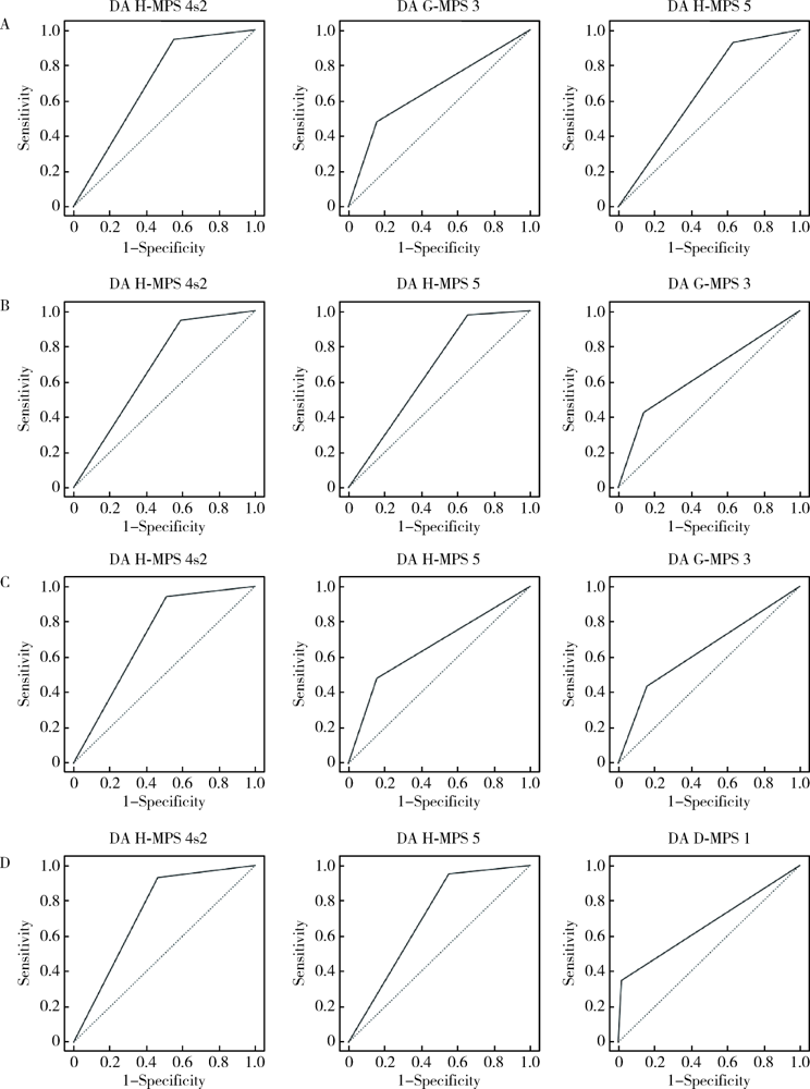
目的: 研究中国人群腭中缝(midpalatal suture,MPS)形态特点分期与Demirjian牙龄间的相关性,以探索牙龄对腭中缝骨化闭合程度的预测能力。方法: 本研究为回顾性研究,收集1 076例样本(女675例,男401例,年龄6.0~20.9岁),观察锥形束CT(cone-beam CT,CBCT)水平位腭中缝区域,记录各样本的MPS分期,同时观察并记录各样本的牙龄。采用Spearman相关系数检验与诊断试验,研究作为有序分类变量的MPS分期与牙龄间的相关关系。结果: (1)左下第二前磨牙:牙龄B~D期者绝大部分处于MPS 1~2期,占95.2%;牙龄E期者MPS 1~2期占85.3%,3期占14.7%;牙龄F期者MPS 3期、1~2期、4s1期分别占45.1%、46.1%和8.8%;牙龄G期者MPS 3期、4s1期、1~2期分别占49.8%、24.9%和18.9%;牙龄H期者大部分处于MPS 4~5期,占80.1%,另16.3%处于3期。(2)左下第二磨牙:牙龄B~D期者绝大部分处于MPS 1~2期,占89.7%;牙龄E期者MPS 1~2期占67.4%,3期占26.1%;牙龄F期者MPS 3期、1~2期、4s1期分别占55.3%、34.2%和10.5%;牙龄G期者MPS 3期、4s1期、1~2期分别占50.7%、24.3%和16.8%;牙龄H期者大部分处于MPS 4~5期,占83.8%;另14.2%处于3期。(3)以牙龄诊断MPS分期,效能较好的配对有:左下第二磨牙牙龄-MPS:H-4s2、H-5、D-1;左下第二前磨牙牙龄-MPS:H-4s2、H-5、G-3。其余配对的诊断效能一般。(4)MPS分期与左下第二磨牙牙龄的Spearman系数最高,为0.68,其次是第二前磨牙,为0.64。(5)若左下第二磨牙或第二前磨牙处于牙龄H期,则该个体很可能超过MPS 4s2期。结论: 除少数诊断效能较好的配对外,牙龄诊断MPS分期的效能总体一般。因此,以牙龄评估腭中缝骨化闭合程度时,应酌情加拍治疗前CBCT,以明确腭中缝骨化闭合状态。
中图分类号:
- R783.5

| [1] |
Bjork A. Sutural growth of the upper face studied by the implant method[J]. Acta Odontol Scand, 1966,24(2):109-127.
doi: 10.3109/00016356609026122 pmid: 5225742 |
| [2] |
Melsen B. Palatal growth studied on human autopsy material[J]. Am J Orthod, 1975,68(1):42-54.
doi: 10.1016/0002-9416(75)90158-x pmid: 1056143 |
| [3] |
Persson M, Thilander B. Palatal suture closure in man from 15 to 35 years of age[J]. Am J Orthod, 1977,72(1):42-52.
doi: 10.1016/0002-9416(77)90123-3 pmid: 267435 |
| [4] |
Haas AJ. The treatment of maxillary deficiency by opening the midpalatal suture[J]. Angle Orthod, 1965,35:200-217.
doi: 10.1043/0003-3219(1965)035<0200:TTOMDB>2.0.CO;2 pmid: 14331020 |
| [5] |
McNamara JA Jr, Baccetti T, Franchi L, et al. Rapid maxillary expansion followed by fixed appliances: a long-term evaluation of changes in arch dimensions[J]. Angle Orthod, 2003,73(4):344-353.
doi: 10.1043/0003-3219(2003)073<0344:RMEFBF>2.0.CO;2 pmid: 12940553 |
| [6] |
Baccetti T, Franchi L, Cameron CG, et al. Treatment timing for rapid maxillary expansion[J]. Angle Orthod, 2001,71(5):343-350.
doi: 10.1043/0003-3219(2001)071<0343:TTFRME>2.0.CO;2 pmid: 11605867 |
| [7] |
Rungcharassaeng K, Caruso JM, Kan JY, et al. Factors affecting buccal bone changes of maxillary posterior teeth after rapid maxillary expansion[J]. Am J Orthod Dentofacial Orthop, 2007,132(4):428.
doi: 10.1016/j.ajodo.2007.02.052 pmid: 17920493 |
| [8] |
Barber AF, Sims MR. Rapid maxillary expansion and external root resorption in man: A scanning electron microscope study[J]. Am J Orthod, 1981,79(6):630-652.
doi: 10.1016/0002-9416(81)90356-0 pmid: 7015868 |
| [9] |
Rinaldi MRL, Azeredo F, de Lima EM, et al. Cone-beam computed tomography evaluation of bone plate and root length after maxillary expansion using tooth-borne and tooth-tissue-borne banded expanders[J]. Am J Orthod Dentofacial Orthop, 2018,154(4):504-516.
doi: 10.1016/j.ajodo.2017.12.018 pmid: 30268261 |
| [10] |
Garib DG, Henriques JF, Janson G, et al. Rapid maxillary expansion: Tooth tissue-borne versus tooth-borne expanders. A computed tomography evaluation of dentoskeletal effects[J]. Angle Orthod, 2005,75(4):548-557.
doi: 10.1043/0003-3219(2005)75[548:RMETVT]2.0.CO;2 pmid: 16097223 |
| [11] | 高璐, 谷岩. 中国人群腭中缝生长发育形态特点分期与其相应生理年龄分布的初步研究[J]. 中华口腔正畸学杂志, 2020,27(2):61-66. |
| [12] |
Demirjian A, Goldstein H, Tanner JM. A new system of dental age assessment[J]. Hum Biol, 1973,45(2):211-227.
pmid: 4714564 |
| [13] |
Demirjian A, Goldstein H. New systems for dental maturity based on seven and four teeth[J]. Ann Hum Biol, 1976,3(5):411-421.
doi: 10.1080/03014467600001671 pmid: 984727 |
| [14] |
Macha M, Lamba B, Avula JSS, et al. Estimation of correlation between chronological age, skeletal age and dental age in children: a cross-sectional study [J]. J Clin Diagn Res, 2017, 11 (9): ZC01-ZC04.
doi: 10.7860/JCDR/2017/25175.10537 pmid: 29207822 |
| [15] |
李萌, 李果, 王虎. Demirjian牙龄推断法及其应用与更新[J]. 国际口腔医学杂志, 2014,41(6):725-729.
doi: 10.7518/gjkq.2014.06.026 |
| [16] |
Perinetti G, Contardo L, Gabrieli P, et al. Diagnostic performance of dental maturity for identification of skeletal maturation phase[J]. Eur J Orthod, 2011,34(4):487-492.
doi: 10.1093/ejo/cjr027 pmid: 21345927 |
| [17] | Proffit WR. Contemporary orthodontics[M]. 6th ed. Philadelphia: Elsevier, 2018. |
| [18] |
Ró$\dot{z}$yło-Kalinowska I, Kolasa-Rᶏczka A, Kalinowski P. Relationship between dental age according to Demirjian and cervical vertebrae maturity in Polish children[J]. Eur J Orthod, 2010,33(1):75-83.
doi: 10.1093/ejo/cjq031 pmid: 20558591 |
| [19] |
Ladewig VM, Capelozza-Filho L, Almeida-Pedrin RR, et al. Tomographic evaluation of the maturation stage of the midpalatal suture in post-adolescents[J]. Am J Orthod Dentofacial Orthop, 2018,153(6):818-824.
doi: 10.1016/j.ajodo.2017.09.019 pmid: 29853239 |
| [20] |
Hajian-Tilaki K. Receiver operating characteristic (ROC) curve analysis for medical diagnostic test evaluation[J]. Caspian J Intern Med, 2013,4(2):627-635.
pmid: 24009950 |
| [21] |
Korbmacher H, Schilling A, Puschel K, et al. Age-dependent three-dimensional micro-computed tomography analysis of the human midpalatal suture[J]. J Orofac Orthop, 2007,68(5):364-376.
doi: 10.1007/s00056-007-0729-7 |
| [22] |
Knaup B, Yildizhan F, Wehrbein H. Age-related changes in the midpalatal suture[J]. J Orofac Orthop, 2004,65(6):467-474.
doi: 10.1007/s00056-004-0415-y |
| [23] | 黄悦勤. 临床流行病学[M]. 4版. 北京: 人民卫生出版社, 2014. |
| [24] | 葛立宏. 儿童口腔医学[M]. 2版. 北京: 北京大学医学出版社, 2013. |
| [1] | 杨刚,胡文杰,曹洁,柳登高. 牙周健康的上颌前牙唇侧嵴顶上牙龈的三维形态分析[J]. 北京大学学报(医学版), 2021, 53(5): 990-994. |
| [2] | 贾鹏程,杨刚,胡文杰,赵一姣,刘木清. 根尖片评估单根牙骨内牙根表面积的准确性[J]. 北京大学学报(医学版), 2018, 50(1): 91-97. |
| [3] | 马静,江久汇. 骨性Ⅱ类和Ⅲ类高角错牙合患者下切牙区的牙槽骨形态分析[J]. 北京大学学报(医学版), 2018, 50(1): 98-103. |
| [4] | 徐筱,徐莉,江久汇,吴佳琪,李小彤,靖无迪. 锥形束CT评判安氏Ⅲ类错牙合上前牙骨开裂与骨开窗的准确性分析[J]. 北京大学学报(医学版), 2018, 50(1): 104-109. |
| [5] | 曹婕1,孟焕新. 锥形束CT用于评估牙槽骨骨缺损的情况和骨再生区域骨密度的变化[J]. 北京大学学报(医学版), 2018, 50(1): 110-116. |
| [6] | 常大桐,周彦恒,刘伟涛. 上颌反复快速扩缩对上气道影响的锥束CT研究[J]. 北京大学学报(医学版), 2017, 49(4): 685-690. |
| [7] | 陈全,张晓1张智勇,高巍,刘文曙,孟甜,陈宇寰,王慧丽. 上颌窦前外侧壁骨内血管孔道位置锥形束CT影像判断分析及其临床应对措施[J]. 北京大学学报(医学版), 2017, 49(3): 540-546. |
| [8] | 赵一姣,王斯维,刘怡,王勇. 基于影像学牙周膜解剖特征快速提取活体牙三维牙根形态的方法[J]. 北京大学学报(医学版), 2017, 49(1): 54-059. |
| [9] | 苏征,白雨豪,侯晓玫. 不同技术对弯曲根管根尖气锁去除效果的锥形束CT研究[J]. 北京大学学报(医学版), 2017, 49(1): 76-080. |
| [10] | 温馥嘉,陈贵,刘怡. 基于锥形束CT的强支抗内收上前牙病例牙根及牙槽骨的形态学分析[J]. 北京大学学报(医学版), 2016, 48(4): 702-708. |
| [11] | 张茗茗,梁宇红,高学军. 根尖X线片和锥形束CT评价根尖周骨病变的比较研究[J]. 北京大学学报(医学版), 2016, 48(3): 539-543. |
| [12] | 王哲,朱榴宁,周琳,伊彪. 锥形束CT融合三维面像评估正颌术后软硬组织的变化[J]. 北京大学学报(医学版), 2016, 48(3): 544-549. |
| [13] | 王斯维,黎敏,杨慧芳,赵一姣,王勇,刘怡. 3种生成大视野锥形束CT数据正中矢状面方法的比较[J]. 北京大学学报(医学版), 2016, 48(2): 330-335. |
| [14] | 黎敏,王斯维,赵一姣,刘怡. 安氏Ⅱ类2分类错牙合上前牙冠根形态的锥形束CT分析[J]. 北京大学学报(医学版), 2016, 48(1): 105-110. |
| [15] | 王莺, 林野, 陈波, 张宇, 邸萍. 即刻种植术后牙槽突骨板改建及美学效果评价[J]. 北京大学学报(医学版), 2016, 48(1): 121-125. |
|
