北京大学学报(医学版) ›› 2022, Vol. 54 ›› Issue (5): 991-999. doi: 10.19723/j.issn.1671-167X.2022.05.028

• 论著 • 上一篇    下一篇

三维超声血管斑块定量分析技术评估颈动脉粥样硬化斑块

邢海英1,2,陈玉辉3,许珂1,2,黄点点1,2,彭清1,2,刘冉1,2,孙葳1,2,黄一宁1,2,*()   

  1. 1. 北京大学第一医院神经内科, 北京 100034
    2. 神经系统小血管病探索北京市重点实验室, 北京 100034
    3. 北京医院神经内科, 北京 100730
  • 收稿日期:2022-06-30 出版日期:2022-10-18 发布日期:2022-10-14
  • 通讯作者: 黄一宁 E-mail:ynhuang@bjmu.edu.cn
  • 作者简介:黄一宁,北京大学第一医院神经内科主任。1987年毕业于中国协和医科大学获硕士学位;1984—2002年在北京协和医院神经科工作,2003年调任现职。长期不间断地在临床工作,经验丰富,并且精通于神经系统疾病常用的检测手段,如神经电生理、神经超声、神经影像学等专业技术,擅长于神经系统急症重症疾病和疑难疾病的诊治,特别是在脑血管病诊断和治疗上有很深的造诣。
    2001年以来,牵头开展了多个国家重大科研项目,如国家十五攻关项目、国家十五攻关专项、卫生部临床重大专项、国家十一五重大专项,以及国家自然科学基金、首都发展基金、中德合作中心项目等。在国际顶级专业杂志(如the Lancet NeurologyNeurologyStroke等)发表多篇文章。现任中华神经科分会常委、脑血管病学组副组长、亚洲急性卒中专家组(ASAP)成员。精通英文,熟悉法文,与国际临床神经病学界有广泛联系。曾被德国乌尔姆大学、法兰克福歌德大学、哥廷根大学、萨拉大学,以及中国的香港中文大学等聘为客座教授、访问教授或访问学者

Evaluation of carotid atherosclerotic plaques by vascular plaque quantification (VPQ) technology of three-dimensional ultrasonography

Hai-ying XING1,2,Yu-hui CHEN3,Ke XU1,2,Dian-dian HUANG1,2,Qing PENG1,2,Ran LIU1,2,Wei SUN1,2,Yi-ning HUANG1,2,*()   

  1. 1. Department of Neurology, Peking University First Hospital, Beijing 100034, China
    2. Department of Neurology, Beijing Key Laboratory of Neurovascular Disease Discovery, Beijing, China
    3. Department of Neurology, Beijing Hospital, Beijing 100730, China
  • Received:2022-06-30 Online:2022-10-18 Published:2022-10-14
  • Contact: Yi-ning HUANG E-mail:ynhuang@bjmu.edu.cn

摘要:

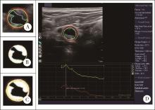
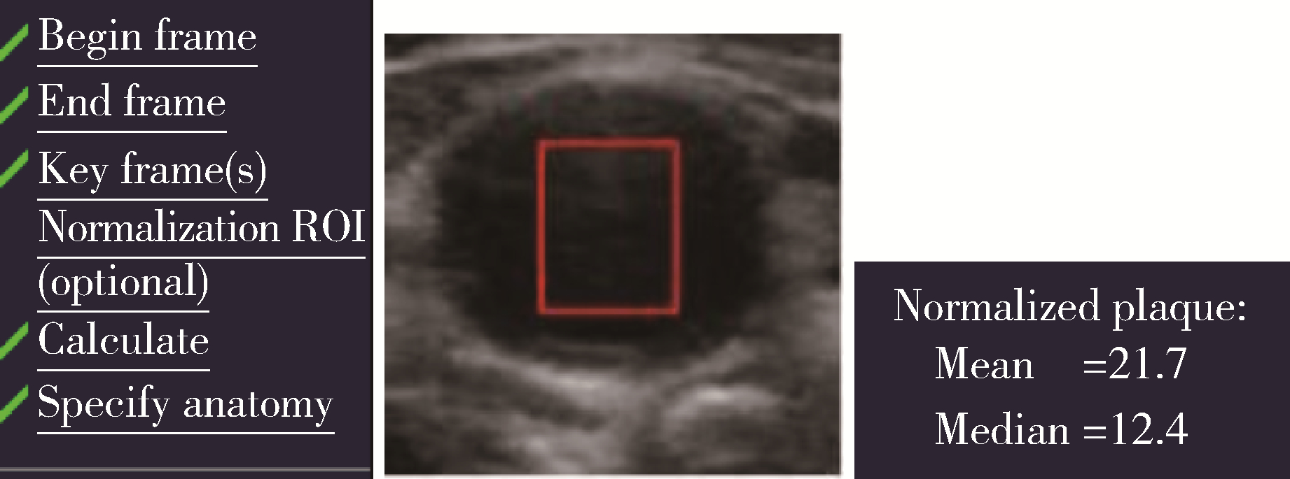
目的: 分析应用三维超声血管斑块定量分析技术(vascular plaque quantification, VPQ)评估颈动脉粥样硬化斑块的可行性并观察他汀类药物对颈动脉粥样硬化斑块的影响。方法: 选择2016年1月至2018年9月于北京大学第一医院神经内科治疗的颈动脉粥样硬化患者, 采用三维超声检查患者的双侧颈动脉, 对存在颈动脉粥样硬化斑块的患者采用VPQ测量颈动脉斑块的灰阶中位数(gray scale median, GSM)及其他血管参数。根据斑块的GSM, 分为低GSM组(GSM < 40)和高GSM组(GSM≥40), 比较两组患者的临床特征和斑块特征, 分析斑块的稳定性。根据患者是否服用他汀类药物, 将患者分为他汀组和非他汀组, 比较两组患者在3个月和2年时颈动脉斑块GSM及其他血管参数的变化。结果: 共入组120例患者, 其中男性79例, 女性41例, 平均年龄(65.39±9.11)岁。低GSM组31例, 高GSM组89例。低GSM组管腔的狭窄程度更重(面积狭窄率41.32%±21.37% vs. 29.79%±17.16%, P < 0.05)。低GSM组的标准化管壁指数(normalized wall index, NWI)显著大于高GSM组(0.61±0.14 vs.0.52±0.12, P < 0.01), 观察了77例患者颈动脉粥样硬化斑块的变化, 其中男51例, 女26例, 年龄(64.96±9.58)岁, 根据是否服用他汀类药物分为他汀组(n=56)和非他汀组(n=21)。与基线相比, 3个月随访时两组患者的颈动脉斑块体积、面积、管腔狭窄度、GSM变化差异均无统计学意义(P >0.05); 2年随访时, 他汀组的GSM增加[中位数10.00(2.00, 28.00)], 而非他汀组的GSM减低[中位数-7.00 (-11.00, 5.50)], 组间差异有统计学意义(P < 0.01)。他汀组的颈动脉斑块体积无明显增加, 而非他汀组的斑块体积略增加, 但两组的变化值差异无统计学意义[他汀组斑块体积增加[中位数0.00 (-30.00, 40.00) mm3, 非他汀组为30.00 (10.00, 70.00) mm3, P>0.05]。结论: 三维超声(three dimensional ultrasound, 3DUS)的VPQ可测量颈动脉斑块的GSM, 低GSM(GSM < 40)组患者的血管狭窄程度更重, 标准化管壁指数更高。VPQ观察到颈动脉粥样硬化斑块的患者服用中强度他汀类药物治疗2年后斑块的灰阶中位数增加, 提示3DUS的VPQ技术可用来观察他汀类药物治疗颈动脉动脉粥样硬化斑块的效果。

关键词: 三维超声, 血管斑块定量分析, 颈动脉斑块, 灰阶中位数, 他汀

Abstract:

Objective: To analyze the feasibility of using vascular plaque quantification (VPQ) to evaluate carotid atherosclerotic plaques and to observe the effect of statins on carotid atherosclerotic plaques. Methods: Patients with carotid plaques from January 2016 to September 2018 in Peking University First Hospital Neurology Department were recruited and underwent three-dimonsional ultrasound (3DUS). Their gray scale median (GSM) and other parameters of carotid plaques were measured with VPQ. The patients were divided into low GSM group (GSM < 40) and high GSM group (GSM≥40). The clinical characteristics and plaque characteristics of the patients in the two groups were compared to analyze the stability of plaques. According to whether taking statins or not, the patients were further divided into statin group and non-statin group, plaque GSM and other parameters of their carotid plaques were measured and the changes of carotid plaques at the end of 3 months and 2 years were observed. Results: A total of 120 patients were enrolled, including 79 males and 41 females, with an average age of (65.39±9.11) years. The patients were divided into low GSM group (31 cases) (GSM < 40) and high GSM group (89 cases) (GSM≥40). The stenosis of the lumen in the low GSM group was more severe (the area stenosis rate was 41.32%±21.37% vs. 29.79%±17.16%, P < 0.05). The nor-malized wall index (NWI) of plaque in low GSM group was significantly higher than that in high GSM group (0.61 ±0.14 vs. 0.52±0.12, P < 0.01). A total of 77 patients, including 51 males and 26 females, aged (64.96±9.58) years, were enrolled to observe the statin effects on carotid plaque. They were divided into statin group (n=56) and non-statin group (n=21) according to whether taking statins or not. At the baseline and 3-month follow-up, there were no significant differences in carotid plaque volume, area, degree of luminal stenosis and GSM between the two groups (P>0.05). At the end of the 2-year follow-up, GSM increased in the statin group [median 10.00 (2.00, 28.00)] but decreased in the non-statin group [median -7.00 (-11.00, 5.50)], with a statistically significant difference between the two groups (P < 0.01). There was no significant increase in carotid plaque volume in the statin group, while there was a slight increase in the non-statin group, but there was no significant difference between the two groups [median increase in plaque volume was 0.00 (-30.00, 40.00) mm3 in the statin group and 30.00 (10.00, 70.00) mm3 in the non-statin group, P>0.05]. Conclusion: The VPQ technology of 3DUS can be used to evaluate carotid atherosclerotic plaques. Patients with low GSM (GSM < 40) have more severe vascular stenosis and higher normalized wall index. VPQ technology can also be used to observe the effect of statins on carotid plaque, the GSM of plaques increase in patients who are taking moderate-intensity statin treatment for two years.

Key words: Three-dimensional ultrasonography, Vascular plaque quantification, Carotid plaque, Gray scale median, Statin

中图分类号: 

  • R543.4

图1

VPQ模式下确定起始帧、关键帧和结束帧"

图2

VPQ测量的斑块各参数"

图3

VPQ标准化GSM的方法"

表1

低GSM组和高GSM组患者基线临床资料比较"

Items Total(n=120) Low GSM group(n=31) High GSM group(n=89) P
Age/years, $\bar x \pm s$ 65.39±9.11 66.1±9.29 65.15±9.09 0.62
Male, n (%) 79 (65.83) 18 (58.06) 61 (68.53) 0.44
Smoking,n (%) 50 (42.37) 10 (32.25) 40 (44.94) 0.25
Hypertension, n (%) 80 (67.80) 18 (58.06) 62 (69.66) 0.29
Diabetes mellitus, n (%) 42 (35.59) 13 (41.94) 29 (32.58) 0.31
ASCVD, n (%) 68 (61.82) 15 (48.38) 53 (62.92) 0.44
Stroke, n (%) 61 (51.70) 14 (45.16) 47 (52.81) 0.52
BMI/(kg/m2), $\bar x \pm s$ 24.64 ±3.31 24.76 ±3.05 24.59 ±3.41 0.80
SBP /mmHg, $\bar x \pm s$ 137.26 ±17.03 139.72 ±16.66 136.45 ±17.16 0.37
DBP /mmHg, $\bar x \pm s$ 74.06 ±12.74 72.45 ±13.47 74.59 ±12.52 0.45
Glucose/(mmol/L), $\bar x \pm s$ 6.13 ±1.73 6.00 ±1.20 6.16 ±1.86 0.61
TG/(mmol/L), $\bar x \pm s$ 1.38 ±0.73 1.41±0.69 1.37 ±0.75 0.79
LDL-C/(mmol/L), $\bar x \pm s$ 1.98 ±0.86 1.86 ±1.05 2.02±0.80 0.45
Antiplatelet, n (%) 99 (83.90) 25 (80.64) 74 (83.14) 0.92
Lipid lowering, n (%) 87 (72.50) 21 (67.74) 66 (74.16) 0.49

表2

低GSM组和高GSM组患者颈动脉斑块特征比较"

Items Total(n=120) Low GSM group(n=31) High GSM group(n=89) P
Plaque volume /mm3, M (P25, P75) 170.0
(100.0, 267.5)
180.0
(120.0, 270.0)
160.0
(95.0, 260.0)
0.29
Plaque area/mm2, $\bar x \pm s$ 15.97 ±11.37 18.02 ±13.13 15.26 ±10.69 0.30
Stenosis ratio/%, $\bar x \pm s$ 32.77 ±18.93 41.32 ±21.37 29.79 ±17.16 0.01
NWI, $\bar x \pm s$ 0.54 ±0.13 0.61 ±0.14 0.52 ±0.12 < 0.01

表3

两组患者基线期和3个月时的颈动脉斑块体积、面积及其变化"

Items Total(n=77) Statin group(n=56) Non-statin group (n=21) P*
Plaque volume /mm3, M (P25, P75)
  Baseline 170.00
(100.00, 230.00)
170.00
(107.50, 235.00)
140.00
(80.00, 245.00)
0.28
  3-month 160.00
(100.00, 260.00)
160.00
(100.00, 230.00)
160.00
(85.00, 275.00)
0.76
  Change 0.00
(-20.00, 20.00)
0.00
(-20.00, 12.50)
0.00
(-25.00, 40.00)
0.47
   P# 0.65 0.46 0.38
Plaque area/mm2
  Baseline, $\bar x \pm s$ 15.12±10.39 16.20±10.85 12.21±8.61 0.13
  3-month, $\bar x \pm s$ 14.35±10.57 14.44±9.93 14.10±12.37 0.90
  Change, M (P25, P75) -0.31 (-3.02, 2.89) -0.83 (-3.35, 1.81) 0.48 (-1.47, 4.10) 0.07
   P# 0.33 0.09 0.32
Stenosis ratio/%
  Baseline, $\bar x \pm s$ 31.69±18.75 33.63±18.52 26.52±18.81 0.14
  3-month, $\bar x \pm s$ 31.30±17.82 31.66±16.52 30.33±21.32 0.77
  Change, M (P25, P75) 0 (-3.50, 5.50) 0.00 (-7.00, 5.00) 2.00 (-1.00, 10.00) 0.10
   P# 0.79 0.52 0.09
NWI
  Baseline, $\bar x \pm s$ 0.54±0.14 0.55±0.14 0.51±0.13 0.25
  3-month, $\bar x \pm s$ 0.54±0.13 0.54±0.13 0.52±0.15 0.61
  Change, M (P25, P75) 0.00 (-0.04, 0.03) 0.00 (-0.05, 0.03) 0.00 (-0.03, 0.04) 0.53
   P# 0.68 0.93 0.31

表4

两组患者基线期和3个月时的颈动脉斑块GSM变化"

Items Total(n=77) Statin group(n=56) Non-statin group(n=21) P*
Baseline, $\bar x \pm s$ 54.20±19.16 55.57±21.03 49.07±7.91 0.06
3-month, $\bar x \pm s$ 55.63±18.38 57.88±18.98 47.20±13.35 0.02
Change, M (P25, P75) 0.00
(-8.00, 11.00)
0.50
(-7.00, 13.00)
-3.00
(-9.00, 6.00)
0.23
P# 0.51 0.33 0.52

表5

两组患者基线期,2年时的颈动脉斑块体积、面积及其变化"

Items Total(n=62) Statin group(n=47) Non-statin group(n=15) P*
Plaque volume /mm3, M (P25, P75)
  Baseline 180.00
(97.50, 312.50)
200.00
(90.00, 320.00)
140.00
(100.00, 280.00)
0.74
  2 years 185.00 (117.50, 302.50) 200.00
(120.00, 300.00)
160.00
(110.00, 360.00)
0.86
  Change 15.00
(-30.00, 50.00)
0.00
(-30.00, 40.00)
30.00
(10.00, 70.00)
0.09
   P# 0.11 0.56 0.02
Plaque area /mm2
  Baseline, $\bar x \pm s$ 15.97±12.63 15.41±10.19 17.72±8.69 0.65
  2 years, $\bar x \pm s$ 13.68±8.82 12.88±8.35 16.19±10.05 0.26
  Change, M (P25, P75) -0.44 (-7.10, 2.58) -0.81 (-8.47, 2.13) 1.46 (-6.17, 5.71) 0.14
   P# 0.13 0.07 0.89
Stenosis ratio/%
  Baseline, $\bar x \pm s$ 31.76±19.80 31.49±18.96 32.60±22.92 0.10
  2 years, $\bar x \pm s$ 31.40±17.25 30.79±17.07 33.33±18.29 0.64
  Change, M (P25, P75) -1.00 (-5.25, 8.00) -1.00 (-4.00, 8.00) -1.00 (-9.00, 9.00) 0.93
   P# 0.95 0.88 0.95
NWI
  Baseline, $\bar x \pm s$ 0.53±0.14 0.53±0.14 0.54±0.16 0.94
  2 years, $\bar x \pm s$ 0.54±0.12 0.53±0.12 0.54±0.13 0.94
  Change, M (P25, P75) -0.01 (-0.05, 0.06) -0.01
(-0.04, 0.06)
-0.02 (-0.06, 0.07) 0.96
   P# 0.87 0.87 0.93

表6

两组患者在基线期和2年时颈动脉斑块GSM变化"

Items Total(n=62) Statin group(n=47) Non-statin group (n=15) P*
Baseline, $\bar x \pm s$ 52.14±17.00 52.72±16.65 50.53±18.36 0.67
2 years, $\bar x \pm s$ 62.55±21.39 67.87±20.62 47.82±16.28 < 0.01
Change, M (P25, P75) 7.00
(-6.00, 21.75)
10.00
(2.00, 28.00)
-7.00
(-11.00, 5.50)
< 0.01
P# < 0.01 < 0.01 0.37
1 Lal BK , Hobson RW , Pappas PJ , et al. Pixel distribution analysis of B-mode ultrasound scan images predicts histologic features of atherosclerotic carotid plaques[J]. J Vasc Surg, 2002, 35 (6): 1210- 1217.
doi: 10.1067/mva.2002.122888
2 Herr JE , Hetu MF , Li TY , et al. Presence of calcium-like tissue composition in carotid plaque is indicative of significant coronary artery disease in high-risk patients[J]. J Am Soc Echocardiogr, 2019, 32 (5): 633- 642.
doi: 10.1016/j.echo.2019.01.001
3 Mujaj B , Bos D , Selwaness M , et al. Statin use is associated with carotid plaque composition: The Rotterdam study[J]. Int J Cardiol, 2018, 260, 213- 218.
doi: 10.1016/j.ijcard.2018.02.111
4 Vergallo R , Crea F . Atherosclerotic plaque healing[J]. N Engl J Med, 2020, 383 (9): 846- 857.
doi: 10.1056/NEJMra2000317
5 Touboul PJ , Hennerici MG , Meairs S , et al. Mannheim carotid intima-media thickness and plaque consensus (2004-2006-2011). An update on behalf of the advisory board of the 3rd, 4th and 5th watching the risk symposia, at the 13th, 15th and 20th European Stroke Conferences, Mannheim, Germany, 2004, Brussels, Belgium, 2006, and Hamburg, Germany, 2011[J]. Cerebrovasc Dis, 2012, 34 (4): 290- 296.
doi: 10.1159/000343145
6 中国成人血脂异常防治指南修订联合委员会. 中国成人血脂异常防治指南(2016年修订版)[J]. 中华心血管病杂志, 2016, 44 (10): 833- 853.
doi: 10.3760/cma.j.issn.0253-3758.2016.10.005
7 Geroulakos G , Ramaswami G , Nicolaides A , et al. Characterization of symptomatic and asymptomatic carotid plaques using high-resolution real-time ultrasonography[J]. Br J Surg, 1993, 80 (10): 1274- 1277.
8 Johri AM , Herr JE , Li TY , et al. Novel ultrasound methods to investigate carotid artery plaque vulnerability[J]. J Am Soc Echocardiogr, 2017, 30 (2): 139- 148.
doi: 10.1016/j.echo.2016.11.003
9 el-Barghouty N , Geroulakos G , Nicolaides A , et al. Computer-assisted carotid plaque characterisation[J]. Eur J Vasc Endovasc Surg, 1995, 9 (4): 389- 393.
doi: 10.1016/S1078-5884(05)80005-X
10 Spanos K , Tzorbatzoglou I , Lazari P , et al. Carotid artery plaque echomorphology and its association with histopathologic characteristics[J]. J Vasc Surg, 2018, 68 (6): 1772- 1780.
doi: 10.1016/j.jvs.2018.01.068
11 Mathiesen EB , Bonaa KH , Joakimsen O . Echolucent plaques are associated with high risk of ischemic cerebrovascular events in carotid stenosis: The tromso study[J]. Circulation, 2001, 103 (17): 2171- 2175.
doi: 10.1161/01.CIR.103.17.2171
12 Hashimoto H , Tagaya M , Niki H , et al. Computer-assisted analysis of heterogeneity on B-mode imaging predicts instability of asymptomatic carotid plaque[J]. Cerebrovasc Dis, 2009, 28 (4): 357- 364.
doi: 10.1159/000229554
13 Huibers A , de Borst GJ , Bulbulia R , et al. Plaque echolucency and the risk of ischaemic stroke in patients with asymptomatic carotid stenosis within the first asymptomatic carotid surgery trial (ACST-1)[J]. Eur J Vasc Endovasc Surg, 2016, 51 (5): 616- 621.
doi: 10.1016/j.ejvs.2015.11.013
14 Zhang M , Zhao Q , Gao Y , et al. Prediction for rupture risk of carotid artery plaques: A comparative study of 3D-GSM and CAS system[J]. Eur J Radiol, 2016, 85 (9): 1659- 1665.
doi: 10.1016/j.ejrad.2016.05.013
15 王秀玲, 张敏郁, 丁桂春, 等. 三维超声灰阶中位数定量评价颈动脉斑块的易损性[J]. 中华医学超声杂志: 电子版, 2017, 14 (1): 23- 28.
doi: 10.3877/cma.j.issn.1672-6448.2017.01.008
16 Urbak L , Sandholt BV , Graebe M , et al. Patients with unstable atherosclerosis have more echolucent carotid plaques compared with stable atherosclerotic patients: A 3-D ultrasound study[J]. Ultrasound Med Biol, 2020, 46 (9): 2164- 2172.
doi: 10.1016/j.ultrasmedbio.2020.04.002
17 Nicolaides AN , Kakkos SK , Kyriacou E , et al. Asymptomatic internal carotid artery stenosis and cerebrovascular risk stratification[J]. J Vasc Surg, 2010, 52 (6): 1486- 1496.
doi: 10.1016/j.jvs.2010.07.021
18 Ruiz-Ares G , Fuentes B , Martinez-Sanchez P , et al. A prediction model for unstable carotid atheromatous plaque in acute ischemic stroke patients: Proposal and internal validation[J]. Ultrasound Med Biol, 2014, 40 (9): 1958- 1965.
doi: 10.1016/j.ultrasmedbio.2014.04.015
19 Biasi GM , Froio A , Diethrich EB , et al. Carotid plaque echolucency increases the risk of stroke in carotid stenting: The imaging in carotid angioplasty and risk of stroke (ICAROS) study[J]. Circulation, 2004, 110 (6): 756- 762.
doi: 10.1161/01.CIR.0000138103.91187.E3
20 Ariyoshi K , Okuya S , Kunitsugu I , et al. Ultrasound analysis of gray-scale median value of carotid plaques is a useful reference index for cerebro-cardiovascular events in patients with type 2 diabetes[J]. J Diabetes Investig, 2015, 6 (1): 91- 97.
doi: 10.1111/jdi.12242
21 国家卫生健康委员会脑卒中防治专家委员会血管超声专业委员会, 中国超声医学工程学会浅表器官及外周血管超声专业委员会, 中国超声医学工程学会颅脑及颈部血管超声专业委员会, 中国超声医学工程学会颅脑及颈部血管超声专业委员会. 头颈部血管超声若干问题的专家共识(颈动脉部分)[J]. 中国脑血管病杂志, 2020, 17 (6): 346- 352.
doi: 10.3969/j.issn.1672-5921.2020.06.013
22 Kadoglou NP , Gerasimidis T , Moumtzouoglou A , et al. Intensive lipid-lowering therapy ameliorates novel calcification markers and GSM score in patients with carotid stenosis[J]. Eur J Vasc Endovasc Surg, 2008, 35 (6): 661- 668.
doi: 10.1016/j.ejvs.2007.12.011
23 Yamaguchi M , Sasaki M , Ohba H , et al. Quantitative assessment of changes in carotid plaques during cilostazol administration using three-dimensional ultrasonography and non-gated magnetic resonance plaque imaging[J]. Neuroradiology, 2012, 54 (9): 939- 945.
doi: 10.1007/s00234-012-1011-2
[1] 巩超,刘秋萍,王佳敏,刘晓非,张明露,杨瀚,沈鹏,林鸿波,唐迅,高培. 社区人群他汀干预策略预防心血管病效果的马尔可夫模型评价[J]. 北京大学学报(医学版), 2022, 54(3): 443-449.
[2] 刘欢,王宏宇,刘金波,赵红薇,周迎燕,李丽红. 颈动脉斑块患者大动脉功能与冠心病和下肢动脉疾病的关系[J]. 北京大学学报(医学版), 2017, 49(6): 1027-1033.
[3] 张静,任景怡,陈红,韩冠平. 他汀类药物可降低不稳定性心绞痛患者血浆中炎症相关microRNAs的表达水平[J]. 北京大学学报(医学版), 2015, 47(5): 761-768.
[4] 欧蒙恩, 张晓, 刘云松, 葛严军, 周永胜. 联合应用基质细胞衍生因子1、辛伐他汀及骨胶原支架进行体内异位成骨[J]. 北京大学学报(医学版), 2015, 47(1): 47-51.
[5] 王艳飞, 贾新未, 赵文萍, 王凤娟, 刘亚宁, 张芳, 张丽敏, 王鸿超. 辛伐他汀后适应对缺血再灌注损伤大鼠TNF-α及NF-κB的影响[J]. 北京大学学报(医学版), 2014, 46(6): 990-992.
[6] 徐璐, 王超, 沈文文, 祁荣. 辛伐他汀纳米脂质体对骨髓间充质干细胞成骨分化的影响[J]. 北京大学学报(医学版), 2014, 46(6): 883-888.
[7] 关静琳, 姜雯 , 王怀宇, 曾辉, 张素敏, 王广, 毛节明. 辛伐他汀对高胆固醇血症患者血管内皮的多重影响[J]. 北京大学学报(医学版), 2014, 46(5): 703-706.
[8] 夏经钢, 曲杨, 胡少东, 许骥, 尹春琳, 徐东. 强化调脂对行非心脏手术的冠状动脉粥样硬化性心脏病患者的心肌保护作用及机制初探[J]. 北京大学学报(医学版), 2014, 46(4): 601-605.
[9] 翟国君, 姚伟娟, 贾子昌, 韩金涛, 赵军, 李选, 董国祥. 静脉人工血管桥内膜增生的形态学特征及洛伐他汀的干预作用[J]. 北京大学学报(医学版), 2012, 44(5): 783-787.
[10] 杨琳, 王莹, 吕天敬, 周利群, 金杰. 临床有效剂量的洛伐他汀对前列腺癌PC3细胞的影响[J]. 北京大学学报(医学版), 2010, 42(4): 391-395.
[11] 宋纯理, 党耕町, 贾弘抖, 郭昭庆, 马庆军. 辛伐他汀促进骨髓基质细胞的成骨分化[J]. 北京大学学报(医学版), 2003, 35(5): 533-536.
[12] 张新超, 张彤, 徐成斌, 金晓峰. 辛伐他汀和西立伐他汀对体外培养血管平滑肌细胞增殖及迁移的影响[J]. 北京大学学报(医学版), 2001, 33(4): 344-346.
[13] 任汉云, 张念先, 徐红, 陆道培. 辛伐他汀选择性抑制慢性髓性白血病细胞增殖并诱导凋亡[J]. 北京大学学报(医学版), 2001, 33(2): 136-139.
Viewed
Full text


Abstract

Cited

  Shared   
  Discussed   
[1] 王军, 肖水芳, 秦永, 王全桂, 陈丽. 以面神经麻痹为首诊表现的Wegener肉芽肿病一例[J]. 北京大学学报(医学版), 2007, 39(4): 434 -436 .
[2] 柳晓辉, 那加, 刘玲玲, 罗斌. 头颈部血管肉瘤3例[J]. 北京大学学报(医学版), 2001, 33(3): 288 -289 .
[3] 张震康. 口腔医学科学研究的重要进展和方向[J]. 北京大学学报(医学版), 2002, 34(2): 97 -98 .
[4] 梁成, 王兴, 伊彪, 李自力, 王晓霞. 骨性颞下颌关节强直伴小颌畸形及阻塞性睡眠呼吸暂停综合征的牵引成骨治疗[J]. 北京大学学报(医学版), 2002, 34(2): 112 -116 .
[5] 张勇, 栾庆先. 牙周维护治疗在保持牙周长期疗效中的作用[J]. 北京大学学报(医学版), 2011, 43(1): 29 -33 .
[6] 夏永华, 刘冬, 张彩凤, 付丹丹, 李敏, 李占国, 田中伟. NF-κB信号通路的阻断对皮肤鳞癌SCL-1细胞凋亡的影响[J]. 北京大学学报(医学版), 2011, 43(2): 179 -182 .
[7] 王学庆, 万有, 于英心, 韩济生. 关节炎大鼠背根神经节细胞的膜电生理学特征[J]. 北京大学学报(医学版), 2001, 33(1): 50 -53 .
[8] 唐志慧, 曾祥龙. 恒牙早期正常骨面型青少年上气道形态和舌骨位置的X线头影测量研究[J]. 北京大学学报(医学版), 2002, 34(2): 140 -143 .
[9] 关志忱, 魏本林, 孟作为. 远程无线排尿日记开发及20例年轻人客观排尿情况报告[J]. 北京大学学报(医学版), 2010, 42(4): 476 -479 .
[10] 张春丽, 王荣福, 李太华, 付占立. 新型有机锗倍半氧化物的抗肿瘤活性及其在荷瘤裸鼠的体内分布[J]. 北京大学学报(医学版), 2008, 40(2): 208 -210 .