Journal of Peking University(Health Sciences) ›› 2019, Vol. 51 ›› Issue (3): 571-578. doi: 10.19723/j.issn.1671-167X.2019.03.029

Previous Articles     Next Articles

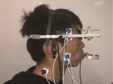
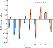
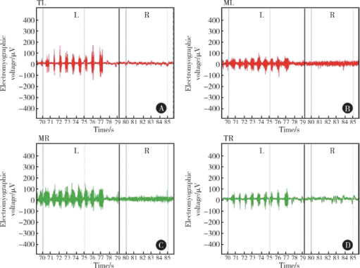
Application of digital mandibular movement record and masticatory muscle electromyography in the evaluation of stomatognathic function in patients with mandibular tumor

Jing WANG,Jun-peng CHEN,Yang WANG,Xiang-liang XU,Chuan-bin GUO()   

  1. Department of Oral and Maxillofacial Surgery, Peking University School and Hospital of Stomatology & National Clinical Research Center for Oral Diseases & National Engineering Laboratory for Digital and Material Technology of Stomatology & Beijing Key Laboratory of Digital Stomatology, Beijing 100081, China
  • Received:2019-03-18 Online:2019-05-10 Published:2019-06-26
  • Supported by:
    Supported by the Fundamental Research Funds for the Central Universities: Peking University Clinical Scientist Program (BMU2019LCKCJ009) and Youth Fund of Peking University School and Hospital of Stomatology (PKUSS20180202)

Abstract: Objective: To study the clinical characteristics of mandibular movement and masticatory muscle function in preoperative and postoperative patients with unilateral mandibular tumors in the region of mandibular body and ramus by combining digital mandibular movement records with electromyography, and to preliminarily explore the relationship and mechanism between movement and masticatory muscle function.Methods: Six preoperative patients with tumor in unilateral body and ramus of mandible were included, and three postoperative patients with unilateral segmental resection and reconstruction of mandibular bone were included. The mandibular movement recording system and surface electromyography system were used to collect the movement trajectory of the patients’ mandibular marginal movement and chewing movement, and the surface electromyography of bilateral masseter and temporalis was recorded concurrently. The surface electromyography of bilateral masseter and temporalis was collected when the patients were at relaxation and at maximal voluntary clenching (MVC). The motion trajectory was observed on the digital virtual model, and the motion amplitude and direction of mandibular marginal movements were analyzed. The characteristics of masticatory electromyogram (EMG) activity in affected and unaffected sides at relaxation, MVC and bilateral mastication were analyzed, and the asymmetry indexes and activity indexes were calculated. Results: The preoperative mean maximum opening of the patients was (35.20±6.87) mm. Three patients had mild mouth opening limitation, and all the patients’ mouth opening trajectory was skewed to the affected side. During lateral movements, the mean range of motion of the affected side [(10.34±1.27) mm] and that of the healthy side [(6.94±2.41) mm] were significantly different. The maximum opening of the postoperative patients was (30.65±17.32) mm, and the mandibular marginal movement characteristics were consistent with those of the patients before surgery. During MVC in the preoperative patients, the median EMG activities of the masseter muscle [44.20 (5.70, 197.90) μV] and the temporalis muscle [42.15 (22.90, 155.00) μV] on the affected side were slightly lower than those of the masseter [45.60 (7.50, 235.40) μV] and the temporalis muscle [63.30 (44.10, 126.70) μV] on the healthy side. In the postoperative patients, individualized changes occurred. Some patients suffered from weakened electromyographic activity on the affected side, while some other ones showed hyperelectromyographic activity on the affected side.Conclusion: Both benign and malignant tumors as well as their surgery can cause abnormal mandibular movements and change of electromyographic activity of bilateral masseter and temporalis muscles.

Key words: Mandibular movement, Electromyography, Head and neck tumor, Rehabilitation and reconstruction, Masticatory muscle

CLC Number: 

  • R782.13

Figure 1

Installation of Zebris jaw movement record system"

Figure 2

The virtual visualization model of mandibular movement"

Table 1

Demographic data of preoperative mandibular tumor patients"

No. Gender Age/years Diagnosis Benign/
malignant
Tumor range Muscle involved
1 F 64 Gingival SCC M Right second premolar to middle ramus Part of temporalis and medial pterygoid
2 F 18 Right mandibular fibrosis B Right second premolar to middle ramus None
3 F 57 AME B Right first molar to upper ramus Part of temporalis
4 F 73 Gingival SCC M Left first premolar to middle ramus Part of masseter and temporalis
5 M 55 AME B Right first molar to sigmoid notch Part of masseter and temporalis
6 F 22 AME B Right first molar to condylar neck Part of masseter and medial pterygoid

Table 2

Demographic data of reconstructed mandibular tumor patients"

No. Gender Age/
years
Diagnosis Postoperation
time
Osteotomy range Muscle involved Reconstruction
method
7 M 24 AME 17 months Right lateral incisor to right condylar neck Part of the masseter, temporalis and medial pterygoid, lateral pterygoid Fibula free flap
8 M 33 AME 9 months Right central incisor to right mandibular angle Part of the masseter, medial pterygoid Iliac crest free flap
9 M 38 AME 10 months Left canine to left condylar neck Part of the masseter, temporalis and medial pterygoid Fibula free flap

Figure 3

Trajectory of the incisal point during maximum opening A,coronal view; B, sagittal view; C, axial view; H, head; F, foot; R, right; L, left; Po, posterior; An, anterior; O, initiation point."

Figure 4

Activity index change from relax to maximal voluntary clenching EMG,electromyography; MVC, maximal voluntary clenching."

Figure 5

Electromyography of both masseter muscle and temporalis muscle during chewing gum on the healthy side and the affected side in postoperative patient no. 8 Right mandibular bone of the patient was the affected side. A, EMG of TL; B, EMG of ML; C, EMG of MR; D, EMG of TR; TL, left temporalis; ML, left masseter; MR, right masseter; TR, right temporalis; L, chewing on left side; R, chewing on right side; EMG, electromyography."

Table 3

Analysis of electromyogram of masticatory muscles"

Muscle and kinetics Preoperative/μV, median (min, max) Postoperative/μV, median (min, max)
Relax
UM 3.90 (2.30, 7.60) 8.00 (5.60, 11.00)
AM 4.50 (2.00, 5.70) 5.20 (4.90, 6.00)
UT 8.15 (4.10, 21.80) 5.60 (3.20, 7.80)
AT 9.85 (5.80, 59.20) 5.90 (5.90, 7.00)
MVC
UM 45.60 (7.50, 235.40) 77.80 (53.50, 95.00)
AM 44.20 (5.70, 197.90) 65.20 (13.70, 103.90)
UT 63.30 (44.10, 126.70) 34.90 (28.80, 165.60)
AT 42.15 (22.90, 155.00) 19.90 (13.90, 371.80)

Table 4

Index table of asymmetric muscle activity"

Patients Total asymmetry index Masseter asymmetry index Temporalis asymmetry index Activity index
Relax
1 -0.08 -0.04 -0.10 -0.38
2 -0.61 0.14 -0.75 -0.67
3 -0.18 0.07 -0.20 -0.85
4 -0.15 -0.14 -0.16 -0.18
5 0.13 -0.16 0.24 -0.47
6 -0.14 0.07 -0.31 -0.07
7 0.07 0.14 -0.03 0.10
8 0.14 0.38 -0.30 0.27
9 0.05 0.04 0.05 -0.16
MVC
1 0.15 0.01 0.32 0.11
2 -0.26 0.02 -0.41 -0.31
3 0.32 0.43 0.24 -0.19
4 0.12 0.28 -0.10 0.13
5 -0.12 -0.23 0.17 0.45
6 0.48 0.14 0.51 -0.82
7 0.24 0.19 0.43 0.53
8 0.52 0.70 0.18 0.31
9 -0.37 -0.32 -0.38 -0.55
[1] 袁道英, 杨佑成, 宋冰 . 下颌骨髁状突骨软骨瘤一例报告[J]. 中华肿瘤防治杂志, 2007,14(22):1759-1760.
[2] Krause I, Eisenstein B, Davidovits M , et al. Maxillomandibular brown tumor: a rare complication of chronic renal failure[J]. Pediatr Nephrol, 2000,14(6):499-501.
doi: 10.1007/s004670050803
[3] Park SR, chung SM, Lim JY , et al. Giant cell tumor of the mandible[J]. Clin Exp Otorhinolaryngol, 2012,5(1):49-52.
doi: 10.3342/ceo.2012.5.1.49
[4] 徐保华, 史宗道 . 下颌髁突原发肿瘤:附12例报告[J]. 实用口腔医学杂志, 2001,17(3):249-250.
[5] Marunick MT, Mathog RH . Mastication in patients treated for head and neck cancer: a pilot study.[J]. J Prosthet Dent, 1990,63(5):566-573.
doi: 10.1016/0022-3913(90)90078-Q
[6] Hidalgo DA . Fibula free flap: a new method of mandible reconstruction[J]. Plast Reconstr Surg, 1989,84(1):71-79.
doi: 10.1097/00006534-198907000-00014
[7] Urken ML, Buchbinder D, Weinberg H , et al. Functional evaluation following microvascular oromandibular reconstruction of the oral cancer patient: a comparative study of reconstructed and nonreconstructed patients[J]. Laryngoscope, 1991,101(9):935-950.
[8] Lee R, Slevin N, Musgrove B , et al. Prediction of post-treatment trismus in head and neck cancer patients[J]. Br J Oral Maxillofac Surg, 2012,50(4):328-332.
doi: 10.1016/j.bjoms.2011.06.009
[9] Kreeft AM, Krap M, Wismeijer D , et al. Oral function after maxillectomy and reconstruction with an obturator[J]. Int J Oral Max Surg, 2012,41(11):1387-1392.
doi: 10.1016/j.ijom.2012.07.014
[10] Akashi M, Shibuya Y, Takahashi S , et al. Four-dimensional computed tomography evaluation of jaw movement following mandibular reconstruction: a pilot study[J]. J Cranio Maxill Surg, 2016,44(5):637-641.
doi: 10.1016/j.jcms.2016.01.027
[11] Haraguchi M, Mukohyama H, Reisberg DJ , et al. Electromyographic activity of masticatory muscles and mandibular movemet during function in marginal mandibulectomy patients[J]. J Med Dent Sci, 2003,50(4):257-264.
[12] Yen CI, Chou PY, Chen CH , et al. Kinematic, kinetic and surface electromyography analysis following zygomatic fracture reconstruction[J]. J Med Biol Eng, 2017,37(4):468-473.
doi: 10.1007/s40846-017-0271-z
[13] Ishida S, Shibuya Y, Kobayashi M , et al. Assessing stomatognathic performance after mandibulectomy according to the method of mandibular reconstruction[J]. Int J Oral Max Surg, 2015,44(8):948-955.
doi: 10.1016/j.ijom.2015.03.011
[14] 樊英显, 孙宏晨, 徐勇忠 . 颞下颔关节紊乱综合征患者咀嚼肌肌电分析[J]. 口腔医学研究, 1994(3):149-150.
[15] Campillo B, Martín C, Palma JC , et al. Electromyographic activity of the jaw muscles and mandibular kinematics in young adults with theoretically ideal dental occlusion: reference values[J]. Med Oral Patol Oral Cir Bucal, 2017,22(3):e383-e391.
[16] 黄超, 许向亮, 孙玉春 , 等. 下颌髁突功能面三维运动轨迹的初步推算和模拟[J]. 中华口腔医学杂志, 2018,53(10):669-673.
[1] Mei-ge LIU,Pu FANG,Yan WANG,Lu CONG,Yang-yi FAN,Yuan YUAN,Yan XU,Jun ZHANG,Dao-jun HONG. Clinical, pathological and genetic characteristics of 8 patients with distal hereditary motor neuropathy [J]. Journal of Peking University (Health Sciences), 2021, 53(5): 957-963.
[2] FANG Shuo-bo,YANG Guang-ju,KANG Yan-feng,SUN Yu-chun,XIE Qiu-fei. Method and accuracy of determining the jaw position of repositioning splint with the aid of digital technique [J]. Journal of Peking University (Health Sciences), 2021, 53(1): 76-82.
[3] Guo-zhong LIN,Zhen-yu WANG,Jing-cheng XIE,Bin LIU,Chang-cheng MA,Xiao-dong CHEN. Clinical study of 21 cases of sacral cysts containing fila terminale [J]. Journal of Peking University (Health Sciences), 2020, 52(3): 582-585.
[4] Shu-dong YAN,Guang-ju YANG,Si-yi MO,Yun LIU,Qiu-fei XIE. Effect of long-term resistance exercise on masseter muscle mechanical hyperalgesia in rats [J]. Journal of Peking University(Health Sciences), 2019, 51(1): 21-27.
[5] LIU Cun-rui, XU Xiao-xiang, CAO Ye, XIE Qiu-fei. Influence of the occlusal interference time on masticatory muscle mechanical hyperalgesia in rats [J]. Journal of Peking University(Health Sciences), 2016, 48(1): 51-56.
Viewed
Full text


Abstract

Cited

  Shared   
  Discussed   
[1] Author. English Title Test[J]. Journal of Peking University(Health Sciences), 2010, 42(1): 1 -10 .
[2] . [J]. Journal of Peking University(Health Sciences), 2009, 41(2): 188 -191 .
[3] . [J]. Journal of Peking University(Health Sciences), 2009, 41(3): 376 -379 .
[4] . [J]. Journal of Peking University(Health Sciences), 2009, 41(4): 459 -462 .
[5] . [J]. Journal of Peking University(Health Sciences), 2010, 42(1): 82 -84 .
[6] . [J]. Journal of Peking University(Health Sciences), 2007, 39(3): 319 -322 .
[7] . [J]. Journal of Peking University(Health Sciences), 2007, 39(3): 333 -336 .
[8] . [J]. Journal of Peking University(Health Sciences), 2007, 39(3): 337 -340 .
[9] . [J]. Journal of Peking University(Health Sciences), 2007, 39(3): 225 -328 .
[10] . [J]. Journal of Peking University(Health Sciences), 2007, 39(4): 346 -350 .