Journal of Peking University(Health Sciences) ›› 2020, Vol. 52 ›› Issue (1): 83-89. doi: 10.19723/j.issn.1671-167X.2020.01.013

Previous Articles     Next Articles

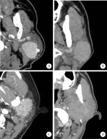
Parotid CT imaging reporting and data system: A preliminary study

Yu-bing LI1,Li-sha SUN2,Zhi-peng SUN1,(),Xiao-yan XIE1,Jian-yun ZHANG3,Zu-yan ZHANG1,Yan-ping ZHAO1,Xu-chen MA1   

  1. 1. Department of Oral Pathology, Peking University School and Hospital of Stomatology & Department of Oral and Maxillofacial Radiology, Beijing 100081, China
    2. Department of Oral Pathology, Peking University School and Hospital of Stomatology & Central Laboratory, Beijing 100081, China
    3. Department of Oral Pathology, Peking University School and Hospital of Stomatology & National Clinical Research Center for Oral Diseases & National Engineering Laboratory for Digital and Material Technology of Stomatology & Beijing Key Laboratory of Digital Stomatology, Beijing 100081, China
  • Received:2019-10-10 Online:2020-02-18 Published:2020-02-20
  • Contact: Zhi-peng SUN E-mail:sunzhipeng@bjmu.edu.cn

Abstract:

Objective: To establish a Parotid Imaging Reporting and Data System (PI-RADS) for CT diagnosis of the parotid gland neoplasms and to investigate the clinical applicable value and feasibility of PI-RADS. Methods: Patients who had been diagnosed with primary parotid gland neoplasms and had received surgical treatments in Peking University School and Hospital of Stomatology during the period of January 2013 to December 2016 were included in this study. The diagnoses were confirmed by the postoperative pathological examinations in all the patients. The CT imaging data of all patients were retrospectively reviewed and analyzed by two readers in consensus. Imaging characteristics related to the parotid neoplasms were extracted and quantified. Based on comprehensive analysis of the imaging characteristics, the probabilities of the benign and malignant neoplasms were evaluated and classified into six grades, PI-RADS 1-6 (PI-RADS 1: normal parotid gland; PI-RADS 2: confidently benign lesions; PI-RADS 3: probably benign lesions without confirmed evidence of malignancy; PI-RADS 4: suspected malignancy without sufficient evidence of malignancy; PI-RADS 5: confidently malignant lesions; PI-RADS 6: lesions with confirmed pathological evidence of malignancy). Results: A total of 897 patients with 1 003 parotid lesions were included. The lesions included 905 benign and 98 malignant lesions. The proportions of the malignancies in PI-RADS 2, PI-RADS 3, PI-RADS 4 and PI-RADS 5 according to the two readers in consensus were 0.4%, 5.7%, 35.5% and 96.7% respectively. The overall Cohen’s Kappa test showed medium consistency between the two independent researchers (κ=0.614, P<0.001, 95%CI: 0.569-0.695). Pearson Chi-square test showed that the proportions of malignancies increased with the diagnostic PI-RADS grades (Cochran-Armitage trend test, Z=-15.579, P<0.001). The results of Pearson Chi-square tests showed significant differences between the grades [PI-RADS 2 and 3 (χ 2=12.048, P=0.001); PI-RADS 3 and 4 (χ 2=75.231, P<0.001); PI-RADS 4 and 5 (χ 2=32.266, P<0.001)]. Conclusion: PI-RADS can be used to evaluate the risk of malignancy and will be helpful to improve the imaging diagnosis and clinical treatment of paro-tid gland neoplasms.

Key words: Parotid neoplasms, Tomography, X-ray computed, Diagnosis, differential, Therapy

CLC Number: 

  • R739.87

Table 1

Correlations between PI-RADS grades of the parotid gland neoplasms and radiologic signs"

Items PI-RADS 2 PI-RADS 3 PI-RADS 3b PI-RADS 4 PI-RADS 5 PI-RADS 6 Benign Malignancy
Size
Small (<2.0 cm) 30 82 7 7 2 0 118 10
Medium (2.0-3.0 cm) 158 453 4 49 14 1 624 55
Large (>3.0 cm) 56 102 1 20 14 3 163 33
Single/multiple
Single on one side 139 584 0 62 19 3 730 77
Multiple on one side 33 25 5 11 9 1 66 18
Multiple on two sides 72 28 7 3 2 0 109 3
Boundary
Absolutely clear 153 153 0 3 0 0 309 0
Majorly clear 68 261 2 21 4 0 345 11
Partially ill-defined 17 182 1 26 3 1 210 20
Majorly ill-defined 6 38 7 18 8 3 37 43
Ill-defined 0 3 2 8 15 0 4 24
Shape
Round 54 269 5 21 7 0 334 22
Oval 154 162 3 13 3 0 322 13
Scalloped 31 182 2 27 6 2 222 28
Irregular 5 24 2 15 14 2 27 35
Dominant density on plain CT
Fat 7 1 0 0 0 0 8 0
Liquid 30 59 0 4 12 0 91 2
Soft-tissue (partial fluid) 66 239 1 36 18 3 311 46
Soft-tissue 141 338 11 36 0 1 495 50
Enhancement degree on first enhanced phase
No significant 25 42 0 3 0 0 68 2
Slightly 10 122 0 12 4 0 140 8
Medium 29 203 5 32 13 1 236 47
Obvious 89 114 7 21 11 3 217 28
Significant 67 53 0 5 1 0 119 7
Enhancement pattern
Gradual 20 294 5 30 11 1 333 28
Fast in-out 124 128 1 23 7 2 254 31
Basically no enhancement 4 4 0 2 1 0 8 3
Gradual in-out 9 51 2 10 2 0 61 13
Enhancement distribution
Membrane enhancement 5 34 0 7 0 0 40 6
Even 152 182 12 24 15 1 346 36
Uneven to even 17 198 0 21 5 1 223 20
Uneven 14 84 0 17 8 2 99 26
No enhancement 32 36 0 4 0 0 72 0
Calcification
No 239 602 12 59 26 4 861 81
Small granular or capsule 5 31 0 10 1 0 38 9
Large irregular 0 4 0 7 3 0 6 8
Cystic percentage
0 177 488 12 49 26 2 683 71
≤25% 19 53 0 10 2 2 72 14
>25%, ≤50% 7 17 0 6 2 0 23 9
>50%, ≤75% 5 33 0 5 0 0 40 3
>75% 36 46 0 6 0 0 87 1
Adjacent structure destruction
No 237 609 7 64 16 3 867 69
Move 5 19 0 5 0 0 26 3
Poor boundary 2 8 5 6 9 0 12 18
Significant 0 1 0 1 5 1 0 8

Figure 1

A characteristic example of a PI-RADS 2 (A),3 (B), 4 (C), 5 (D) tumor A, a parotid gland neoplasm showing all regular and well-defined oval boundary, characteristic enhancement feature of benign nature (enhancement phase 1), indicating a PI-RADS 2 tumor. It was proved as Warthin tumor. B, a parotid gland neoplasm showing basically regular and well-defined round boundary, with mild enhancement (enhancement phase 3), indicating a PI-RADS 3 tumor. It was proved as pleomorphic adenoma. C, a parotid gland neoplasm showing seemingly well-defined boundary, with mild enhancement (enhancement phase 1), indicating a PI-RADS 4 tumor. It was proved as mucoepidermoid carcinoma. D, a paro-tid gland neoplasm showing all poorly-defined boundary, with erosion into adjacent structures and mild enhancement (enhancement phase 2), indicating a PI-RADS 5 tumor. It was proved as adenoid cystic carcinoma."

Table 2

The diagnostic criteria of PI-RADS"

PI-RADS Criteria
1: Normal parotid gland There are definitely no masses in the parotid gland
2: More likely benign (1) Multiple bilateral or unilateral masses presented regular arc border and oval contour with enhancement and without enlarged or necrotic lymph nodes around the parotid gland and neck; (2) A single tumor may also be considered if it is typical (e.g. oval, with obvious and uniform enhancement in the first enhancement phase, with smooth clear border or a low-density fat envelope)
3: Indeterminate (1) Single, round, no enhancement or uniform enhancement, most of the boun-dary clear, for example majorly clear or partially ill-defined; (2) Granular with uneven enhancement but definite boundary; (3) Multiple without enhancement, and without enlarged or necrotic lymph nodes around the parotid gland and neck; (4) Small tumors, that means, all tumors diameter less than 1 cm are included, regardless of their enhancement features
3b: Inflammation including Sjögren
syndrome and IgG4
In multiple gland, lesions are diffuse, with enhancement, without necrotic lymph nodes around the neck
4: Probably malignant (1) Single lesion, the shape is slightly irregular, part of the boundary is indistinctly (the boun-dary marked 3 or 4) or the membrane enhancement with villous edges; (2) The enhancement type is medium enhancement even slightly enhancement, solid lesion with little or without cystic degeneration; (3) Soft tissue density or with partial fluid especially with calcification, a small amount of unilateral diffuse; (4) Have large masses of irregular calcification with swelling lymph nodes but without necrosis lymph nodes
5: Highly suggestive malignancy (1) Necrotic lymph nodes are seen in the neck; (2) The boundary is unclear, the shape is irregular; (3) The border of the surrounding muscles, bones or fat tissues are not clear, which means destruction
6: Already had malignant diagnosis Had pathological result to pronounced it is a malignant neoplasm
7: Unsatisfied illustration on CT The density of neoplasm and the parotid is too nearly to find out what the neoplasm is really like. But couldn’t exclude that a neoplasm may exist

Table 3

Correlations between pathological results of the parotid gland neoplasms and PI-RADS grades"

Items PI-RADS 2 PI-RADS 3 PI-RADS 3b PI-RADS 4 PI-RADS 5 PI-RADS 6 PI-RADS total
Warthin tumor 176 72 0 5 0 0 253
Pleomorphic adenoma 4 367 0 23 1 0 395
Basal cell adenoma 11 81 0 2 0 0 94
Myoepithelioma 0 3 0 0 0 0 3
Oncocytoma 4 4 0 0 0 0 8
Cystadenoma 0 6 0 0 0 0 6
Keratocystoma 0 0 0 1 0 0 1
Cyst 28 14 0 0 0 0 42
Lipoma 4 1 0 0 0 0 5
Schwannoma 0 9 0 0 0 0 9
Vascular malformation 2 9 0 2 0 0 13
Eosinophilic lymphogranuloma 0 1 2 0 0 0 3
Inflammation 13 31 9 12 0 0 65
Calcified epithelioma 1 1 0 0 0 0 2
Non-sebaceous lymphoadenoma 0 0 0 1 0 0 1
Lymphoepithelial lesions 0 0 0 3 0 0 3
Nodular fasciitis 0 1 0 0 0 0 1
Myofibromatosis 0 1 0 0 0 0 1
Mucoepidermoid carcinoma 1 6 0 6 6 1 20
Adenocarcinoma, NOS 0 2 0 3 4 0 9
Acinic cell carcinoma 0 13 0 2 0 0 15
Salivary duct carcinoma 0 0 0 1 3 0 4
Adenoid cystic carcinoma 0 4 0 0 3 0 7
Polymorphous adenocarcinoma 0 3 0 5 1 0 9
Uncertainty adenocarcinoma 0 0 1 2 5 0 8
Epithelial-myoepithelial carcinoma 0 1 0 0 0 0 1
Clear cell carcinoma 0 0 0 0 1 0 1
Oncocytic carcinoma 0 0 0 2 0 0 2
Sarcoma 0 1 0 0 0 1 2
Lymphoepithelial carcinoma 0 1 0 2 2 2 7
Malignant lymphoma 0 4 0 3 4 0 11
Malignant melanoma 0 0 0 1 0 0 1
Metastatic solitary fibroma 0 1 0 0 0 0 1
Total 244 637 12 76 30 4 1 003
[1] 张震康, 俞光岩 . 口腔颌面外科学[M]. 2版. 北京: 北京大学医学出版社, 2013.
[2] Abdullah A, Rivas FF, Srinivasan A . Imaging of the salivary glands[J]. Semin Roentgenol, 2013,48(1):65-74.
[3] 俞光岩, 高岩, 孙勇刚 . 口腔颌面部肿瘤[M]. 北京: 人民卫生出版社, 2002.
[4] Seethala RR, Stenman G . Update from the 4th edition of the World Health Organization classification of head and neck tumours: Tumors of the salivary gland[J]. Head Neck Pathol, 2017,11(1):55-67.
[5] White SC, Pharoah MJ . Oral radiology principles and interpretation[M]. 3rd ed. St. Louis, Missouri: Mosby Elsevier, 2014.
[6] 马绪臣 . 口腔颌面医学影像学[M]. 北京: 北京大学医学出版社, 2014.
[7] 马绪臣, 李铁军 . 口腔颌面部疾病CT诊断与鉴别诊断[M]. 北京: 北京大学医学出版社, 2019.
[8] Horvath E, Majlis S, Rossi R , et al. An ultrasonogram reporting system for thyroid nodules stratifying cancer risk for clinical ma-nagement[J]. J Clin Endocrinol Metab, 2009,94(5):1748-1751.
[9] Barentsz JO, Richenberg J, Clements R , et al. ESUR prostate MR guidelines 2012[J]. Eur Radiol, 2012,22(4):746-757.
[10] Abdel Razek AA, Ashmalla GA, Gaballa G , et al. Pilot study of ultrasound parotid imaging reporting and data system (PIRADS): Inter-observer agreement[J]. Eur J Radiol, 2015,84(12):2533-2538.
[11] 马大权, 俞光岩 . 唾液腺病学[M]. 2版. 北京: 人民卫生出版社, 2014.
[12] Kim H, Kim SY, Kim YJ , et al. Correlation between computed tomography imaging and histopathology in pleomorphic adenoma of parotid gland[J]. Auris Nasus Larynx, 2018,45(4):783-790.
[13] Ito FA, Jorge J, Vargas PA , et al. Histopathological findings of pleomorphic adenomas of the salivary glands[J]. Med Oral Patol Oral Cir Bucal, 2009,14(2):E57-61.
[14] 黄敏娴, 马大权, 俞光岩 , 等. 复发性涎腺多形性腺瘤的临床与病理分析[J]. 现代口腔医学杂志, 2008,22(1):1-4.
[1] Guang-yan MENG,Yun-xiao ZHANG,Yu-xin ZHANG,Yan-ying LIU. Clinical characteristics of central nervous system involvement in IgG4 related diseases [J]. Journal of Peking University (Health Sciences), 2021, 53(6): 1043-1048.
[2] Li ZHAI,Nan QIU,Hui SONG. Multicentric reticulohistiocytosis: A case report [J]. Journal of Peking University (Health Sciences), 2021, 53(6): 1183-1187.
[3] Gang YANG,Wen-jie HU,Jie CAO,Deng-gao LIU. Three-dimensional morphology analysis of the supraosseous gingival profile of periodontally healthy maxillary anterior teeth [J]. Journal of Peking University (Health Sciences), 2021, 53(5): 990-994.
[4] LI Xin-fei, PENG Yi-ji, YU Xiao-teng, XIONG Sheng-wei, CHENG Si-da, DING Guang-pu, YANG Kun-lin, TANG Qi, MI Yue, WU Jing-yun, ZHANG Peng, XIE Jia-xin, HAO Han, WANG He, QIU Jian-xing, YANG Jian, LI Xue-song, ZHOU Li-qun. Three dimensional nephrometry system for partial nephrectomy: Our initial exploration [J]. Journal of Peking University (Health Sciences), 2021, 53(3): 613-622.
[5] LIAO Xu-he,WANG Rong-fu,LIU Meng,CHEN Xue-qi,XIONG Yan,NONG Lin,YIN Lei,ZHANG Bing-ye,DU Yu-jing. Semiquantitative parameters of 18F-FDG PET/CT, gene mutation states of epidermal growth factor receptor and anaplastic lymphoma kinase in prognosis evaluation of patients with lung adenocarcinoma [J]. Journal of Peking University (Health Sciences), 2021, 53(2): 246-254.
[6] MENG Yuan,ZHANG Li-qi,ZHAO Ya-ning,LIU Deng-gao,ZHANG Zu-yan,GAO Yan. Three-dimentional radiographic features of 67 maxillary radicular cysts [J]. Journal of Peking University (Health Sciences), 2021, 53(2): 396-401.
[7] ZHOU Jing,LIU Yi. Cone-beam CT evaluation of temporomandibular joint in skeletal class Ⅱ female adolescents with different vertical patterns [J]. Journal of Peking University (Health Sciences), 2021, 53(1): 109-119.
[8] GAO Lu,GU Yan. Chinese morphological stages of midpalatal suture and its correlation with Demirjian dental age [J]. Journal of Peking University (Health Sciences), 2021, 53(1): 133-138.
[9] YUAN Yuan,LANG Ning,YUAN Hui-shu. CT spectral curve in differentiating spinal tumor metastasis and infections [J]. Journal of Peking University (Health Sciences), 2021, 53(1): 183-187.
[10] LI Peng,PIAO Mu-zi,HU Hong-cheng,WANG Yong,ZHAO Yi-jiao,SHEN Xiao-jing. Radiography study on osteotome sinus floor elevation with placed implant simultaneously with no graft augmentation [J]. Journal of Peking University (Health Sciences), 2021, 53(1): 95-101.
[11] Qi KANG,Ji-xin ZHANG,Ying GAO,Jun-qing ZHANG,Xiao-hui GUO. Analysis of diagnosis and treatment of 100 patients with Hürthle cell adenoma [J]. Journal of Peking University (Health Sciences), 2020, 52(6): 1098-1101.
[12] Gong CHENG,Xia ZHANG,Fei YANG,Jia-yu CHENG,Yan-ying LIU. Angioimmunoblastic T-cell lymphoma with fever, arthritis and skin pigmentation: A case report [J]. Journal of Peking University (Health Sciences), 2020, 52(6): 1150-1152.
[13] Shuang HU,Li-ping YANG. Expression pattern of different serotypes of adeno-associated viral vectors in mouse retina [J]. Journal of Peking University (Health Sciences), 2020, 52(5): 845-850.
[14] Cai-nan LUO,Zheng-fang LI,Li-jun WU,Hai-juan CHEN,Chun-mei YANG,Wen-hui XU,Xiao-ling LIU,Wei TANG,Ping QIAO,Baihetiya Rena. Multicenter performance of the different classification criteria for rheumatoid arthritis [J]. Journal of Peking University (Health Sciences), 2020, 52(5): 897-901.
[15] Huan-bin YU,Wen-jie WU,Xiao-ming LV,Yan SHI,Lei ZHENG,Jian-guo ZHANG. 125I seed brachytherapy for recurrent salivary gland carcinoma after external radiotherapy [J]. Journal of Peking University (Health Sciences), 2020, 52(5): 919-923.
Viewed
Full text


Abstract

Cited

  Shared   
  Discussed   
[1] . [J]. Journal of Peking University(Health Sciences), 2009, 41(4): 456 -458 .
[2] . [J]. Journal of Peking University(Health Sciences), 2009, 41(2): 125 -128 .
[3] . [J]. Journal of Peking University(Health Sciences), 2009, 41(2): 135 -140 .
[4] . [J]. Journal of Peking University(Health Sciences), 2009, 41(2): 158 -161 .
[5] . [J]. Journal of Peking University(Health Sciences), 2009, 41(2): 217 -220 .
[6] . [J]. Journal of Peking University(Health Sciences), 2009, 41(1): 52 -55 .
[7] . [J]. Journal of Peking University(Health Sciences), 2009, 41(3): 297 -301 .
[8] . [J]. Journal of Peking University(Health Sciences), 2009, 41(5): 505 -515 .
[9] . [J]. Journal of Peking University(Health Sciences), 2009, 41(5): 599 -601 .
[10] . [J]. Journal of Peking University(Health Sciences), 2009, 41(5): 516 -520 .