
根尖预备直径对下前磨牙牙根抗折强度的影响
[田诗雨1 , 白伟2 , 梁宇红1, △  ]
]
]
|
|


目的 观察不同根尖预备直径对下前磨牙牙根抗折强度的影响。方法 收集因正畸新鲜拔除的牙根发育完成、无牙根折裂的单根下前磨牙,单根管、根管弯曲度小于10°、距根尖5 mm处根管颊舌径与近远中径之比小于2,初锉≤15#。离体牙截冠后,保留13 mm牙根,按重量以随机区组分组法分为6组( n=10), 根管预备至不同主锉号数:A组,空白对照组,不进行根管预备;B组,40#主锉组;C组,45#主锉组;D组,50#主锉组;E组,55#主锉组;F组,60#主锉组。预备采用不锈钢手用K锉,以步退法进行预备,步退4号,每次步退1 mm,预备锥度为0.05,预备过程中冲洗液采用蒸馏水。将牙根用义齿基托树脂包埋,制作试件,使用万能试验机垂直加载,直至牙根折裂,记录抗压载荷(N)和折裂的类型。用单因素方差分析法、Tukey检验比较各实验组牙根抗压载荷,卡方检验比较不同实验组的牙根折裂类型。结果 根管预备后, 5个预备实验组平均抗压载荷均明显低于未经根管预备的空白对照组,其中50#主锉组[(1 027±128)N]、55#主锉组[(994±150)N]、60#主锉组[(983±166)N]的平均抗压载荷降低明显,与空白对照组[(1 444±155)N]、40#主锉组[(1 339±131)N]、45#主锉组[(1 287±144)N]相比差异有统计学意义( P<0.05)。试件折裂类型分析显示各实验组无明显差别,发生于颊舌向折裂的最高(55%), 近远中向折裂率为13%,复合折裂率为32%。结论 根尖预备大于50#后,下前磨牙牙根抗折强度明显下降。
Objective: To compare the fracture resistance of roots of mandibular premolar with diffe-rent apical preparation diameters.Methods: Sixty single-rooted single canal permanent mandibular premolar teeth extracted newly for orthodontic reason without immatureness, fracture or cracks were selected, with a curvature less than 10°, and internal length: short diameter of less than 2 at a level 5 mm from the apex. All the teeth were decoronated, leaving roots 13 mm in length. The initial apical file size for the teeth was ≤15#. The roots were assigned to 6 groups based on weights with random block design. Group A: blank control group, no instrumentation was performed. Groups B-F: the master apical file (MAF) was 40#, 45#, 50#, 55# and 60#, respectively. In the five experimental groups the roots were instrumented using hand files with step-back technique at 1 mm increments, resulting in a taper of 0.05. The irrigant used was distilled water. After mounted in acrylic resin, all the teeth were subject to vertical loading using an Instron testing machine until fractured. The occurrence of fractures was detected when the applied load suddenly decreased. The fracture load values and fracture modes were recorded. One-way ANOVA and post-hoc Tukey test were used to determine the difference of fracture load values between the groups ( P<0.05). Chi-square tests were used to compare the modes of root fracture.Results: Five experimental groups exhibited lower fracture load values than that of control group [(1 444±155) N]. The mean fracture load values for roots instrumented to an apical diameter of 50# [(1 027±128) N], 55# [(994±150) N] and 60# [(983±166) N] were significantly lower than that of control group and 40# group [(1 339±131) N] and 45# [(1 287±144) N] ( P<0.05). Buccal-lingual fracture, mesio-distal fracture and compound fracture occurred 55%, 13% and 32%, respectively. No difference of fracture mode was detected in the six groups.Conclusion: The fracture resistance reduced significantly when the roots were instrumented to an apical diameter of 50# or larger.
根管预备是根管治疗的关键环节, 目标在于清理根管系统内的感染[1]。研究证实, 根管预备尺寸越大, 切削受感染的根管壁牙本质越多, 越有利于清除根管系统的细菌及其代谢产物、牙髓组织和碎屑, 充分的机械预备也利于冲洗, 提高化学清创的作用[2, 3]。但与此同时, 随着预备尺寸增大, 预备风险增加, 其中一个风险就是随着切削组织量增多, 根管壁变薄, 有可能影响牙根负荷能力, 甚至发生治疗后折裂[4]。不同个体、不同牙位的自然解剖条件不同, 适度安全的机械预备尺寸也不尽相同, 研究表明, 同样预备至50#/0.10时, 尖牙的抗折强度不变, 但下前磨牙的抗折强度下降了30%[5]。
适度安全的机械预备尺寸尤其是根尖预备直径一直是临床医生关注的热点问题, Weine[6]建议根尖预备主锉号数比初锉大3号, 一直沿用至今。但凭手感确定的初锉, 往往因为阻挡或根尖处根管形态不规则而号数偏小[7], 按大3号的预备标准可能导致预备不足[8]。学者们基于人群平均根尖大小推荐不同牙位的根尖预备直径, 但即使针对一个牙位, 推荐的尺寸亦有不同, 比如Trope等[9]建议下颌前磨牙(单根管)应预备至60#, 而Tronstad[10]建议预备至40#~70#, Hecker等[11]则认为单根管前磨牙至少预备到60#~70#, 才能使根尖区得到充分预备。
本实验通过比较不同根尖预备直径的下前磨牙牙根抗压载荷, 寻找下前磨牙适度安全的机械预备尺寸, 为临床医生提供参考。
经患者知情同意, 选取因正畸拔除的新鲜单根下前磨牙, 储存于蒸馏水中。在立体显微镜(ZOOM-630E, 上海长方光学仪器有限公司)下观察, 选取牙根发育完成、无牙根折裂的牙, 拍摄近、远中向和颊舌向根尖片, 选择单根管、距影像学根尖5 mm处根管颊舌径与近远中径之比小于2、根管弯曲度小于10° (Schneider法)、未经根管治疗的牙。于釉牙本质界或其根方处将离体牙截冠后, 保留13 mm牙根, 初锉≤ 15#, 不锈钢K锉疏通根管至15#。用电子天平(先行者, OHAUS, 南京华璧科学仪器有限公司)测量牙根重量(g)。
将60个牙根按重量顺序排列, 分为10个区组, 每个区组6个牙根。将各区组的6个牙根随机分到6个实验组(n=10), A组为空白对照组, 不进行根管预备; 其他实验组按根尖预备主锉(master apical file, MAF)不同分为:B组, MAF 40#; C组, MAF 45#; D组, MAF 50#; E组, MAF 55#; F组, MAF 60#。
于根尖孔可见15# K锉尖处减1 mm作为工作长度, 使用不锈钢K锉(M-Access, 瑞士登士柏)以步退法预备, 步退4号, 每次步退1 mm, 锥度0.05, 用主锉回锉。每次更换锉针后及预备完毕后均用2 mL蒸馏水冲洗根管。
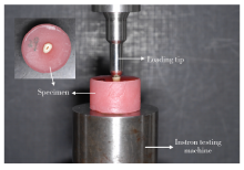
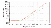
用义齿基托树脂(Meliodent仿生型, 德国贺利士古莎齿科有限公司)包埋固定牙根, 暴露牙根冠方2 mm。将直径为8 mm半球状不锈钢加压头固定在万能试验机(美国Instron)夹头上, 将样本置于载物台, 使加压头与牙根表面垂直, 以0.5 mm/min速度加压(图1)。加载力值陡然下降判定为折裂(图2), 加载力值陡然下降前的最大值记录为该牙的抗压载荷(N), 观察牙根折裂线的位置及方向, 将折裂分为颊舌向、近远中向、复合折裂3种类型(图3)。
使用SPSS Statistics 20.0软件(美国IBM)对实验数据进行统计处理。用单因素方差分析法比较各实验组牙根重量, 用单因素方差分析法和Tukey检验比较各实验组抗压载荷强度, 用卡方检验比较实验组的牙根折裂类型分布。P< 0.05为差异具有统计学意义。
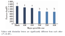
各实验组牙根重量差异无统计学意义(P> 0.05)。各实验组平均抗压载荷分别为:空白对照组(1 444± 155) N、40#主锉组(1 339± 131) N、45#主锉组(1 287± 144) N、50#主锉组(1 027± 128) N、55#主锉组(994± 150) N、60#主锉组(983± 166) N。其中, 大号预备的主锉组(50#主锉组、55#主锉组、60#主锉组)的平均抗压载荷明显低于未经根管预备的空白对照组、40#主锉组和45#主锉组(P< 0.05, 图4); 40#主锉组、45#主锉组平均抗压载荷较空白对照组虽有所降低, 但差异无统计学意义(P> 0.05); 50#主锉组、55#主锉组、60#主锉组之间平均抗压载荷差异亦无统计学意义(P> 0.05)。各实验组牙根折裂类型分布差异无统计学意义(P> 0.05), 试件颊舌向折裂率最高(55%), 近远中向折裂率为13%, 复合折裂率为32%。
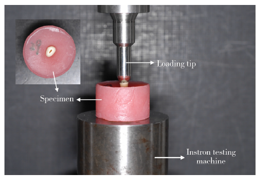 | 图1 静态载荷实验装置图Figure 1 Setting diagram of static strength testing |
本研究严格选取因正畸新鲜拔除的完整下前磨
牙, 并对根管的形态、弯曲度做了入选限定, 尽量避免因牙根解剖条件不同而影响实验结果。离体牙储存液和根管预备冲洗液均选用蒸馏水, 以避免化学药物对牙本质机械性能造成的影响[12, 13]。本研究在牙根长度一致的基础上根据牙根重量进行分组[14], 保证了各组样本硬组织量均衡, 减少了因牙根壁硬组织量不同对实验结果造成的影响。
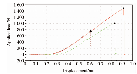 | 图2 位移-载荷曲线Figure 2 Displacement-load curve |
 | 图3 静态载荷实验牙根折裂类型Figure 3 Root fracture patterns in static strength testing |
 | 图4 实验组牙根抗压载荷Figure 4 Fracture resistance for groups |
研究牙根抗折强度的常用实验方法主要有静态载荷与循环载荷:静态载荷指以一个恒定的方向持续向样本施加载荷; 循环载荷在多个方向上反复对样本施加一定载荷, 能够更好地模拟实际的咀嚼情况, 但样本有可能长时间不折裂, 实验耗时较长, 而静态载荷耗时短、成本低, 因此成为广泛使用的方法[15]。本实验仅研究根管预备对牙根抗折强度的影响, 截去牙冠, 采用了静态载荷法, 通过静态载荷实验得到的抗压载荷数值也不宜直接与牙齿在口腔中行使咀嚼功能的咬合力相比较。
静态载荷实验中选用的加压头主要分为两种:一种为侧方加压器[16], 一种为不同直径球状加压头[5, 17]。不同形状的加压头, 牙根受力方式不同, 但对实验结果的影响并无报告, 本研究选择了直径8 mm 的半球状加压头。测试过程中加压速度不同, 亦可能影响实验结果, 有研究认为, 随着加压速度降低, 实验测得的抗折强度增高[18], 文献中最常采用的加压速度是0.5 mm/min或1 mm/min[16, 19], 本研究选取了0.5 mm/min的加压速度。
本研究结果显示, 根管预备后, 牙根抗折强度呈现不同程度的下降, 这和以往研究的结论相同[5, 19, 20, 21]。下前磨牙预备到50#/0.05或更大号后, 牙根抗折强度明显降低, 提示50#~70#的建议预备号数有可能过大。预备到45#/0.05时, 距根尖孔距离1 mm、2 mm、3 mm处的根管直径分别为0.45 mm、0.50 mm、0.55 mm, 预备到50#/0.05时分别为0.50 mm、0.55 mm、0.60 mm, 而临床常用的旋转镍钛预备器械, 如使用ProTaperUniversal预备到F3时, 距根尖孔1 mm、2 mm、3 mm处根管直径分别为0.30 mm、0.39 mm、0.48 mm, 预备到F4时分别为0.40 mm、0.46 mm、0.52 mm, 以上的直径均小于根尖预备到45#/0.05时相应部位的直径, 提示预备到F2、F3还是安全的。而预备到F5时, 距根尖孔 1 mm、2 mm、3 mm的根管直径分别为0.50 mm、0.55 mm、0.60 mm, 相当于本研究步退法预备到50#/0.05, 提示临床医师应慎重考虑安全预备, 牙根的抗折强度可能会有明显的变化。
随着预备增大, 切削组织量增多, 根管壁变薄, 为了寻找导致牙根抗折强度显著下降的根管壁危险厚度, 陈君等[22]制作根管壁厚度不同的试件, 研究表明根管壁去除越多, 横截面根管壁面积越小, 牙根的最大断裂载荷就越小, 但临床医师不易衡量预备到特定号数后剩余根管壁厚度的变化。分析预备号数增大导致牙根抗折强度降低的原因, 首要因素是剩余牙体组织量的减少, 此外, 也有研究发现预备器械在预备过程中可能会对牙根造成微创伤[23, 24], 从而影响牙根抗力。本研究预备到50#/0.05或更大号后, 牙根抗折强度降低, 但牙根折裂类型分布无明显改变, 仍以颊舌向折裂最多, 这与既往研究结论一致[16]。有限元实验发现拉应力集中于牙根颊舌侧, 尽管颊舌壁较近远中壁厚, 折裂还是易发于颊舌侧[25]。
综上, 本研究发现将下前磨牙预备到50#/0.05后牙根抗折强度明显下降, 提示确定根尖预备主锉时要考虑到安全性的要求, 根管预备锥度对牙根抗折强度的影响有待进一步研究和探讨。
The authors have declared that no competing interests exist.
| [1] |
|
| [2] |
|
| [3] |
|
| [4] |
|
| [5] |
|
| [6] |
|
| [7] |
|
| [8] |
|
| [9] |
|
| [10] |
|
| [11] |
|
| [12] |
|
| [13] |
|
| [14] |
|
| [15] |
|
| [16] |
|
| [17] |
|
| [18] |
|
| [19] |
|
| [20] |
|
| [21] |
|
| [22] |
|
| [23] |
|
| [24] |
|
| [25] |
|