
深层桡尺远侧韧带对桡尺远侧关节稳定作用的生物力学研究
[荣艳波, 田光磊, 陈山林 ]
]
]
|
|


目的 明确掌、背侧深层桡尺韧带对桡尺远侧关节的稳定作用。方法 将14具新鲜冰冻上肢标本随机分为2组,解剖显露桡尺远侧韧带,分别标记掌、背侧深层韧带,解剖游离出旋前圆肌及旋后肌,在桡骨Lister结节处垂直骨面置一枚克氏针,前臂中立位时在尺骨远端同一水平位置平行置一枚克氏针作为标记点,用自制的夹具将标本固定于生物力学仪器上,使肘关节屈曲90°,固定尺骨,使桡骨可围绕尺骨自由旋转。于旋前圆肌加载50 N的力模拟前臂主动旋前,于旋后肌加载60 N的力模拟主动旋后,分别切断掌、背侧深层桡尺韧带,测量桡骨相对于尺骨的位移。结果 切断掌侧深层韧带后,前臂旋前时桡骨相对于尺骨的位移明显改变( t=5.591, P=0.001),旋后时无明显改变( t=0.433, P=0.680)。切断背侧深层韧带后,前臂旋前时桡骨相对于尺骨的位移无明显改变( t=1.000, P=0.356),旋后时明显改变( t=-6.225, P=0.001)。结论 单独切断掌侧深层桡尺韧带会造成桡尺远侧关节旋前时不稳定,单独切断背侧深层桡尺韧带会造成桡尺远侧关节旋后时不稳定。
Objective: To evaluate the role of the deep radioulnar ligament in the stability of the distal radioulnar joint (DRUJ).Methods: In the study, 14 fresh cadaver upper extremities were randomly divided into two groups. After exposuring the palmar and dorsal deep distal radioulnar ligament, one group was marked as palmar deep radioulnar ligament, and the other group was marked as dorsal deep radioulnar ligament. The pronator teres and the supinator were exposed. A Kirschner wire perpendicular to the bone on Lister tubercle of radius was inserted, then another Kirschner wire on the same level of ulnar inserted when the forearm was in neural position, which was kept parallel to the first Kirschner wire. These specimens were mounted on a specially designed jig which held the limb rigidly, keeping the elbow fle-xion and the ulnar fixation. The radius could freely rotate around the ulnar. Then 50 N force on the pronator teres was applied to simulate the active pronation, and 60 N force on the supinator to simulate the active supination. The active pronation was stimulated, and the displacement of the distal radius was measured with respect to the ulna. The active supination was atimulated, and the displacement of the distal radius was measured with respect to the ulna. The palmar deep radioulnar ligament in one group was cut, then the displacement of the distal radius measured with respect to the ulna when the forearm was in pronation and supination. The dorsal deep radioulnar ligament in the other group was cut, and the displacement of the distal radius measured with respect to the ulna when the forearm was in pronation and supination.Results: After resection of the palmar deep radioulnar ligament, the displacement of the distal radius with respect to the ulna was statistically significantly different when the forearm was in pronation ( t=5.591, P=0.001), but there was no difference when the forearm was in supination ( t=0.433, P=0.680). After resection of the dorsal deep radioulnar ligament, the displacement of the distal radius with respect to the ulna was not different when the forearm was in pronation ( t=1.000, P=0.356), but there was statistically significant difference when the forearm was in supination ( t=6.225, P=0.001).Conclusion: DRUJ is unstable when the forearm is in pronation after resection of the palmar deep ra-dioulnar ligament, and DRUJ is unstable when the forearm is in supination after resection of the dorsal deep radioulnar ligament.
桡尺远侧关节(distal radioulnar joint, DRUJ)结构复杂而功能重要, 其损伤日益受到临床医师的关注。DRUJ的不稳定通常引起疼痛、握力下降等症状[1], 其稳定结构包括关节囊, 掌、背侧桡尺韧带, 三角纤维软骨复合体, 尺侧腕伸肌腱下腱鞘, 尺腕韧带, 前臂骨间膜等[2, 3, 4, 5, 6, 7, 8]。解剖学研究表明, 三角纤维软骨掌、背侧缘增厚移行为桡尺远侧韧带, 分别发自于桡骨乙状切迹掌、背侧缘, 在乙状切迹与尺骨茎突之间的中部分为浅、深两层, 浅层韧带止于尺骨茎突基底部, 深层韧带止于尺骨茎突下陷窝[4, 5, 9]。桡尺远侧韧带为DRUJ的主要稳定结构[9, 10], 许多研究已经评估了其作用, 但仍然存在争议。
1985年, Af Ekenstam等[11]的研究指出, 切除背侧桡尺远侧韧带, DRUJ旋后时不稳; 切除掌侧桡尺远侧韧带, DRUJ旋前时不稳。1991年, Schuind等[3]报道了完全相反的观点。1993年, Acosta等[12]的试验结果表明旋后时掌侧韧带长度增加, 旋前时背侧韧带长度增加。1994年, Hagert[13]回顾了前人的研究结论后指出, 两种观点均只说明了掌、背侧桡尺韧带的部分结构的功能, 然而, 此后相关的争论并未停止。本研究旨在通过生物力学研究明确掌、背侧深层桡尺韧带对关节的稳定作用。
14具新鲜冰冻上肢标本均从肘关节上方10 cm处截断, 通过DRUJ应力试验[10], 排除以前存在的DRUJ不稳定, 所有14具标本均不存在DRUJ不稳定。本研究获得北京积水潭医院伦理委员会批准(积伦科审字第201703-24号)。
所有标本均密封在双层塑料袋中, 贮藏于-28 ℃冰柜中。实验前将标本于室温下解冻, 解剖显露桡尺远侧韧带, 分别标记掌、背侧深层韧带(图1), 解剖游离出旋前圆肌及旋后肌备用。在桡骨Lister结节处垂直骨面置一枚克氏针, 前臂旋转中立位时在尺骨远端同一水平位置平行置一枚克氏针作为标记点。将实验标本平均分为两组, 一组切断掌侧深层韧带, 另一组切断背侧深层韧带。
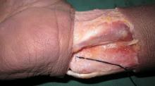
 | 图1 掌侧深层桡尺韧带(系线处)Figure 1 Palmar deep radioulnar ligament (mark with the suture) |
用自制的力学夹具将标本固定于MTS 858 Mini Bionix®型生物力学仪上, 使肘关节屈曲90° , 固定尺骨, 使桡骨可围绕尺骨自由旋转(图2)。通过预试验, 于旋前圆肌加载50 N的力模拟旋前圆肌收缩, 可使前臂达到最大的旋前位, 于旋后肌加载 60 N的力模拟旋后肌收缩, 可使前臂达到最大的旋后位。试验中于旋前圆肌加载50 N的力模拟前臂主动旋前运动, 使前臂处于最大旋前位, 测量桡骨相对于尺骨的位移, 撤消旋前圆肌加载; 于旋后肌加载60 N的力模拟前臂主动旋后运动, 使前臂处于最大旋后位, 测量桡骨相对于尺骨的位移, 撤消旋后肌加载。根据分组, 切断掌侧或背侧深层桡尺韧带, 再次模拟旋前、旋后运动, 测量桡骨相对于尺骨的位移(图3)。
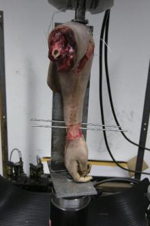
 | 图2 用自制的力学夹具将标本固定于MTS 858 Mini Bionix®型生物力学仪上, 使肘关节屈曲90° , 固定尺骨, 使桡骨可围绕尺骨自由旋转Figure 2 The specimen was mounted on a specially designed jig and connected to MTS 858 Mini Bionix®, keep the elbow flexion and the ulnar fixation, the radius can free rotate around the ulnar |
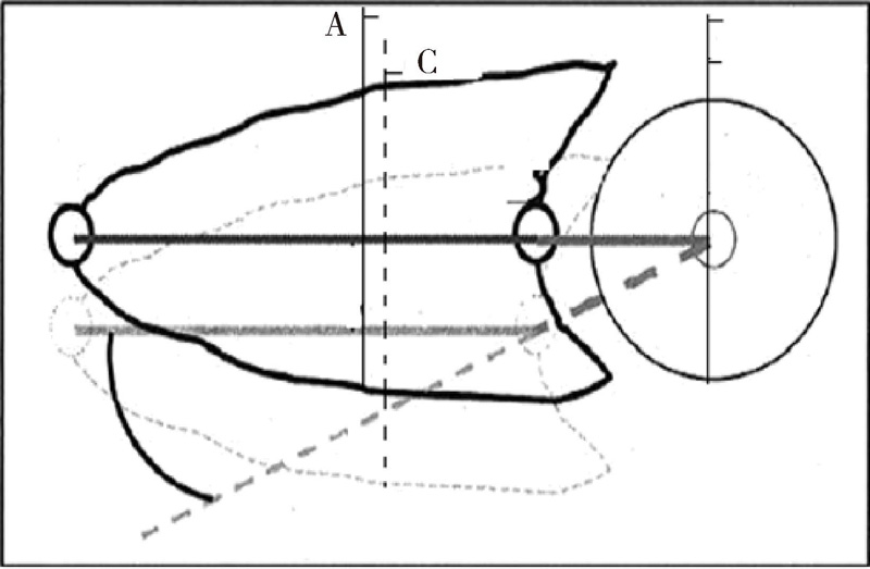 | 图3 使用游标卡尺测量桡骨相对于尺骨的位移ACFigure 3 Measure the transition AC between radius and ulnar with sliding gauge A, the marked point, on the Kirschner wire inserted the Lister tubercle of radius, the forearm in neural position; C, the marked point, on the Kirschner wire inserted the Lister tubercle of radius, the forearm in pronation position. |
将所得各组实验数据用SPSS 16.0统计软件进行分析, 计量资料以均数± 标准差表示, 进行单样本K-S检验各组数据符合正态分布, 两组间比较使用配对t检验, P< 0.05为差异有统计学意义。
单样本K-S检验表明各组数据均符合正态分布, 进行两样本配对t检验, 表1结果表明, 切断掌侧深层韧带后, 旋前时桡骨相对于尺骨的位移发生明显改变(t=5.591, P=0.001), 旋后时无明显改变(t=0.433, P=0.680)。切断背侧深层韧带后, 旋前时桡骨相对于尺骨的位移无明显改变(t=1.000, P=0.356), 旋后时明显改变(t=-6.225, P=0.001)。
| 表1 韧带切断前后桡骨相对于尺骨的位移比较 Table 1 Comparison the displacement of the distal radius with respect to the ulna between the before and after cutting the ligament |
以往有许多研究已经评估了桡尺远侧韧带对DRUJ的稳定作用, 但仍然存在争议。Af Ekenstam等[11]通过解剖研究5具新鲜标本, 首次指出切除背侧桡尺远侧韧带DRUJ旋后时不稳, 切除掌侧桡尺远侧韧带DRUJ旋前时不稳。然而6年后, Schuind等[3]报道了完全相反的观点, 该研究通过立体摄影测量技术显示掌侧韧带在前臂旋后时紧张, 背侧韧带在前臂旋前时紧张。Acosta等[12]测量出桡尺远侧韧带长度的变化, 结果说明旋后时掌侧韧带长度增加, 旋前时背侧韧带长度增加。Hagert[13]回顾了前人的研究结论后指出, 两种观点均只说明了掌、背侧桡尺韧带的部分结构的功能, 提出掌、背侧桡尺韧带分为浅、深层结构, 并指出Af Ekenstam等[11]的研究只讨论了深层韧带的功能, 而Schuind等[3]的研究只论述了浅层韧带的功能。Hagert[13]明确提出了前臂旋前时背侧浅层韧带、掌侧深层韧带紧张, 而旋后时掌侧浅层韧带、背侧深层韧带紧张。
然而, 此后相关争论并没有停止。Kihara等[4]在生物力学研究中模拟前臂主动旋转, 先后切断背侧韧带+1/2三角纤维软骨(triangular fibrocartilage, TFC)或掌侧韧带+1/2 TFC, 剩余的韧带+1/2 TFC, 旋前方肌+前臂骨间膜远侧部分, 剩余的前臂骨间膜等结构, 试验说明, 当前臂骨间膜断裂时, 旋前时主要稳定结构为背侧桡尺韧带, 而旋后时为掌侧韧带, 但当前臂骨间膜完整时, 单独切断远侧桡尺韧带不会引起关节不稳定。
Van der Heijden等[14]通过解剖测量掌、背侧桡尺韧带长度在前臂被动旋转运动中的变化, 认为旋前时背侧韧带紧张, 旋后时掌侧韧带紧张。Stuart等[7]进行的生物力学试验先后切断掌侧桡尺韧带、背侧桡尺韧带、尺腕韧带、尺侧腕伸肌下腱鞘、前臂骨间膜远侧部分、前臂骨间膜近侧部分, 研究结果认为, 防止尺骨相对于桡骨背侧脱位的主要结构为掌侧桡尺韧带, 防止尺骨相对于桡骨掌侧脱位的主要为背侧韧带, 其次为掌侧韧带及前臂骨间膜。Ward等[6]的试验结果显示, 旋前时背侧韧带紧张、掌侧韧带松弛, 旋后时则相反; 切除其他稳定结构后, 桡尺远侧韧带可维持关节稳定, 切除背侧韧带旋前不稳, 切除掌侧韧带旋后不稳。
黄继锋等[15]的试验结果表明, 切除背侧韧带+后侧1/2 TFC后, DRUJ在旋前位不稳定, 切除掌侧韧带+前侧1/2 TFC后, DRUJ在旋后位不稳定, 全切除后, 则引起DRUJ各方向明显的不稳定。Haugstvedt等[16]的生物力学试验结果表明, 浅层韧带与深层韧带对关节的稳定作用无明显差异。Ditano等[17]的研究结果表明, 旋前时背侧韧带张力大于掌侧韧带, 旋后时掌侧韧带张力大于背侧韧带。Gofton等[18]的生物力学试验结果表明, 切除掌、背侧桡尺远侧韧带不会引起关节不稳, 切除其他稳定结构后, 桡尺远侧韧带可维持关节稳定。Watanabe等[19]的研究不支持上述结果, 其认为切除掌、背侧关节囊就会引起关节不稳。周祖彬等[20]的研究指出, 切断背侧桡尺韧带, 尺骨相对桡骨的背侧位移明显增加, 切断掌侧桡尺韧带, 尺骨相对桡骨的掌、背侧位移都明显增加, 而掌、背侧桡尺韧带都切断会导致DRUJ明显不稳。Haugstvedt等[21]分别先后切断桡尺远侧韧带的浅、深层止点, 发现切断韧带后在无加载情况下无明显不稳, 加载情况下深层韧带对关节的稳定作用大于浅层韧带。Xu等[22]应用CT扫描7位健康受试者, 所得CT图像通过软件进行三维重建, 根据尺、桡骨与桡尺远侧韧带的解剖关系, 在三维图像中虚拟掌、背侧桡尺远侧韧带的位置, 并测量其在前臂旋转过程中的长度变化, 结果表明旋前时背侧浅层、掌侧深层韧带紧张, 旋后时掌侧浅层、背侧深层韧带紧张。
早期的研究多切除多种DRUJ的稳定结构, 或同时切断掌、背侧深层及浅层韧带, 本研究只单独切断掌侧或背侧深层韧带, 而保持其他所有结构的完整性, 结果表明, 单独切除掌侧深层桡尺韧带会造成DRUJ旋前不稳定, 尺骨头向背侧脱位; 单独切除背侧深层桡尺韧带会造成DRUJ旋后不稳定, 尺骨头向掌侧脱位, 这可能与浅层韧带在尺骨茎突成锐角, 而深层韧带在茎突下隐窝成钝角有关。前臂旋转运动中, 尺骨相对固定, 桡骨以桡骨小头中心与尺骨茎突下隐窝连线为轴围绕尺骨旋转[10], 而深层韧带止于尺骨茎突下隐窝。韧带的这一解剖关系造成前臂旋前时, 背侧浅层、掌侧深层韧带紧张而掌侧浅层、背侧深层松弛, 旋后时则为相反情况。
以前的试验研究已证实, 腕关节屈伸位置变化对DRUJ稳定结构切断后DRUJ的稳定性无影响[7], 因此本研究中未固定腕关节及手的位置。
由本研究结果可见, 掌、背侧深层韧带为DRUJ的主要稳定结构, 单独损伤, 即会出现DRUJ不稳定, 因此建议临床上仔细检查深层远侧桡尺韧带的完整性, 及时发现或排除问题, 有针对性地进行治疗。
The authors have declared that no competing interests exist.
| [1] |
|
| [2] |
|
| [3] |
|
| [4] |
|
| [5] |
|
| [6] |
|
| [7] |
|
| [8] |
|
| [9] |
|
| [10] |
|
| [11] |
|
| [12] |
|
| [13] |
|
| [14] |
|
| [15] |
|
| [16] |
|
| [17] |
|
| [18] |
|
| [19] |
|
| [20] |
|
| [21] |
|
| [22] |
|