


CXCL16/CXCR6在类风湿关节炎成纤维样滑膜细胞中的表达及其在滑膜细胞增殖中的作用
[张霞1, 2 , 赵金霞1 , 孙琳1 , 刘湘源1, △  ]
]
]
|
|


目的:探讨趋化因子CXCL16及其受体CXCR6在类风湿关节炎(rheumatoid arthritis,RA)患者成纤维样滑膜细胞(fibroblast-like synoviocytes,FLS)中的表达及其对FLS增殖的作用。方法:纳入RA 8例、骨关节炎(osteoarthritis,OA)7例、健康对照3例,采用组织块贴壁方法体外分离培养人膝关节FLS,第3~5代细胞用于后续研究。Western blot方法检测CXCL16及其受体CXCR6在3组FLS的表达水平;不同浓度的重组人CXCL16(0、10、50、100、200 μg/L)刺激3组FLS后,细胞活性检测试剂盒-8(cell counting kit,CCK-8)检测FLS增殖水平;Western blot方法检测重组人CXCL16刺激后RA-FLS中pAKT/AKT水平;ELISA法检测重组人CXCL16刺激后RA-FLS培养上清中TNF-ɑ、IL-6、MMP-3、RANKL水平。结果:RA-FLS中CXCL16、CXCR6蛋白表达水平均明显高于OA组和对照组( P<0.05),OA组和对照组间差异无统计学意义;重组人CXCL16(50、100、200 μg/L)刺激后RA-FLS增殖能力明显高于非刺激组( P均<0.05),OA组和对照组FLS经CXCL16刺激后细胞增殖水平无明显变化( P>0.05);200 μg/L CXCL16刺激后,RA-FLS表达pAKT/AKT明显增高;CXCL16(50、100、200 μg/L)刺激后,RA-FLS培养上清中IL-6和RANKL表达明显增加( P < 0.05),而MMP-3、TNF-ɑ水平无明显变化。结论:CXCL16及其受体在RA-FLS中表达增高,重组人CXCL16可促进RA-FLS增殖活化、分泌炎性因子增加,提示CXCL16参与了RA的滑膜炎症。
Objective:It has been found that serum CXCL16 concentration in rheumatoid arthritis (RA) patients are significantly higher than those in osteoarthritis (OA) and normal subjects, and are positively correlated with disease activity and bone erosion. However, how is CXCL16 involved in the pathogenesis of RA is unclear. To evaluate the expression of CXCL16 and its receptor CXCR6 in fibroblast-like synoviocytes (FLS) of rheumatoid arthritis (RA) patients, and to explore the role of CXCL16 in the proliferation of RA-FLS.Methods:FLS were isolated from knee synovial tissues obtained from 8 patients of RA, 7 osteoarthritis (OA) and 3 normal controls. The diagnosis of RA was in line with the 1987 American Rheumatology Association (ACR) RA classification criteria, osteoarthritis met the 1996 ACR revised knee osteoarthritis classification criteria. Control synovium were obtained from trauma caused knee joint injury in healthy individuals who required surgery. Human knee FLS were cultured by tissue explants adherent method.FLS between passages 3 and 5 were used in the experiment. Expression of CXCL16 and its receptor CXCR6 were performed in Western blot analysis. FLS proliferation follo-wing stimulation with TNF-α and different concentrations of CXCL16 was examined by cell counting kit-8 (CCK-8). Expression of phosphorylated AKT (pAKT) in RA-FLS stimulated by CXCL16 was quantified by Western blot. Different concentrations of recombinant human CXCL16 were added to the culture medium of RA-FLS. After 48 h culture, supernantants were collected, and TNF-α, IL-6, RANKL and MMP3 in culture supernatants of RA-FLS were determined by enzyme-linked immunosorbent assays (ELISA) operated following the kit instructions.Results:Expression of CXCL16 and CXCR6 in RA-FLS was significantly higher than that of OA and controls ( P<0.05), but no significant difference was found between OA-FLS and control FLS. Proliferation of RA-FLS was markedly up-regulated after stimulation of CXCL16 ( P <0.05). In the case of the CXCL16 stimulated OA-FLS and control FLS, the FLS proliferation remained basically unchanged. Expression of phosphorylated AKT in RA-FLS increased remarkably in condition of CXCL16 (50,100, 200 μg/L) stimulation. The levels of IL-6 and RANKL in culture supernatants of RA-FLS were obviously increased under CXCL16 (200 μg/L) stimulation, while TNF-α and MMP-3 levels in the culture supernatants remained unchanged after CXCL16 (200 μg/L) stimulation.Conclusion:This study shows that the expression of CXCL16 and its receptor was highly elevated in RA-FLS. Recombinant CXCL16 promoted RA-FLS proliferation and activation in vitro. All these indicate that CXCL16 play an important role in the pathogenesis of RA, anti-CXCL16 treatment may help to relieve inflammation and bone damage of RA patients. However, due to the limitations of this study, the role of CXCL16 and its receptors in RA-FLS remains to be elucidated by further research.
类风湿关节炎(rheumatoid arthritis, RA)是一种主要累及滑膜关节的自身免疫性疾病, 其病理改变主要包括慢性滑膜炎、血管翳形成、骨质破坏等[1, 2], 成纤维样滑膜细胞(fibroblast-like synoviocyte , FLS)在维持正常的关节平衡中起着重要的作用, RA中FLS表现为失去接触抑制的锚着非依赖性生长, 有异常增殖和侵蚀特性, 分泌多种炎性细胞因子, 并与局部浸润的炎性细胞相互作用, 共同参与了RA的关节破坏过程[3, 4]。
趋化因子CXCL16是2000年发现的一种炎性趋化因子, 主要表达于单核巨噬细胞、树突状细胞、内皮细胞[5]。CXCR6是CXCL16的唯一受体, 主要表达于T细胞、单核巨噬细胞、成纤维细胞[6]。有研究发现RA患者血清中趋化因子CXCL16的水平较骨关节炎和正常人明显升高, 并且与疾病活动度和骨侵蚀呈正相关, 应用肿瘤坏死因子拮抗剂治疗RA, 随着病情的缓解, RA患者体内CXCL16水平也明显降低, 提示CXCL16在RA发病中起重要作用[7], 但目前有关CXCL16参与RA发病的分子机制尚不明确。本研究拟探讨CXCL16(受体CXCR6)在RA-FLS的表达及其在FLS增殖活化中的作用, 为进一步了解CXCL16在RA发病中的作用提供实验依据。
滑膜标本取自2010年6月至2012年1月于北京大学第三医院运动医学科行关节置换术或滑膜切除术的住院患者, 其中RA 8例, 诊断符合1987年美国风湿病协会(ACR)的RA分类标准。骨关节炎(osteoarthritis, OA )7例, 诊断符合1996年ACR修订的膝骨关节炎分类标准。3例对照者为外伤致关节韧带损伤需行手术治疗而无基础疾病者。遵循程序符合负责人体试验委员会所制定的伦理学标准, 本研究开始前已经北京大学第三医院伦理委员会审查批准, 所有患者均签署知情同意书。
DMEM培养液、胎牛血清购自美国HyClone公司, 胰蛋白酶购自美国Gibco公司, 96孔板、培养瓶、培养皿购自美国Corning公司, 蛋白变性裂解液RIPA、BCA检测试剂盒购自北京普利莱基因技术有限公司, CCK-8试剂盒购自日本同仁化学研究所, RANKL ELISA试剂盒购自加拿大GBD公司, 人重组CXCL16蛋白购自美国Peproteche公司, TNF-ɑ 、MMP-3 ELISA试剂盒购自美国BD公司, CXCL16抗体、CXCR6抗体购自美国Abcom公司, β -actin抗体购自北京康为世纪公司, AKT抗体、pAKT抗体、PI3激酶抑制剂LY294002购自美国Cell Signaling公司, IL-6 ELISA试剂盒购自美国RD公司。
1.3.1 成纤维样滑膜细胞的分离培养 无菌获取膝关节滑膜组织, 剔除脂肪及纤维组织后, PBS冲洗2~3次, 在完全1640培养液中, 将其反复剪成细小组织块。将组织块接种到25 cm2细胞培养瓶中, 培养瓶倒置(即组织块贴壁的一面朝上), 加入含15%(体积分数)胎牛血清的DMEM 培养液3 mL后, 放置于37 ℃、5% (体积分数)CO2恒温培养箱内, 组织块不接触培养液孵育4 h后培养瓶组织块贴壁的一面朝下放置, 使组织块浸入培养液中, 培养瓶继续置于37 ℃、5%CO2(体积分数)恒温培养箱内孵育, 48 h后换液。5~7 d可见原代细胞贴壁, 适时胰蛋白酶消化传代, 培养至3~5代细胞用于后续研究。
1.3.2 Western blot法检测蛋白质表达水平 RIPA裂解液裂解细胞提取蛋白质后用BCA法定量, 取等量蛋白与上样缓冲液混合后, 上样到10%(质量分数) SDS-PAGE凝胶, 电泳分离, 转膜到PVDF膜(200 mA, 2 h)。用封闭液封闭2 h, 加入一抗反应液(用质量分数5%脱脂奶粉1 ∶ 1 000稀释兔抗人CXCL16抗体、兔抗人CXCR6抗体、鼠抗人β -actin抗体), 4 ℃孵育过夜。TBS-T洗膜3次, 每次10 min, 加入质量分数5%脱脂奶粉1 ∶ 10 000稀释羊抗兔或羊抗鼠荧光二抗, 室温孵育1 h, 再以TBS-T洗膜3次, 扫描图像并进行分析。
1.3.3 细胞活性检测试剂盒-8(cell counting kit, CCK-8)检测细胞增殖情况 CCK-8试剂盒是检测细胞增殖、细胞毒性的试剂盒, 为MTT法的替代方法, 其检测灵敏度高, 操作简便。具体步骤如下:用含有2%(体积分数)FBS的DMEM培养液调节滑膜细胞密度为3× 10⁴/mL, 按100 μ L/孔接种于96孔板中, 将培养板置于37 ℃、5%(体积分数)CO2培养箱中预培养24 h后, 加入TNFα (10 μ g/L)或不同终(质量)浓度的重组人CXCL16(10、50、100、200 μ g/L), 37 ℃、5%(体积分数)CO2环境下继续培养48 h时。按试剂盒说明书用CCK-8法检测细胞增殖情况。每孔加入10 μ L CCK-8工作液, 于37 ℃、5%(体积分数)CO2孵育箱中培养2 h, 用酶标仪450 nm波长检测光密度值。各组设6个复孔, 实验重复2次。CXCL16和TNFα 浓度选择参考了已经发表的相关研究[8, 9]。
1.3.4 Western blot检测CXCL16对RA-FLS中AKT蛋白磷酸化水平的影响 RA-FLS按照1× 10⁸/mL接种于75 cm2细胞培养瓶中, 分别加入不同终(质量)浓度的重组蛋白CXCL16(0、50、100、200 μ g/L), 同时设置抑制剂组[CXCL16(200 μ g/L)+PI3K抑制剂(50 mmol/L)]。培养30 min后收集各组细胞, PI3K抑制剂(50 mmol/L)在加CXCL16刺激之前1 h加入培养基。将定量好的细胞总蛋白与SDS上样缓冲液混合, 100 ℃水浴煮3~5 min备用。配好分离胶及浓缩胶后SDS-PAGE电泳, 浓缩胶80 V 30 min, 分离胶120 V 90 min。250 mA转膜 2 h后用5%(质量分数)BSA封闭1 h, 依次加一抗(兔抗人AKT抗体、兔抗人pAKT抗体、鼠抗人β -actin抗体), 4 ℃孵育过夜、洗涤, 加荧光二抗(羊抗兔或羊抗鼠)室温孵育1 h、洗涤。扫描图像并分析。
1.3.5 ELISA方法检测细胞培养上清中细胞因子水平 收集细胞增殖实验中RA-FLS加入不同浓度的重组人CXCL16(0、10、50、100、200 μ g/L)在37 ℃、5%(体积分数)CO2环境下培养48 h后的培养基, 1 000 r/min离心5 min, 取上清。ELISA方法检测TNF-ɑ 、IL-6、RANKL、MMP-3水平, 操作步骤按照试剂盒说明书进行。
实验数据用SPSS 20.0软件进行统计学分析, 计量资料以均数± 标准差表示, 两组计量资料的比较采用t检验, 多组间计量资料比较采用方差分析, 组间两两比较采用q检验。P< 0.05认为差异有统计学意义。
采用Western blot方法检测体外培养的FLS中CXCL16和CXCR6表达水平, 发现RA-FLS中CXCL16和CXCR6表达水平均明显高于OA和对照组(Con), OA组和对照组中二者的表达水平差异无统计学意义(图1)。RA、OA、对照组CXCL16/β -actin依次为0.55± 0.28、0.28± 0.17和0.09± 0.01(F = 5.72, P < 0.05); CXCR6/β -actin依次为0.62± 0.12、0.26± 0.15和0.12± 0.03(F=48.43, P< 0.05)。
 | 图1 CXCL16和CXCR6蛋白在3组FLS的表达情况 A, the expression of CXCL16 and CXCR6 was up-regulated in RA-FLS, compared with OA-FLS and control FLS; B and C, the histograph showed relative protein level of CXCL16/β -actin and CXCR6/β -actin; * P< 0.05. Con, control; OA, osteoarthritis; RA, rheumatoid arthritis.Figure 1 The expression of CXCL16 and CXCR6 in three groups of FLS |
分别用10、50、100、200 μ g/L重组人CXCL16刺激3组FLS, 48 h后采用CCK-8法检测CXCL16对3组FLS增殖的影响, 结果显示50 μ g/L CXCL16刺激后RA-FLS增殖就明显增加, 而OA和对照组FLS经CXCL16刺激后细胞增殖与非刺激时相比差异无统计学意义(图2A)。
 | 图2 不同浓度CXCL16刺激后各组FLS增殖水平 A, the proliferation of RA-FLS was significantly up-regulated under CXCL16 stimulation; B, RA-FLS proliferated under stimulation of CXCL16(200 μ g/L) or TNF-α (10 μ g/L); * P < 0.05 compared with CXCL16 in 0 μ g/L concentration; # P < 0.01 compared with CXCL16 in 0 μ g/L concentration. Con, control; OA, osteoarthritis; RA, rheumatoid arthritis.Figure 2 Proliferation of FLS under different concentration of CXCL16 |
比较CXCL16与TNF-α 刺激后RA-FLS增殖水平的差异, 同样采用CCK-8法检测, 显示CXCL16(200 μ g/L)刺激后RA-FLS的增殖能力低于TNF-α (10 μ g/L)刺激组(P< 0.05, 图2B)。
采用Western blot法检测不同浓度CXCL16(50, 100, 200 μ g/L)刺激后RA-FLS中pAKT的表达水平, 发现200 μ g/L CXCL16刺激后RA-FLS中pAKT表达明显高于非刺激组(pAKT/AKT 0.24± 0.09 vs. 0.14 ± 0.06, t = 2.62, P = 0.02)。加入PI3K抑制剂(LY294002 50 mmol/L)后, 再给予CXCL16(200 μ g/L)刺激时AKT蛋白的磷酸化水平明显减低(图3)。
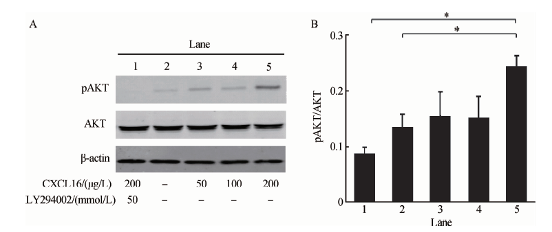 | 图3 CXCL16刺激后RA-FLS中pAKT的表达 A, the expression of pAKT was up-regulated under the stimulation of CXCL16 (50, 100, 200 μ g/L; lane 3, 4 and 5), and down-regulated in LY294002 (50 mmol/L) pre-stimulated RA-FLS; B, the histograph showed the relative pAKT/AKT level; * P < 0.05.Figure 3 pAKT level in CXCL16 stimulated RA-FLS |
ELISA方法检测不同浓度重组人CXCL16刺激后RA-FLS培养上清中TNF-ɑ 、IL-6、MMP-3、RANKL的水平, 结果发现不同浓度CXCL16刺激后RANKL和IL-6表达呈剂量依赖性增加(P< 0.05), 而MMP-3和TNF-ɑ 水平差异无统计学意义(图4)。
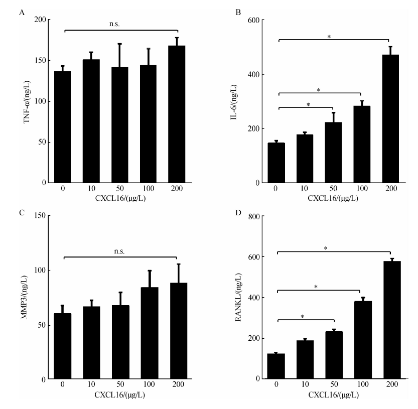 | 图4 不同浓度CXCL16刺激后RA-FLS培养上清炎症因子表达水平 * P< 0.05; n.s., no significent.Figure 4 Inflammatory cytokines in supernatants of CXCL16 stimulated RA-FLS |
趋化因子是由机体细胞合成并分泌的小分子蛋白, 在免疫应答和炎性反应中发挥重要作用, 目前的多项研究表明趋化因子在RA的发生发展中起重要作用。
CXCL16属于CXC趋化因子家族, 以膜结合型和可溶型两种形式存在, 膜结合型主要发挥细胞间黏附分子的作用[10], 裂解金属蛋白酶-10 (ADAM-10)可把膜结合型CXCL16裂解为可溶型[11], 后者可诱导CXCR6+ T细胞迁移到病变部位[12, 13], 还通过AKT、ERK等信号通路参与血管平滑肌细胞、牙龈成纤维样细胞的增殖[14, 15], 从而参与多种炎性疾病的病理过程。
T细胞增殖活化、滑膜细胞过度增殖均为RA的重要特点, 因此CXCL16/CXCR6在类风湿关节炎中的作用也备受关注。2005年van der Voort等[16]观察到RA滑液可刺激体外培养的滑膜巨噬细胞分泌CXCL16。Nanki等[17]用免疫组织化学的方法检测到RA滑膜组织表达CXCL16, 滑膜T细胞表达CXCR6增加, 应用抗CXCL16单克隆抗体可减轻胶原诱导关节炎小鼠的关节炎症和骨破坏, 提示CXCL16/CXCR6在RA的发生发展中起关键作用。随后有研究发现RA滑液中高浓度CXCL16可激活MAPK通路趋化T淋巴细胞聚集到关节炎症部位[18, 19]。本研究用免疫印迹的方法检测发现RA-FLS高表达CXCL16及其受体CXCR6, 明显高于OA和对照组, 进一步提示CXCL16/CXCR6参与了RA关节炎症反应。
鉴于RA关节炎症的显著特点为滑膜细胞过度增殖, 本研究通过CCK-8法观察不同浓度的重组人CXCL16对RA-FLS增殖是否有影响, 发现随着CXCL16刺激浓度的增加, RA-FLS增殖水平递增, 200 μ g/L 重组人CXCL16刺激后RA-FLS增殖明显增加。而OA和对照组FLS接受刺激后增殖水平差异无统计学意义。TNF-α 可促进RA-FLS增殖, 参与了RA滑膜炎症发生发展, 抗TNF治疗对RA疗效显著。本研究中200 μ g/L CXCL16刺激RA-FLS增殖的作用低于10 μ g/L TNF-α , 这一结果提示炎症状态下表达增加的CXCL16可促进RA-FLS增殖, 但其促RA-FLS增殖的作用没有TNF-α 强。
PI3K/AKT通路参与了许多炎性细胞的活化、细胞增殖过程, 用Western blot方法检测到高浓度CXCL16刺激后RA-FLS表达pAKT/AKT增加, 表明CXCL16可能是通过PI3K/AKT通路来活化成纤维样滑膜细胞。
RA的发生发展与多种炎症因子密切相关, 为进一步了解CXCL16在RA滑膜炎症中的作用, 本研究采用酶联免疫吸附方法检测CXCL16刺激后RA-FLS分泌炎症因子水平, 发现CXCL16刺激后RA-FLS培养上清中RANKL和IL-6水平随着CXCL16浓度增加而增加。RANKL属于肿瘤坏死因子配体家族, 通过激活NF-κ B、AKT、MAPK等途径, 参与破骨细胞活化及分化过程[20, 21]。RA-FLS表达RANKL增加, 促进破骨细胞大量产生, 造成关节骨质破坏。IL-6能诱导T细胞活化、FLS增殖, 还可刺激B细胞成熟, 促进类风湿因子等自身抗体的产生, 在RA关节局部和全身炎症反应中起重要作用[22]。CXCL16作用于RA的成纤维样滑膜细胞后, 促使成纤维样滑膜细胞表达RANKL和IL-6, 提示CXCL16及其受体CXCR6参与了RA滑膜炎症和骨破坏的发生, 并可能参与促进了RA相关自身抗体的产生。
本研究的局限性:体外培养人膝关节成纤维样滑膜细胞, 离体后FLS的生长增殖过程可能与体内不同, 体内FLS的增殖受更多因素调控, 且本研究纳入的样本数较少。
综上所述, CXCL16及其受体不仅在RA-FLS表达增加, 还可活化AKT信号通路, 促进RA-FLS增殖活化、表达炎症因子增加, 抗CXCL16治疗可能有助于缓解RA患者滑膜炎症和骨破坏。但由于本研究存在一定局限性, 关于RA-FLS中CXCL16及其受体的表达及作用机制仍有待于更深入的基础研究予以阐明。
The authors have declared that no competing interests exist.
| [1] |
|
| [2] |
|
| [3] |
|
| [4] |
|
| [5] |
|
| [6] |
|
| [7] |
|
| [8] |
|
| [9] |
|
| [10] |
|
| [11] |
|
| [12] |
|
| [13] |
|
| [14] |
|
| [15] |
|
| [16] |
|
| [17] |
|
| [18] |
|
| [19] |
|
| [20] |
|
| [21] |
|
| [22] |
|