水通道蛋白4在阿尔兹海默病脑组织间液引流改变中的作用
刘娥1, 张艺璇1, 孙琳琳1, 滕泽2, 王艾博2, 韩鸿宾2, 闫军浩1,
1. 北京大学基础医学院人体解剖学与组织胚胎学系, 北京 100191
2. 北京市磁共振成像设备与技术重点实验室, 北京 100191
摘要
目的:通过磁共振成像(magnetic resonance imaging, MRI)示踪剂钆喷酸葡胺(gadolinium-diethylene triamine pentacetic acid, Gd-DTPA)在脑组织间隙(extracellular space,ECS)内扩散的特征,反映大鼠阿尔兹海默病(Alzheimer’s disease,AD)脑组织间液(interstitial fluid,ISF)引流的改变及水通道蛋白4(aquaporin-4,Aqp4)在AD中发挥的作用。方法:将野生型SD大鼠(300~350 g)和Aqp4基因敲除(Aqp4-/-)SD大鼠(300~350 g)分别分为Sham组(10只)、AD组(10只)、Aqp4-/--Sham组(10只)、Aqp4-/--AD组(10只)。Sham组和Aqp4-/--Sham组连续6周每天腹腔注射生理盐水,AD组和Aqp4-/--AD组连续6周每天腹腔注射D-半乳糖。将示踪剂Gd-DTPA(10 mmol/L,2 μL)注射到大鼠海马区,分别于0.5 h、1 h、1.5 h、2 h、3 h 进行MRI扫描,观察示踪剂在海马区的动态分布,并测量示踪剂的扩散速率 D*值、清除速率 k' 值和半衰期 t1/2值。结果:Sham组示踪剂在ECS 内的扩散率 D*值为(2.66±0.36)×10-6 mm2/s,AD组 D*值(2.72±0.62)×10-6 mm2/s ,Aqp4-/--Sham组 D*值(2.75±0.47)×10-6 mm2/s,Aqp4-/--AD组 D*值(2.802±0.55)×10-6 mm2/s,4组之间差异无统计学意义(One-Way ANOVA, P>0.05)。Sham组海马区的示踪剂清除速率 k' 为(4.57±0.14)×10-4/s,AD组 k' 为(3.68±0.22)×10-4/s,Aqp4-/--Sham组 k' 为(3.17±0.16)×10-4/s,Aqp4-/--AD组 k' 为(2.59±0.19)×10-4/s,4组之间两两比较差异均具有统计学意义(one-way ANOVA, P<0.05),4组之间的示踪剂清除半衰期 t1/2值依次延长,Sham组海马区的 t1/2为(0.67±0.12) h,AD组 t1/2为(0.88±0.08) h,Aqp4-/--Sham组 t1/2 为(1.12±0.15) h,Aqp4-/--AD组 t1/2 为(1.58±0.11) h,两两比较差异均具有统计学意义(One-Way ANOVA, P<0.05)。结论:AD的退行性病变使脑内ISF引流减慢,Aqp4的缺失使AD大鼠脑内ISF引流明显减慢,Aqp4对清除脑组织间隙的代谢废物具有重要作用。
关键词: 水通道蛋白4; 阿尔兹海默病; 组织间液; 磁共振
中图分类号:R445 文献标志码:A 文章编号:1671-167X(2018)02-0203-04
Role of aquaporin-4 in the change of interstitial fluid drainage in Alzheimer’s disease
LIU E1, ZHANG Yi-xuan1, SUN Lin-lin1, TENG Ze2, WANG Ai-bo2, HAN Hong-bin2, YAN Jun-hao1,
1. Department of Anatomy and Embryology, School of Basic Medical Sciences, Peking University, Beijing 100191, China
2. Beijing Key Lab of Magnetic Resonance Imaging Device and Technique, Beijing 100191, China
△ Corresponding author’s e-mail, yjh@bjmu.edu.cn
Abstract
Objective:To observe the characteristics of the interstitial fluid (ISF) drainage in the Alzheimer’s disease (AD) rats through magnetic resonance imaging (MRI) tracer gadolinium-diethylene triamine pentacetic acid (Gd-DTPA)spread in the brain extracellular space (ECS) and to discuss the role of aquaporin-4 (Aqp4) in the AD.Methods:Wild type SD rats (300-350 g) and Aqp4 gene knock out (Aqp4-/-) SD rats (300-350g) were divided into Sham group, AD group, Aqp4-/--Sham group and Aqp4-/--AD group. Sham group and Aqp4-/--Sham group were injected with saline by intraperitoneal each day for 6 weeks, and the AD group and Aqp4-/--AD group were injected with D-galactose by intraperitoneal each day for 6 weeks. MRI tracer Gd-DTPA (10 mmol/L, 2 μL) was injected into the hippocampus of the rats. MRI scan was performed at the end of 0.5 h, 1.5 h, 1 h, 2 h, and 3 h to observe the dynamic distribution of the Gd-DTPA in the hippocampus and the diffusion rate D*, clea-rance rate k' and half-life t1/2 measured.Results:The diffusion rate D* in Sham group was (2.66±0.36)×10-6 mm2/s, the diffusion rate D* in AD group was (2.72±0.62)×10-6 mm2/s, the diffusion rate D* in Aqp4-/--Sham group was (2.75±0.47)×10-6 mm2/s, the diffusion rate D* in Aqp4-/--AD group was (2.802±0.55)×10-6 mm2/s, and there was no statistically significant dif-ference in the four groups (One-Way ANOVA, P>0.05).The clearance rate k' in Sham group was (4.57±0.14)×10-4/s, the clearance rate k' in AD group was (3.68±0.22)×10-4/s, the clea-rance rate k' in Aqp4-/--Sham group was (3.17±0.16)×10-4/s, the clearance rate k' in Aqp4-/--AD group was (2.59±0.19)×10-4/s, and there was significant difference in the four groups (One-Way ANOVA, P<0.05). The half-life t1/2 in Sham group was (0.67±0.12) h, the half-life t1/2 in AD group was (0.88±0.08) h, the half-life t1/2 in Aqp4-/--Sham group was (1.12±0.15) h, the half-life t1/2 in Aqp4-/--AD group was (1.58±0.11) h, and there was significance difference in the four groups(one-way ANOVA, P<0.05).Conclusion:The ISF drainage is slow after AD and the loss of Aqp4 in the AD makes the ISF drainage obviously slow down, Aqp4 plays an important role in AD to remove the metabolism of waste out of the brain.
Key words: Aquaporin-4; Alzheimer’s disease;; Interstitial fluid; Magnetic resonance imaging

阿尔兹海默病( Alzheimer’ s disease, AD)是一种严重的中枢神经系统退行性疾病, 主要表现为认知和记忆能力降低, 在发达国家65岁以上的人群中, 约13%的老年人遭受着阿尔兹海默病带来的痛苦[1, 2], 但迄今为止AD的发病机制尚未完全阐明, AD的治疗也不理想。有毒蛋白聚体淀粉样蛋白-β (amyloid beta, Aβ )斑块和高度磷酸化Tau 蛋白缠结是AD的重要病理特征。由于Aβ 产生和清除之间的稳态被打破, 导致Aβ 累积, 继而Aβ 清除能力下降是导致AD的关键因素[3], 因此, 如何加速脑内组织间液(interstitial fluid, ISF)的流动, 以保证有效清除Aβ 蛋白是减缓或停止AD的有效策略。

水通道蛋白4(aquaporin-4, Aqp4)高度表达于星型胶质细胞足突上, 有报道称其能够促进ISF的流动, 有利于脑脊液(cerebrospinal fluid, CSF)中营养物质输送进入脑实质和将代谢废物清除出脑[4], Aqp4的缺失会使ISF引流减慢。最新的研究表明, 改善AD模型中的Aqp4的表达和极性分布, 会显著加快ISF中Aβ 的清除[5], 可见Aqp4对于Aβ 的清除具有重要的作用, 但动物活体状态下观察AD的ISF引流改变却从未有过报道。基于目前的研究现状, 我们认为AD后脑ISF引流减慢, Aqp4的缺失会使AD的脑ISF引流更加缓慢。

本研究拟通过磁共振成像(magnetic resonance imaging, MRI)观察示踪剂钆喷酸葡胺(gadolinium-diethylene triamine pentacetic acid, Gd-DTPA)在脑组织间隙(extracellular space, ECS)内扩散的特征, 间接反映AD模型大鼠脑ISF引流的改变, 并通过比较示踪剂在ECS中的扩散和清除速率, 探讨Aqp4在AD模型大鼠脑内ISF引流中的作用。

1 资料与方法
1.1 实验动物

本研究获得北京大学医学部实验动物伦理审查委员会的批准, 所用SD大鼠和Aqp4基因敲除(Aqp4-/-)SD大鼠均来自北京大学医学部实验动物科学部。实验动物体重为300~350 g, 将野生型SD大鼠和Aqp4-/-大鼠随机分为4组:Sham组(10只)、AD组(10只)、Aqp4-/--Sham组(10只)、Aqp4-/--AD组(10只)。

1.2 AD模型制备

参照文献[6, 7, 8, 9], 本研究中D-半乳糖粉末在无菌生理盐水中溶解配制成3%(质量分数)浓度的溶液, AD组和Aqp4-/--AD组每天腹腔注射D-半乳糖溶液(5 mL/kg), Sham组和Aqp4-/--Sham组每天注射无菌生理盐水(5 mL/kg), 均连续注射6周。

1.3 MRI扫描

对大鼠进行编号, 经腹腔注射戊巴比妥钠(50 mg/kg)麻醉。采用 Siemens Magnetom Trio 3.0T 超导MRI扫描仪, 用腕线圈快速采集大鼠颅脑磁化准备梯度回波序列(three-dimensional magnetization prepared rapid acquisition gradient echo sequences, 3D-MP-RAGE)T1加权图像, 扫描参数:TR 1 500 ms, TE 3.7 ms, 翻转角9° , TI 900 ms, 视野267 mm, 矩阵512× 512, 分辨率 0.5 mm× 0.5 mm× 0.5 mm, 获取每只大鼠的T1WI 零时刻扫描图像[10]。然后将大鼠固定于鼠脑立体定位仪(美国Stoelting公司), 切开大鼠头皮, 分离骨膜, 暴露前囟, 依照《大鼠脑立体定位图谱》定位海马区, 颅骨钻孔, 缓慢进针, 用微量注射泵(瑞士Hamilton公司)在海马区域注射Gd-DTPA(10 mmol/L)2 μ L(0.2 μ L/min), 注射完毕后再次行MRI扫描, 于0.5 h、1 h、1.5 h、2 h、3 h 连续观察示踪剂的扩散特征。应用自主开发的软件测量扩散速率D* 、清除速率k'和清除半衰期(half life, t1/2)[11]

1.4 统计学分析

采用 SPSS 18.0 软件, 实验数据采用均值± 标准差来表示, 组间数据比较采用单因素方差分析(One-Way analysis of variance, One-Way ANOVA)的方法, 两两组间比较采用Tukey’ s Multiple Comparison Test的方法, P< 0.05 表示差异有统计学意义。

2 结果

AD组和Aqp4-/--AD组大鼠在连续6周注射D-半乳糖溶液后行动缓慢, 对声音和触碰反应迟钝, Sham组和Aqp4-/--Sham组大鼠未出现异常, 说明AD模型成功。

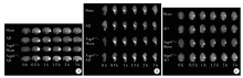
在Gd-DTPA注射进入海马区后0.5 h、1 h、1.5 h、2 h、3 h, 分别于冠状位(图1A)、矢状位(图1B)和水平位(图1C)连续观察示踪剂的扩散特征, 结果显示, Sham组、AD组、Aqp4-/--Sham组和Aqp4-/--AD组海马示踪剂的扩散速率D* 差异无统计学意义(F=1.351, P=0.286 3)。

图1 海马区冠状位、矢状位、水平位示踪剂扩散分布Figure 1 Distribution of the tracer in hippocampus in coronary sections, sagittal sections, and horizontal sections
A, intensity of tracer signal was gradually weakened, which suggested that tracer in the interstitial fluid was gradually cleared with the extension of observation time. The tracer clearance of AD group, Aqp4 -/--Sham group and Aqp4-/--AD group was decreased in coronary sections. B, tracer clearance in hippocampus of AD, Aqp4 -/--Sham and Aqp4-/--AD group got slower compared with sham group in sagittal sections, the rate of clearance in Aqp4-/--AD group was the slowest. C, tracer clearance in hippocampus of AD group, Aqp4 -/--Sham group and Aqp4 -/--AD group was slower compared with Sham group in horizontal sections and the rate of clearance in Aqp4-/--AD group was the slowest.AD, Alzheimer’ s disease ; Aqp4, aquaporin-4.

示踪剂Gd-DTPA在AD组大鼠海马内的清除速率k'明显低于Sham组, Aqp4-/--Sham组清除速率也明显低于Sham组, Aqp4-/--AD组的示踪剂清除速率最慢, 4组之间差异具有统计学意义(F=277.2, P< 0.001)。

示踪剂Gd-DTPA在Sham组、AD组、Aqp4-/--Sham组和Aqp4-/--AD组的清除半衰期t1/2依次延长, 4组之间差异具有统计学意义(F=212.9, P< 0.001, 表1)。

表1 大鼠海马区示踪剂平均扩散速率D* 、清除速率k't1/2的比较 Table 1 Comparisons of D* , k' and t1/2 of the tracer in the hippocampus
3 讨论

本研究中, 我们将MRI示踪剂Gd-DTPA注射进入各组大鼠的海马, 通过MRI观察其流动和清除情况, 结果表明AD模型大鼠脑内ISF引流减慢, Aqp4的缺失使大鼠脑内ISF引流明显下降, 加重了AD大鼠脑内ISF引流损伤。

在ECS内流动的ISF含有细胞代谢及信号传递的各种物质, 是维持细胞内环境的重要组成部分, 能够将营养物质带入脑内并将代谢废物(包括Aβ 蛋白)引流出脑[12]。本研究中应用的示踪剂Gd-DTPA几乎不被细胞吸收, 并且能够缩短周围水分子的T1加权像弛豫时间, 可以间接反映其所在ISF的流动及代谢特征[13]

本研究中应用的D-半乳糖致老化型大鼠AD模型是目前广泛接受的AD模型, 病变大鼠表现出接近自然老化的神经退行性变, 如Aβ 蛋白的产生、胆碱能退行性变、神经元凋亡和星形胶质细胞增生等[14]。本研究中发现, 示踪剂Gd-DTPA在AD模型大鼠海马内的流动和清除显著减慢, 我们推测其可能由于AD大鼠脑内ECS中有大量Aβ 蛋白沉积, 致使流动阻力增加; 同时, AD后神经元的凋亡和星形胶质细胞的增生等病变, 致使ESC空间构象严重变形, ECS内的迂曲度明显增加, 使得ISF的流动速度明显下降, 清除减慢且半衰期延长。

Aqp4是脑内表达量最多的水通道蛋白, 高度表达于星形胶质细胞足突, 能够促进ISF中水和部分离子的流动[15]。通常认为, 高度敏感和高代谢率的神经元和胶质细胞需要一个能将ISF中的代谢物质快速清除的细胞外环境[16], 但脑内并没有组织结构上的淋巴管, 也缺乏与外周系统类似的清除ISF溶质的独立路径。有研究表明, 主要由Aqp4组成的类淋巴系统(glymphytic system)是Aβ 蛋白清除的一个重要途径, 约40%~80%的大蛋白分子和溶质通过此途径清除[17]。类似于外周淋巴清除系统, 营养物质随着CSF沿软膜动脉和穿通动脉的血管周围间隙(virchow-robin space, VRS)进入, 再进入动脉管周间隙并通过Aqp4的作用进入ECS, 被神经细胞摄取, ECS中的废物随着ISF沿静脉管周间隙运输出脑实质, 因此, Aqp4的正常生理结构及功能对脑内ISF的流动和清除均有重要意义[18, 19]。另外, 有研究表明, Aqp4缺失后组织间隙变大、细胞水肿从而ECS迂曲度增加, 增加了ISF的流动阻力[20]。本研究的结果表明, 在Aqp4缺失后, 海马内的ISF流动和清除明显下降, 也进一步印证了这个观点。

综上所述, 本实验中我们发现, AD大鼠的脑内ISF引流系统明显受损, 而Aqp4对正常大鼠和AD大鼠脑内ISF引流均具有重要的意义, 在清除Aβ 蛋白层面来说, 对AD的产生、发展和转归均具有重要的意义。可以预见, 加强脑内Aqp4主导的类淋巴系统引流功能将有望成为未来治疗AD的新靶点。

The authors have declared that no competing interests exist.

参考文献
[1] 张琳琳, 宋宛珊, 王凯, . 阿尔茨海默病发病机制及药物治疗研究进展[J]. 世界中医药, 2017, 12(5): 1200-1203. [本文引用:1]
[2] 宋昕, 洪羽蓉, 胡秋莹. 阿尔兹海默病发病原因及机制的研究进展[J]. 临床和实验医学杂志, 2015, 14(10): 871-873. [本文引用:1]
[3] Iliff JJ, Wang M, Zeppenfeld DM, et al. Cerebral arterial pulsation drivesparavascular CSF-interstitial fluid exchange in the murine brain[J]. J Neurosci, 2013, 33(46): 18190-18199. [本文引用:1]
[4] Iliff JJ, Wang M, Liao Y, et al. A paravascular pathway facilitates CSF flow through the brain parenchyma and the clearance of interstitial solutes, including amyloid beta[J]. Sci Transl Med, 2012, 4(147): 111. [本文引用:1]
[5] He XF, Liu DX, Zhang Q, et al . Voluntary exercise promotes glymphatic clearance of amyloid beta and reduces the activation of astrocytes and microglia in aged mice[J]. Front Mol Neurosci, 2017, 10: 144. [本文引用:1]
[6] Liu L, Lu Y, Kong H, et al. Aquaporin-4 deficiency exacerbates brain oxidative damage and memory deficits induced by long-term ovarian hormone deprivation and D-galactose injection[J]. Int J Neuropsychopharmacol , 2012, 15(1): 55-68. [本文引用:1]
[7] Zhang XL, An LJ, Bao YM, et al. D-galactose administration induces memory loss and energy metabolism disturbance in mice: Protective effects of catalpol[J]. Food Chem Toxicol, 2008, 46(8): 2888-2894. [本文引用:1]
[8] 赵波, 张新宇, 付学锋. 阿尔兹海默病动物模型的研究进展[J]. 神经解剖杂志, 2012, 28(1): 102-104. [本文引用:1]
[9] 白杨, 辛随成. 阿尔兹海默病动物模型的研究进展[J]. 实验动物科学, 2013, 30(6): 61-65. [本文引用:1]
[10] 韩鸿宾, 李凯, 闫军浩. 应用MRI 示踪法在体研究大鼠脑组织间液的生物学特点[J]. 中国科学, 2012, 42(8): 622-628. [本文引用:1]
[11] 韩鸿宾. 应用磁共振成像技术定量测量活体大鼠脑细胞外间隙的扩散参数[J]. 北京大学学报(医学版), 2012, 44(5): 770-775. [本文引用:1]
[12] Lei Y, Han H, Yuan F, et al. The brain interstitial system: Ana-tomy, modeling, in vivo measurement, and applications[J]. Prog Neurobiol, 2017, 157: 230-246. [本文引用:1]
[13] 和清源, 韩鸿宾, 许方婧伟, . Gd-DTPA磁共振成像示踪法对脑细胞外间隙成像与定量分析的初步研究[J]. 北京大学学报(医学版), 2010, 42(2): 188-191. [本文引用:1]
[14] Arshavsky YI. Alzheimer disease and cellular mechanisms of memory storage[J]. J Neuropathol Exp Neurol, 2014, 73(3): 192-205. [本文引用:1]
[15] Papadopoulos MC, Verkman AS. Aquaporin-4 and brain edema[J]. Pediatr Nephrol, 2007, 22(6): 778-784. [本文引用:1]
[16] Loukas M, Bellary SS, Kuklinski M, et al. The lymphatic system: a historical perspective[J]. Clin Anat, 2011, 24(7): 807-816. [本文引用:1]
[17] Nedergaard M. Garbage truck of the brain[J]. Science, 2013, 340(6140): 1529-1530. [本文引用:1]
[18] Tarasoff-Conway JM, Carare RO, Osorio RS, et al. Clearance systems in the brain-implications for Alzheimer disease[J]. Nat Rev Neurol, 2015, 11(8): 457-470. [本文引用:1]
[19] Mendelsohn AR, Larrick JW. Sleep facilitates clearance of metabolites from the brain: glymphatic function in aging and neurodege-nerative diseases[J]. Rejuvenation Res, 2013, 16(6): 518-523. [本文引用:1]
[20] Papadopoulos MC, Manley GT, Krishna S, et al. Aquaporin-4 facilitates reabsorption of excess fluid in vasogenic brain edema[J]. FASEB J, 2004, 18(11): 1291-1293. [本文引用:1]