
胸椎管狭窄症术后脑脊液漏继发皮下积液的治疗
[王永强, 刘晓光△  , 姜亮, 韦峰, 于淼, 吴奉梁, 党礌, 周华, 刘忠军]
, 姜亮, 韦峰, 于淼, 吴奉梁, 党礌, 周华, 刘忠军]
|
|


目的: 探讨胸椎管狭窄症术后脑脊液漏继发皮下积液的治疗方法。方法: 回顾分析2005年1月至2014年12月北京大学第三医院骨科脊柱组完成手术的胸椎管狭窄症患者,其中术中、术后发生脑脊液漏者186例,选取其中继发皮下积液且规律随访的11例患者作为研究对象,治疗方法依据患者脑脊液漏严重程度以及胸脊髓病恢复情况决定。采取日本骨科协会(Japanese Orthopedic Association, JOA)改良11分评分评价患者术前及术后的脊髓功能,将手术前后的JOA评分进行统计学分析。结果: 11例患者均获得随访,其中后纵韧带骨化合并黄韧带骨化者6例,均接受椎管后壁切除及“涵洞塌陷法”360°脊髓环形减压术,单纯黄韧带骨化行椎管后壁切除术者5例。随访时间30~131个月,平均(85±34)个月。术前症状持续时间3个月至8年,中位数18个月;术后引流管拔出时间2~6 d,平均(4.2±1.1) d;围手术期出现发热患者10例,最高体温(37.3~39.7) ℃,其中高热患者2例,延长抗生素使用时间。10例患者采取保守治疗,随访发现脑脊液漏全部吸收,其中拔出引流管后加压包扎伤口者8例,局部穿刺抽液后加压包扎伤口者2例;1例患者因保守治疗无效,继发脑脊液假性囊肿而接受再次手术治疗。11例患者的JOA评分从术前的(3.8±1.6)分升高到末次随访时的(8.9±1.2)分,神经功能改善率为70.8%。没有患者出现切口感染、切口不愈合及颅内感染等并发症。结论: 胸椎管狭窄症术后脑脊液漏继发皮下积液者多数可以采取保守治疗,只有出现脑脊液假性囊肿压迫脊髓时需要采取手术治疗。
, JIANG Liang, WEI Feng, YU Miao, WU Feng-liang, DANG Lei, ZHOU Hua, LIU Zhong-jun
Objective: To investigate the treatment strategy for subcutaneous fistula secondary to cerebrospinal fluid leakage (CSFL) in thoracic spinal stenosis (TSS) cases.Methods: In the study, 186 CSFL cases diagnosed with TSS and operated in general spine group of Department of Orthopedics, Peking University Third Hospital from January 2005 to December 2014 were retrospectively reviewed, of which eleven had subcutaneous fistula secondary to CSFL and were regularly followed up. Treatment strategy for subcutaneous fistula depended on the severity of CSFL and the recovery rate of thoracic myelopathy. Japanese Orthopedic Association(JOA) score was utilized to evaluate the neurologic status of these patients preoperatively and postoperatively. Statistical analysis was conducted between preoperative and postoperative JOA scores.Results: All of the 11 patients were regularly followed up for at least 24 months. Six of them had ossification of the posterior longitudinal ligament (OPLL) combined with ossification of ligamentum flavum (OLF), all of them undertook “cave-in” 360°circumferential decompression of the spinal cord with instrumentation. Five cases had OLF only, and received En bloc resection of lamina and OLF and fixation. The follow-up period ranged from 30 months to 131 months, and averaged at (85±34) months. Preoperative symptoms lasted from 3 months to 8 years, and the median was 18 months. Drainages were placed for 2-6 days, and averaged at (4.2±1.1) days. Ten cases appeared with fever during the perioperative period, the maximum body temperature was (37.3-39.7) ℃. Prolonged antibiotics were applied in two cases with high fever. Ten cases were treated with conservative methods, CSFL were completely absorbed during the follow-up time, of which compressive dressing was utilized in 8 cases, and punctures combined with compressive dressing were used in 2 cases. For only 1 case, conservative therapy failed and reoperation was required because of neurological deterioration arising from CSF pseudocyst. For these 11 cases, preoperative JOA score arose from (3.8±1.6) preoperatively to (8.9±1.2) at the end of the final follow-up, the recovery rate was 70.8%. No infection of wound or central nerve system were noticed, and neither were unhealing wound.Conclusion: Most TSS cases with subcutaneous fistula secondary to CSFL could be cured by conservative methods, and reoperation is required only if myelopathy caused by cerebrospinal fluid pseudocyst is identified.
胸椎管狭窄症(thoracic spinal stenosis, TSS)引起的脊髓损害非常严重, 可以导致截瘫及二便障碍。胸椎后纵韧带骨化(ossification of the posterior longitudinal ligament, OPLL)及胸椎黄韧带骨化(ossification of ligamentum flavum, OLF)是最常见的致病因素[1]。北京大学第三医院采用“ 揭盖式” 椎管后壁切除术治疗胸椎黄韧带骨化, 采用“ 涵洞塌陷法” 360° 脊髓环形减压术治疗胸椎黄韧带骨化合并后纵韧带骨化, 取得了满意疗效[2], 但术中、术后脑脊液漏发生率较高, 达到32.3%[3]。脑脊液漏若处理不当, 可能继发脑脊液假性囊肿、切口愈合不良甚至颅内感染等并发症[4, 5]。目前关于胸椎管狭窄症术后脑脊液漏的报道较少, 而且缺乏直观的影像学资料观察脑脊液漏的转归情况。本研究收集了北京大学第三医院骨科脊柱专业组完成的胸椎管狭窄症术后脑脊液漏合并皮下积液的患者资料, 通过总结其临床表现及疾病转归, 期望为同行对胸椎术后脑脊液漏的治疗选择提供一些参考。
回顾2005年1月至2014年12月北京大学第三医院骨科脊柱专业组手术治疗的胸椎管狭窄症出现脑脊液漏的186例患者, 所有患者手术后出院前均复查胸椎磁共振(magnetic resonance imaging, MRI)。纳入标准:(1)术中或术后明确存在脑脊液漏者; (2)术后MRI观察到皮下积液者, 而无关积液量大小; (3)随访时规律复查MRI者; (4)至少2年随访者。最终选取11例患者作为研究对象。男5例, 女6例, 年龄35~66岁, 平均(51.5± 9.9)岁, 术前症状持续时间3个月至8年, 中位数18个月。术前常规行胸椎正侧位X线片、CT平扫及重建以及MRI检查。
本研究11例患者中OPLL合并OLF者6例, 均接受“ 涵洞塌陷法” 360° 脊髓环形减压术[2], 单纯OLF行“ 揭盖式” 椎管后壁切除术[6]者5例。手术采取全身麻醉, 俯卧位后正中入路。“ 揭盖式” 椎管后壁切除术使用高速磨钻沿双侧关节突内外缘的中线磨透椎板全层、关节突及骨化的黄韧带, 直至硬脊膜侧壁外露, 分离骨化的黄韧带与硬脊膜间的粘连, 将椎板及骨化韧带整体切除。“ 涵洞塌陷法” 360° 脊髓环形减压术去除椎管后壁后, 在OPLL节段去除关节突, 沿椎弓根至椎体用高速磨钻、刮匙磨刮, 至椎体后壁水平后, 自椎体后壁两侧深层斜向内挖去椎体后1/3的松质骨。分离OPLL与硬膜的粘连, 粘连严重或者存在硬膜骨化者可以切除部分硬脊膜。向腹侧压塌OPLL, 从脊髓侧后方将其取出。
对于存在硬膜骨化的患者, 减压时要求将骨化的硬膜一并切除, 硬膜缺损范围大、形状不规则, 通常难以修补。本组患者术中均采取明胶海绵覆盖缺损的硬脊膜, 并逐层严密关闭切口, 放置常压引流。
术后嘱患者平卧或侧卧, 记录切口引流液的颜色及引流量。满足以下条件者拔除常压引流:(1)24 h引流量逐渐减少并且小于50 mL; (2)术后引流量持续较多, 但引流液为淡血性, 且引流放置时间已超过48 h。拔除引流管后, 用3-0线将引流管口深缝1~2针, 嘱患者俯卧, 同时用1 kg的沙袋或者盐袋持续压迫引流管口24 h, 患者可间断采取侧卧位休息。若出现切口或引流管口渗液, 或者切口周围肿胀, 则局部穿刺抽液后再加压包扎5~7 d。如果临床怀疑脑脊液假性囊肿压迫脊髓产生症状, 则再次手术清除囊肿。术后常规应用广谱抗生素48 h预防感染, 并维持水电解质平衡, 对症处理低颅压症状, 对高热患者延长抗生素使用时间。术后常规复查胸椎MRI。
所有患者均有完整随访资料, 包括胸椎正侧位X线片以及胸椎MRI检查。神经功能评价采取日本骨科协会(Japanese Orthopedic Association, JOA)评分, 从患者住院病历获取术前JOA评分, 在末次随访资料中获取末次随访时神经功能。神经功能改善率=(术后评分-术前评分)/(11-术前评分)× 100%。
以均数± 标准差或中位数的形式表示数据, 将手术前、后的JOA评分采用配对t检验进行比较。所有数据均采用SPSS 23.0 软件进行分析, P< 0.05为差异有统计学意义。
本组患者术前症状持续时间3个月至8年, 中位数18个月; 手术时间100~350 min, 平均(227± 66) min; 出血量400~3 400 mL, 平均(1 655± 927) mL。手术切除OLF节段数2~8个, 平均(5.2± 2.0)个; 切除OPLL节段数1~3个, 平均(2.3± 0.8)个。硬膜损伤的原因均为硬膜骨化, 其中硬膜破口位于脊髓背侧者6例, 其中5例为单纯OLF患者, 1例为OPLL合并OLF患者; 硬膜破口位于脊髓腹侧者5例。
引流管拔除时间为术后2~6 d, 平均(4.2± 1.1) d; 术后最大引流量410~780 mL, 平均(602± 114) mL; 拔除引流管时患者引流量为50~780 mL, 平均(386± 229) mL; 其中拔管时引流量低于80 mL者3例, 破口均位于脊髓背侧; 10例患者出现发热, 均为拔管后出现, 体温37.5~39.7 ℃, 平均(38.1± 0.9) ℃, 其中低热37~38 ℃5例, 中热38~39 ℃ 3例, 高热39~40 ℃ 2例。高热患者使用抗生素抗炎治疗后好转, 低热及中热患者均未使用抗生素, 而是通过物理降温等对症治疗后好转。
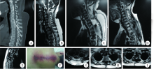
8例患者拔除引流后未见切口周围肿胀, MRI提示皮下积液量较少, 加压包扎24 h后下床活动; 3例患者拔除引流后出现切口周围肿胀, MRI提示皮下积液量较多, 但均无渗液, 予局部穿刺抽液后分别加压包扎5、7、7 d(图1)。
 | 图1 1例接受保守治疗的脑脊液漏继发皮下积液患者的影像学资料Figure 1 Conservative treatment for one patient with subcutaneous fistula secondary to cerebrospinal fluid leakageA, reconstructive computed tomography reveals C7-T3 continuous OPLL and T2-4 OLF; B, sagittal MRI demonstrated severe decompression of the spinal cord; C, postoperative MRI shows circumferential decompression of T1-2 OPLL and subcutaneous fistula secondary to CSFL, the patient undergoes C7-T5 laminectomy and T1-2 circumferential decompression via a posterior approach; D-E, MRI at 3 months’ and 6 months’ follow-up, CSF is absorbed gradually after aspiration. F, image of the swelling incision 2 weeks after surgery; G-I, axial sections of MRI reveal the absorption of CSFL from immediately postoperatively to 3 month and 6 month follow-up, respectively. Residual CSF could be detected. OPLL, ossification of posterior longitudinal ligament; OLF, ossification of ligamentum flavum; CSFL, cerebrospinal fluid leakage; CSF, cerebrospinal fluid; MRI, magnetic resonance imaging. |
随访时间30~131个月, 平均(85± 34)个月。随访发现10例患者脑脊液漏全部吸收, 所需时间为6个月。1例拔除引流管后出现切口周围肿胀的患者, 虽经穿刺抽液并加压包扎, 但在随访过程中发现脑脊液假性囊肿出现分隔未完全吸收, 患者症状改善不明显, 再次接受手术治疗(图2)。11例患者的JOA评分从术前的(3.8± 1.6)分升高到末次随访时的(8.9± 1.2)分, t=-13.8, P< 0.01, 神经功能改善率为70.8%。没有患者出现切口感染、切口不愈合或颅内感染等并发症。
 | 图2 1例因脑脊液假性囊肿而接受再次手术患者的影像学资料Figure 2 Reoperation for one patient presented with pseudocyst of cerebrospinal fluidA, preoperative MRI shows T3-7 OPLL and T3-4 OLF; B, 3 months postoperative MRI reveals formation of pseudocyst of CSF; C, pseudocyst of CSF is not absorbed at 6 months’ follow-up; D, axial section of MRI demonstrates subcutaneous fistula; E, axial section of MRI at 3 months’ follow-up; F, compression of the spinal cord resulting from the pseudocyst of CSF could be detected on the axial section of MRI at 6 months’ follow-up. The patient undergoes reoperation to remove the pseudocyst, and the JOA score increases from 2 preoperatively to 8 at the final follow-up. MRI, magnetic resonance imaging; OPLL, ossification of posterior longitudinal ligament; OLF, ossification of ligamentum flavum; CSF, cerebrospinal fluid; JOA, Japanese Orthopedic Association. |
胸椎手术硬脊膜损伤合并脑脊液漏的概率明显高于颈腰椎手术, 文献报告为3.8%~50.0%[3, 7, 8, 9, 10, 11]。北京大学第三医院研究发现胸椎管狭窄症术后脑脊液漏的发生率为32.3%[3]。本研究手术治疗的胸椎管狭窄症患者中, 发生脑脊液漏者186例, 其中合并皮下积液者11例(5.9%), 可见胸椎管狭窄症术后脑脊液漏合并皮下积液的发生率并不高, 而且本研究11例患者中只有3例表现出切口周围肿胀, 另外8例均为术后常规复查胸椎MRI时发现合并皮下积液, 其发生较为隐匿。如果皮下积液的量较大, 可能会影响切口的愈合, 甚至继发中枢神经系统感染, 本研究中未出现严重并发症的原因可能是病例数太少。随着超声骨刀等先进设备的应用, 越来越多的医院将具备开展胸椎手术的条件, 因此如何处理胸椎术后脑脊液漏继发皮下积液, 是我们无法回避的问题。
本研究中, 硬膜损伤的原因均为硬膜骨化, 其中硬膜破口位于脊髓背侧者6例, 位于脊髓腹侧者5例。脊髓腹侧的硬膜损伤难以术中修补, 即便是位于脊髓背侧的硬膜破口, 因其范围较大, 形状很不规则, 修补的难度也很大。本组患者术中采用明胶海绵覆盖, 术后患者均出现脑脊液漏, 引流量较大, 但有3例OLF患者引流量逐渐减少至80 mL以下, 提示术中对硬膜漏口的封堵可能有效, 成功率为27.3%。孙垂国等[7]报道的胸椎OLF手术并发脑脊液漏的修复成功率为34.8%, Sun等[11]报告266例胸椎OLF手术并发脑脊液漏的修复成功率为23.5%, 均明显低于腰椎术后脑脊液漏患者的修复成功率。Papavero 等[12]采取术中“ 10步法” 修复技术, 报告腰椎术后硬膜损伤的修复成功率为100%; McMahon等[13]报告脊柱手术硬膜损伤的修复成功率为92.1%, 但该研究的104例患者中仅有15例为胸椎手术。
对于胸椎术后脑脊液漏, 最有效的手段是预防其发生, 术前制定周密的手术计划以及术中仔细操作分离粘连是必不可少的, 但胸椎OPLL或OLF常合并硬膜骨化, 在揭盖切除椎管后壁或分离脊髓腹侧OPLL时, 非常容易损伤硬膜, 而且为了保证减压效果, 有时需要主动切除骨化的硬膜[2]。对于已经发生硬膜损伤的病例, 很多学者主张术中一期修复硬膜[14], 缝合技术多种多样[13, 15], 修复材料包括胶原蛋白基质[16]、水凝胶[17]、纤维蛋白胶[18]等, 但本研究中硬膜的破口范围大、形状不规则, 术中难以通过缝合或使用硬脊膜替代材料来进行修复, 而覆盖明胶海绵的有效率较低。还有学者采用腰大池置管引流治疗脊柱术后脑脊液漏获得满意的疗效, 满意率可以达到100%, 并且可以通过鞘内注射抗生素治疗颅内或者伤口深部感染[19]。但持续腰大池引流作为一项有创操作, 费用昂贵, 可能出现低颅压性头痛、脑疝、继发性出血等并发症, 而且如果脑脊液中混杂红细胞可能造成腰大池引流管的堵塞。总体而言, 我们认为腰大池引流是治疗胸椎管狭窄症术后脑脊液漏的有效方法, 其满意率优于本组保守治疗的病例, 但花费较多, 临床工作中可以根据具体情况酌情选择。
本研究发现, 少量皮下积液者采用加压包扎等保守治疗方法即可治愈; 对于3例大量皮下积液、切口周围明显肿胀者, 穿刺抽液联合加压包扎后, 有2例患者脑脊液漏完全吸收, 1例患者皮下积液吸收但深层的积液未完全吸收, 并且形成脑脊液假性囊肿压迫脊髓, 患者的症状恢复不满意从而需要二次手术(图2)。该例需再次手术的患者, 术中可见脑脊液假性囊肿形成分隔, 硬膜破口已愈合, 找不到假性囊肿与硬膜下腔相连的通道, 也没有发现脑脊液持续漏出。我们分析脑脊液漏合并皮下积液的可能原因包括:(1)腰背筋膜层缝合不够严密; (2)腰背筋膜缝合严密, 但漏出的脑脊液量太大, 筋膜层深方压力太高, 而患者筋膜层及肌肉力量薄弱, 难以阻挡脑脊液流注到皮下; 我们考虑后者可能性更大。10例患者保守治疗有效, 可能是由于加压包扎增加了患者切口对抗脑脊液漏的阻力[20], 筋膜层深方的脑脊液漏不再继续流注到皮下, 而皮下的脑脊液漏缓慢完全吸收。即便是接受二次手术的病例, 其皮下积液也完全吸收, 这间接地说明患者的筋膜层缝合比较严密。本研究中, 患者术后平均拔除引流管时间为(4.2± 1.1) d, 10例患者拔管后出现发热。我们考虑若引流液中血性成分不多, 延长引流时间可能会增加感染风险, 所以倾向于早期拔除引流管; 发热可能为拔管后产生的吸收热。但Fang等[20]主张延长引流管放置时间至7~10 d, 使筋膜层有足够的愈合时间。本组患者的JOA评分改善率为70.8%, 与采取相同术式的文献报告的改善率相当[21, 22], Desai 等[23]报告389例腰椎退变性滑脱患者, 发现术中发生脑脊液漏与否并不影响患者术后疗效。
本组11例脑脊液漏合并皮下积液患者保守治疗的成功率为90.9%, 因此我们主张对于胸椎术后脑脊液漏合并皮下积液的患者首选加压包扎或者穿刺抽液联合加压包扎等保守治疗。若脑脊液假性囊肿形成并压迫脊髓, 则需要二次手术清除脑脊液假性囊肿。
(本文编辑:刘淑萍)
The authors have declared that no competing interests exist.
| [1] |
|
| [2] |
|
| [3] |
|
| [4] |
|
| [5] |
|
| [6] |
|
| [7] |
|
| [8] |
|
| [9] |
|
| [10] |
|
| [11] |
|
| [12] |
|
| [13] |
|
| [14] |
|
| [15] |
|
| [16] |
|
| [17] |
|
| [18] |
|
| [19] |
|
| [20] |
|
| [21] |
|
| [22] |
|
| [23] |
|