促结缔组织增生型成釉细胞瘤的CT影像特点
孙崇珂1,张建运2,孙志鹏1,△( ),傅开元1,赵燕平1,张祖燕1,马绪臣1
),傅开元1,赵燕平1,张祖燕1,马绪臣1
Computed tomographic features of desmoplastic ameloblastoma of the jaw
Chong-ke SUN1,Jian-yun ZHANG2,Zhi-peng SUN1,△(),Kai-yuan FU1,Yan-ping ZHAO1,Zu-yan ZHANG1,Xu-chen MA1
),Kai-yuan FU1,Yan-ping ZHAO1,Zu-yan ZHANG1,Xu-chen MA1
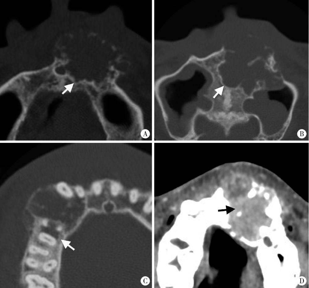
图1. 促结缔组织增生型成釉细胞瘤的边界形态特征
Figure 1. The boundary features of DA
A-B, DA shows scalloped shape with short sclerosed border on axial CT images (white arrows); C, Ill-defined border, infiltration into surrounding bone (white arrow) and locally destroyed cortex can be observed in DA; D, DA shows as solid mass on CT (black arrow).