中国人群遗传性周围神经病的致病基因分布
Genetic distribution in Chinese patients with hereditary peripheral neuropathy
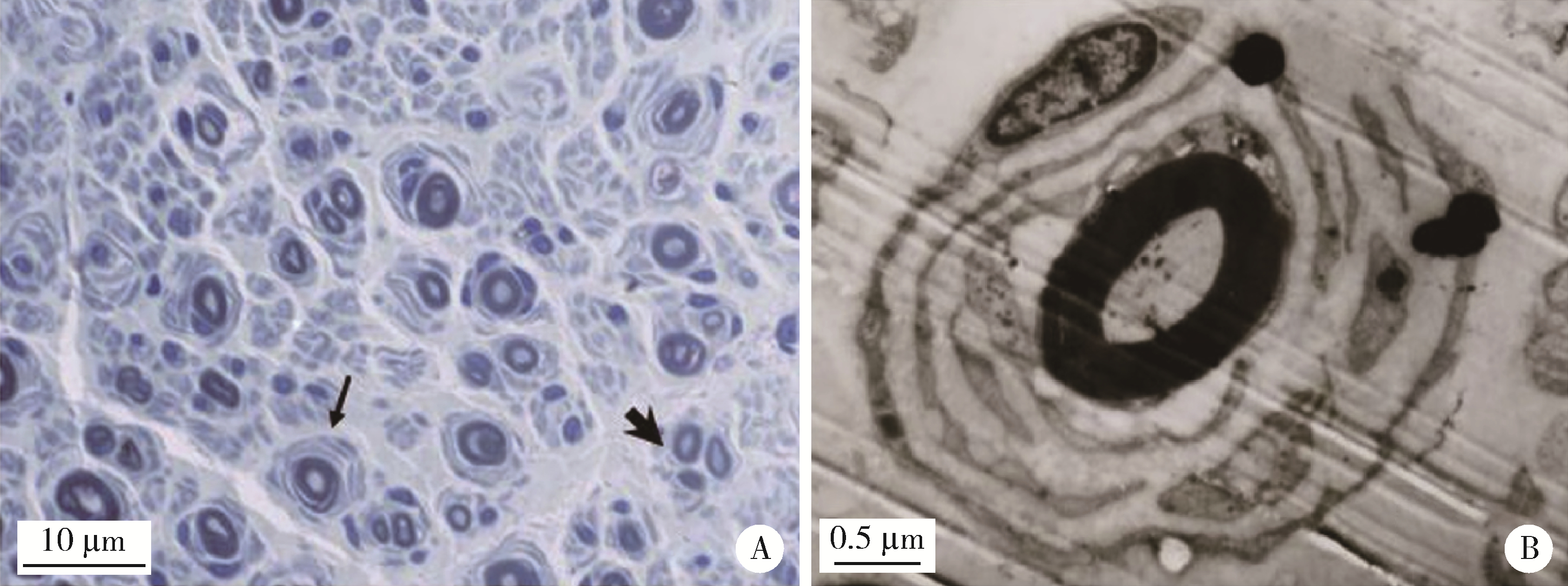
A, the number of normal myelinated nerve fibers is reduced, and some myelin are lost and regenerated, showing a typical "onion" like structure (thin arrow), and occasionally regenerative cluster like structure (thick arrow, semi-thin sections, toluidine blue staining ×200); B, the concentric round collagen layer structure formed by Schwann cell proliferation, namely the "onion" structure (electron micrography). CMT, Charcot-Marie-Tooth.