Journal of Peking University(Health Sciences) ›› 2018, Vol. 50 ›› Issue (6): 998-1003. doi: 10.19723/j.issn.1671-167X.2018.06.010
• Article • Previous Articles Next Articles
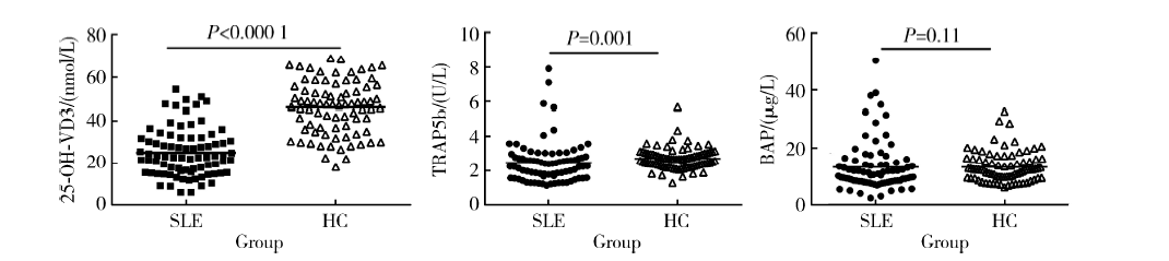
Study of bone mineral density and serum bone turnover markers in newly diagnosed systemic lupus erythematosus patients
Hai-hong YAO1,Su-mei TANG1,Zhi-min WANG1,Xia ZHANG1,Xu-yong CHEN1,Li GAO1,Jing LIU1,Yi-jun DAI1,Zhao-heng HU2,Xue-wu ZHANG( ),Zhan-guo LI1
- 1. Department of Rheumatology and Immunology, 2. Department of Endocrinology, Peking University People’s Hospital,Beijing 100044, China
CLC Number:
- R593.241


| [1] | Hochberg MC . Updating the American College of Rheumatology revised criteria for the classification of systemic lupus erythematosus[J]. Arthritis Rheum, 1997,40(9):1725. |
| [2] |
Guo Q, Fan P, Luo J , et al. Assessment of bone mineral density and bone metabolism in young male adults recently diagnosed with systemic lupus erythematosus in China[J]. Lupus, 2017,26(3):289-293.
doi: 10.1177/0961203316664596 pmid: 27522093 |
| [3] |
Boyanov M, Robeva R, Popivanov P . Bone mineral density changes in women with systemic lupus erythematosus[J]. Clin Rheumatol, 2003,22(4-5):318-323.
doi: 10.1007/s10067-003-0743-0 pmid: 14579164 |
| [4] |
Coimbra IB, Costallat LT . Bone mineral density in systemic lupus erythematosus and its relation to age at disease onset, plasmatic estradiol and immunosuppressive therapy[J]. Joint Bone Spine, 2003,70(1):40-45.
doi: 10.1016/S1297-319X(02)00009-X pmid: 12639616 |
| [5] |
Almehed K , Forsblad dH, Kvist G, et al. Prevalence and risk factors of osteoporosis in female SLE patients-extended report[J]. Rheumatology (Oxford), 2007,46(7):1185-1190.
doi: 10.1093/rheumatology/kem105 pmid: 17500075 |
| [6] |
Furukawa M, Kiyohara C, Tsukamoto H , et al. Prevalence of and risk factors for low bone mineral density in Japanese female patients with systemic lupus erythematosus[J]. Rheumatol Int, 2011,31(3):365-376.
doi: 10.1007/s00296-009-1244-5 pmid: 20020143 |
| [7] |
Cramarossa G, Urowitz MB, Su J , et al. Prevalence and associa-ted factors of low bone mass in adults with systemic lupus erythematosus[J]. Lupus, 2017,26(4):365-372.
doi: 10.1177/0961203316664597 pmid: 27522094 |
| [8] |
Compeyrot-Lacassagne S, Tyrrell PN, Atenafu E , et al. Prevalence and etiology of low bone mineral density in juvenile systemic lupus erythematosus[J]. Arthritis Rheum, 2007,56(6):1966-1973.
doi: 10.1002/art.22691 pmid: 17530722 |
| [9] |
Alsufyani KA, Ortiz-Alvarez O, Cabral DA , et al. Bone mineral density in children and adolescents with systemic lupus erythematosus, juvenile dermatomyositis, and systemic vasculitis: relationship to disease duration, cumulative corticosteroid dose, calcium intake, and exercise[J]. J Rheumatol, 2005,32(4):729-733.
doi: 10.1097/01.rhu.0000158687.22004.18 pmid: 15801032 |
| [10] |
Lilleby V, Lien G, Frey FK , et al. Frequency of osteopenia in children and young adults with childhood-onset systemic lupus erythematosus[J]. Arthritis Rheum, 2005,52(7):2051-2059.
doi: 10.1002/(ISSN)1529-0131 |
| [11] |
Ureña P, Hruby M, Ferreira A , et al. Plasma total versus bone alkaline phosphatase as markers of bone turnover in hemodialysis patients[J]. J Am Soc Nephrol, 1996,7(3):506-512.
doi: 10.1046/j.1464-410X.1996.96837.x pmid: 8704118 |
| [12] |
Shidara K, Inaba M, Okuno S , et al. Serum levels of TRAP5b, a new bone resorption marker unaffected by renal dysfunction, as a useful marker of cortical bone loss in hemodialysis patients[J]. Calcif Tissue Int, 2008,82(4):278-287.
doi: 10.1007/s00223-008-9127-4 pmid: 18421493 |
| [13] |
Keronen S, Martola L, Finne P , et al. Bone histomorphometry and indicators of bone and mineral metabolism in wait-listed dialysis patients[J]. Clin Nephrol, 2016,85(3):127-134.
doi: 10.5414/CN108709 pmid: 26833298 |
| [14] |
Dhillon VB, Davies MC, Hall ML , et al. Assessment of the effect of oral corticosteroids on bone mineral density in systemic lupus erythematosus: a preliminary study with dual energy X ray absorptiometry[J]. Ann Rheum Dis, 1990,49(8):624-626.
doi: 10.1136/ard.49.8.624 |
| [15] |
Breslin LC, Magee PJ, Wallace JM , et al. An evaluation of vitamin D status in individuals with systemic lupus erythematosus[J]. Proc Nutr Soc, 2011,70(4):399-407.
doi: 10.1017/S0029665111001613 pmid: 21861947 |
| [16] |
Bartels LE, Hvas CL, Agnholt J , et al. Human dendritic cell antigen presentation and chemotaxis are inhibited by intrinsic 25-hydroxy vitamin D activation[J]. Int Immunopharmacol, 2010,10(8):922-928.
doi: 10.1016/j.intimp.2010.05.003 pmid: 20483384 |
| [17] |
Jirapongsananuruk O, Melamed I, Leung DY . Additive immunosuppressive effects of 1, 25-dihydroxy vitamin D3 and corticosteroids on TH1, but not TH2, responses[J]. J Allergy Clin Immunol, 2000,106(5):981-985.
doi: 10.1067/mai.2000.110101 pmid: 11080724 |
| [18] |
Correale J, Ysrraelit MC, Gaitán MI . Immunomodulatory effects of vitamin D in multiple sclerosis[J]. Brain, 2009,132(Pt 5):1146-1160.
doi: 10.1093/brain/awp033 pmid: 19321461 |
| [19] |
Ruiz-Irastorza G, Gordo S, Olivares N , et al. Changes in vitamin D levels in patients with systemic lupus erythematosus: Effects on fatigue, disease activity, and damage[J]. Arthritis Care Res (Hoboken), 2010,62(8):1160-1165.
doi: 10.1002/acr.20186 pmid: 20235208 |
| [1] | Peng WANG,Hua WU,Ying CHE,Dong-wei FAN,Jue LIU,Li-yuan TAO. Evaluation of screening accuracy on osteoporosis self-assessment tool for Asians and its cut-off value in healthy physical examination population [J]. Journal of Peking University(Health Sciences), 2019, 51(6): 1085-1090. |
| [2] | CAO Jie, MENG Huan-xin. Evaluation of using cone beam computed tomography as a regular test before and after periodontal regenerative surgery#br# [J]. Journal of Peking University(Health Sciences), 2018, 50(1): 110-116. |
| [3] | WANG Yu, HAO Yan-jie, DENG Xue-rong, LI Guang-tao, GENG Yan, ZHAO Juan, ZHOU Wei, ZHANG Zhuo-li. Risk factors for bone mineral density changes in patients with rheumatoid arthritis and fracture risk assessment [J]. Journal of Peking University(Health Sciences), 2015, 47(5): 781-786. |
| [4] | WANG Yu, GENG Yan, DENG Xue-rong, ZHANG Zhuo-li. Relationship between wrist bone mineral density and synovitis, erosion by ultrasonography in female rheumatoid arthritis patients [J]. Journal of Peking University(Health Sciences), 2015, 47(5): 774-780. |
|
||