Journal of Peking University(Health Sciences) ›› 2019, Vol. 51 ›› Issue (3): 525-529. doi: 10.19723/j.issn.1671-167X.2019.03.022

Previous Articles     Next Articles

Quantitative evaluation of image quality of megavoltage computed tomography for guiding helical tomotherapy

Yu-liang HUANG1,Chen-guang LI1,Kai MAO2,Jian-an WU2,Tian-tian DAI2,Yuan-yuan HAN2,Hao WU1,Hai-yang WANG1,Yi-bao ZHANG1△()   

  • Received:2019-03-14 Online:2019-06-18 Published:2019-06-26
  • Supported by:
    Supported by the Capital’s Funds for Health Improvement and Research (2018-4-1027), Beijing Natural Science Foundation (7172048), the National Natural Science Foundation of China (11505012, 11505300), the Fundamental Research Funds for the Central Universities: Peking University Medicine Fund of Fostering Young Scholars’ Scientific & Technological Innovation (BMU2017PY028), Sichuan Science and Technology Program (2018HH0099), Beijing Municipal Administration of Hospitals Incubating Program (PX2019042, PX2016060), and Beijing Municipal Administration of Hospitals’ Youth Programme (QML20171104)

Abstract: Objective: To quantitatively analyze image quality of two sets of phantom (CatPhan504 and Cheese) Megavoltage computed tomography (MVCT) images acquired by Helical Tomotherapy with three scanning modes (Fine, Normal and Coarse), and to explore and validate a semi-automatic quality assurance procedure for MVCT images of Helical Tomotherapy.Methods: On Helical Tomotherapy, CatPan504 and Cheese phantoms were scanned with three pitch levels (Fine, Normal, Coarse: 4 mm, 8 mm, 12 mm/circle) respectively. Pylinac, Matlab and Eclipse were used to calculate and compare spatial resolution, noise level and low contrast resolution of images obtained under three scanning modes respectively. The spatial resolution can be evaluated by the blurring of line-pair CT value in the images of CatPhan504’s CTP528 module. The noise level can be evaluated by the integral non-uniformity in the images of Cheese’s uniformity module. the low contrast resolution can be evaluated by contrast-to-noise ratio of both phantoms’ plug-in module, or visibility of the region of interest (Supra-Slice) in the images of CatPhan504’s CTP515 module. Results: Analyses on CatPhan504’s line pair module(CTP528 module) showed that the first three line pairs(the gap size are 0.500 cm, 0.250 cm and 0.167 cm respectively) could be clearly observed but blurring began to occur from the fourth line pair(the gap size is 0.125 cm) under Coarse mode. Meanwhile, the first four line pairs were all observable under the Normal and Fine modes. Integral non-integrity index(the value negatively correlated with the noise level) were 0.155 7, 0.136 8 and 0.122 9 for Coarse, Normal and Fine modes respectively. None of the Supra-Slice in CatPhan504’s CTP515 module could be observed under three imaging modes. Low contrast contrast-to-noise ratio of Cheese phantom was similar under three modes and the insert visibility exhibited nearly linear growth with the increasing difference between CT average value of the insert material and background.Conclusion: Superiority and inferiority of three image modes in terms of the three image quality index was not consistent. Evaluation results above could provide reference for more rational decision on scanning modes selection of helical tomotherapy, which was based on image visualization demands in clinical practice. The proposed method could also provide guidance for similar image quality assessment and periodic quality assurance.

Key words: Helical tomotherapy, Megavoltage computed tomography, Image Quality, Quality Assurance

CLC Number: 

  • R815

Figure 1

The internal structure of CTP528 module in CatPhan504 and the gap size of each line pair"

Figure 2

CatPhan504 CTP528 module line pair area, CT value as a function of sample point’s serial number A, coarse; B, normal; C, fine."

Table 1

Image noise evaluation results of Cheese phantom"

Group Location
Upper left Lower left Upper right Lower right Integral
non-uniformity/
(×10-2)
Coarse 17.933 18.819 13.692 17.105 15.77
Normal 17.627 19.878 15.905 16.289 13.68
Fine 19.236 20.944 16.361 17.454 12.29

Figure 3

Contrast-to-noise ratio of CatPhan504 phantom LDPE, low density polyethylene; PMP, polymethylpentene."

Figure 4

Contrast-to-noise ratio of cheese phantom"

[1] Langen KM, Papanikolaou N, Balog J , et al. QA for helical tomotherapy: report of the AAPM task group 148[J]. Med Phys, 2010,37(9):4817-4853.
doi: 10.1118/1.3462971
[2] Yadav P, Tolakanahalli R, Rong Y , et al. The effect and stability of MVCT images on adaptive tomotherapy[J]. J Appl Clin Med Phys, 2010,11(4):4-14.
doi: 10.1120/jacmp.v11i4.3229
[3] Maggiulli E, Fiorino C, Passoni P , et al. Characterisation of rectal motion during neo-adjuvant radiochemotherapy for rectal cancer with image-guided tomotherapy: implications for adaptive dose escalation strategies[J]. Acta Oncol, 2012,51(3):318-324.
doi: 10.3109/0284186X.2012.666358
[4] 丛小虎, 徐寿平, 解传滨 , 等. Tomotherapy系统五年质控结果及其模式的转变[J]. 中国医疗器械杂志, 2016,40(5):380-385.
[5] 李正贤, 赵晶晶, 马永乐 , 等. 医科达XVI锥形束CT头颈部扫描角度优化[J]. 中国医学物理学杂志, 2017,34(11):1106-1109.
[6] Meeks SL, Harmon FJ, Langen K , et al. Performance characterization of megavoltage computed tomography imaging on a helical tomotherapy unit[J]. Med Phys, 2005,32(8):2673-2681.
doi: 10.1118/1.1990289
[7] 刘明娜, 王谦, 杨新 , 等. 图像质量客观评价方法在 CT 图像中的应用[J]. 生物医学工程学杂志, 2011,28(2):357-364.
[8] Zarb F, Rainford L, Mcentee MF . Image quality assessment tools for optimization of CT images[J]. Radiography, 2010,16(2):147-153.
doi: 10.1016/j.radi.2009.10.002
[9] Klein EE, Hanley J, Bayouth J , et al. Task group 142 report: quality assurance of medical accelerators[J]. Med Phys, 2009,36(9):4197-4212.
doi: 10.1118/1.3190392
[10] Zaila A, Adili M, Bamajboor S . Pylinac: a toolkit for performing TG-142 QA related tasks on linear accelerator[J]. Phys Med, 2016,32(3):292-293.
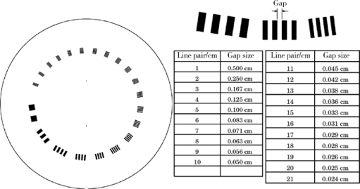
[11] David J . Goodenough: catphan 504 manual[M]. New York: The Phantom Laboratory, 2012: 7.
[12] Kerns JR. Pylinac Documentation: Realease 2.2.1[EB/OL]. ( 2018 -12-15)[2019-01-18]. https://buildmedia.readthedocs.org/media/pdf/pylinac/v2.2.1/pylinac.pdf .
[13] Cheng HC, Wu VW, Liu ES , et al. Evaluation of radiation dose and image quality for the Varian cone beam computed tomography system[J]. Int J Radiat Oncol Biol Phys, 2011,80(1):291-300.
doi: 10.1016/j.ijrobp.2010.06.014
[14] Gulliksrud K, Stokke C, Martinsen AC , et al. How to measure CT image quality: variations in CT-numbers, uniformity and low contrast resolution for a CT quality assurance phantom[J]. Phys Med, 2014,30(4):521-526.
doi: 10.1016/j.ejmp.2014.01.006
[15] Alahbabi SS, Bradley DA, Nisbet A , et al. Tomotherapy evaluation for head and neck cases using two types of phantoms[J]. Radiat Phys Chem, 2014,95:323-325.
doi: 10.1016/j.radphyschem.2013.04.022
[16] Verdun FR, Racine D, Ott JG , et al. Image quality in CT: from physical measurements to model observers[J]. Phys Med, 2015,31(8):823-843.
doi: 10.1016/j.ejmp.2015.08.007
[1] LI Yuan, ZHENG Gang, LIN Hong. Evaluation method with radiographic image quality indicator for internal defects of dental casting metallic restoration [J]. Journal of Peking University(Health Sciences), 2014, 46(6): 963-968.
Viewed
Full text


Abstract

Cited

  Shared   
  Discussed   
[1] Author. English Title Test[J]. Journal of Peking University(Health Sciences), 2010, 42(1): 1 -10 .
[2] . [J]. Journal of Peking University(Health Sciences), 2009, 41(2): 188 -191 .
[3] . [J]. Journal of Peking University(Health Sciences), 2009, 41(3): 376 -379 .
[4] . [J]. Journal of Peking University(Health Sciences), 2009, 41(4): 459 -462 .
[5] . [J]. Journal of Peking University(Health Sciences), 2009, 41(6): 682 -686 .
[6] . [J]. Journal of Peking University(Health Sciences), 2010, 42(1): 82 -84 .
[7] . [J]. Journal of Peking University(Health Sciences), 2007, 39(3): 319 -322 .
[8] . [J]. Journal of Peking University(Health Sciences), 2007, 39(3): 333 -336 .
[9] . [J]. Journal of Peking University(Health Sciences), 2007, 39(3): 337 -340 .
[10] . [J]. Journal of Peking University(Health Sciences), 2007, 39(3): 225 -328 .