Journal of Peking University (Health Sciences) ›› 2021, Vol. 53 ›› Issue (4): 808-810. doi: 10.19723/j.issn.1671-167X.2021.04.032

Previous Articles     Next Articles

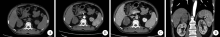
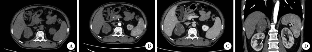
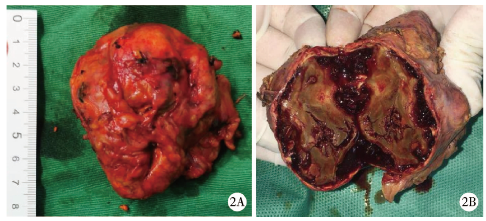
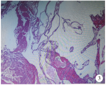
Adrenal cavernous hemangioma: A case report and literature review

KANG Wen-yu1,WANG Lu2,QIU Min2,Δ(),ZHANG Fan2,GUO Wei1,Δ(),QIANG Ya-yong1,TUO Peng-fei1,ZONG You-long1,LIU Lei-lei1,WANG Shuai-shuai1   

  1. 1. Department of Urology, Yan’an City Hospital of Traditional Chinese Medicine, Yan’an 716000, Shaanxi, China
    2. Department of Urology, Peking University Third Hospital, Beijing 100191, China
  • Received:2021-03-16 Online:2021-08-18 Published:2021-08-25
  • Contact: Min QIU,Wei GUO E-mail:qiumin@bjmu.edu.cn;15756560@qq.com

Abstract:

A 57-year-old male patient was referred to our department with complaints of his right adrenal gland occupancy and hypertension about 6 months. When admitted to the hospital, the blood pressure was about 160/100 mmHg, and the heart rate was 110 beats/min. He was no obvious obesity, acne, abnormal mood, without weakness of limbs, acral numbness, palpitation and headache. He presented with type 2 diabetes for more than 3 years, with oral administration of metformin enteric coated tablets and subcutaneous injection of insulin glargine to control blood glucose, and satisfied with blood glucose control. Enhanced CT showed that: the right adrenal gland showed a kind of oval isodense, slightly hypodense shadow, the edge was clear, lobular change, the size was about 5.8 cm×5.4 cm, uneven density, there were nodular and strip calcification, round lipid containing area and strip low density area, and the CT value of solid part was about 34 HU. Enhanced scan showed heterogeneous nodular enhancement in the solid part of the right adrenal gland, nodular enhancement could be seen inside. The CT values of solid part in arterial phase, venous phase and delayed phase were 45 HU, 50 HU and 81 HU, respectively. Considering from the right adrenal gland, cortical cancer was more likely. No obvious abnormality was found in his endocrine examination. After adequate preoperative preparation, retroperitoneal laparoscopic adrenalectomy was performed under general anesthesia. During the operation, the 6 cm adrenal tumor was closely related to the inferior vena cava and liver, and after careful separation, the tumor was completely removed and normal adrenal tissue was preserved. The operation lasted 180 min and the blood loss was 100 mL, and the blood pressure was stable during and after the operation. There was no obvious complication. The results of pathological examinations were as follows: the size of the tumor was 7.5 cm×6.0 cm×3.5 cm, soft, with intact capsule and grayish-red cystic in section. Pathological diagnosis: (right adrenal gland) cavernous hemangioma, secondary intravascular thrombosis, old hemorrhagic infarction with calcification and ossification. After 6 months of observation, no obvious complications and tumor recurrence were found. In summary, cavernous hemangioma of adrenal gland is a rare histopathological change. Its essence is a malformed vascular mass. Blood retention is the cause of thrombosis and calcification in malformed vessels. The imaging findings were inhomogeneous enhancement of soft tissue masses, and the adrenal function examination showed no obvious abnormalities. Retroperitoneal laparoscopic surgery is feasible after adequate preoperative preparation. It is difficult to diagnose the disease preoperatively and needs to be confirmed by postoperative pathology.

Key words: Adrenal gland neoplasms, Cavernous hemangioma, Adrenalectomy

CLC Number: 

  • R736.6

Figure 1

CT findings of adrenal cavernous hemangioma A, plain scan of the right adrenal gland showed a kind of elliptic isopensity and slightly lower density shadow with clear edge and lobulated change; B, enhanced arterial level: uneven progressive nodular enhancement of the right adrenal mass, with no enhancement in the low-density area; C, delayed phase level: right adrenal mass still had enhancement; D, coronal scanning: a large, well-defined adrenal mass with calcification in the right adrenal gland."

Figure 2

The right adrenal mass specimen was completely resected 2A, the tumor size was 7.5 cm×6.0 cm×3.5 cm, and the capsule was complete; 2B, the profile of the mass was cystic, grayish-red and soft;"

Figure 3

Histopathological examination of the right adrenal mass (HE ×10) 3, adrenal tumor with large, thin-walled, cystic dilated vessels, followed by intravascular thrombosis, old hemorrhagic infarction with calcification and ossification."

[1] Feo CV, De Troia A, Pedriali M, et al. Adrenal cavernous hemangioma: a case report [J]. BMC Surg, 2018, 18(1):103-107.
doi: 10.1186/s12893-018-0429-9
[2] Degheili JA, Abou HN, El-Moussawi M, et al. Adrenal cavernous hemangioma: A rarely perceived pathology-case illustration and review of literature [J]. Case Rep Pathol, 2019, 2019:8463890.
doi: 10.1155/2019/8463890 pmid: 31949968
[3] Noh JJ, Choi SH, Hwang HK, et al. Adrenal cavernous heman-gioma: a case report with review of the literature [J]. JOP, 2014, 15(3):254-257.
[4] Johnson CC, Jeppesen FB. Hemangioma of the adrenal [J]. J Urol, 1955, 74(5):573-577.
doi: 10.1016/S0022-5347(17)67320-8
[5] Hashimoto A, Yoshino H, Yoshikawa F, et al. Giant cavernous hemangioma of the adrenal gland in an elderly patient [J]. Intern Med, 2018, 57(9):1317-1319.
doi: 10.2169/internalmedicine.9711-17
[6] Agrusa A, Romano G, Salamone G, et al. Large cavernous hemangioma of the adrenal gland: Laparoscopic treatment. Report of a case [J]. Int J Surg Case Rep, 2015, 16:150-153.
doi: 10.1016/j.ijscr.2015.09.040
[1] Wen-cheng AN,Hui-xian YAN,Zheng-zhao DENG,Fang CHEN,Xiao-hong OU,Hong-xin JIN,Wei HUANG. Chronic kidney disease after adrenalectomy in a patient with primary aldosteronism [J]. Journal of Peking University (Health Sciences), 2021, 53(6): 1201-1204.
[2] Lei LIU,Guo-liang WANG,Lu-lin MA,Min LU,Cheng LIU,Xiao-fei HOU. Diagnosis and surgical treatment of massive adrenal area tumor with tumor thrombus [J]. Journal of Peking University(Health Sciences), 2019, 51(4): 684-688.
[3] FAN Hua, LI Han-zhong, XU Wei-feng, JI Zhi-gang, ZHANG Yu-shi. Surgery treatment of ectopic adrenocorticotrophic hormone syndrome [J]. Journal of Peking University(Health Sciences), 2017, 49(4): 652-656.
[4] QIU Min, ZONG Ya-nan, LU Jian, MA Lu-lin, ZHENG Qing, GUO Xiang-yang. Rocuronium anesthesia induced anaphylactic shock: a case report [J]. Journal of Peking University(Health Sciences), 2015, 47(5): 885-887.
[5] WU Chu-Jun, QIU Min, MA Lu-Lin. Progress in diagnosis and treatment of adrenal metastases tumor [J]. Journal of Peking University(Health Sciences), 2015, 47(4): 728-731.
Viewed
Full text


Abstract

Cited

  Shared   
  Discussed   
[1] Author. English Title Test[J]. Journal of Peking University(Health Sciences), 2010, 42(1): 1 -10 .
[2] . [J]. Journal of Peking University(Health Sciences), 2009, 41(2): 188 -191 .
[3] . [J]. Journal of Peking University(Health Sciences), 2009, 41(3): 376 -379 .
[4] . [J]. Journal of Peking University(Health Sciences), 2009, 41(4): 459 -462 .
[5] . [J]. Journal of Peking University(Health Sciences), 2010, 42(1): 82 -84 .
[6] . [J]. Journal of Peking University(Health Sciences), 2007, 39(3): 319 -322 .
[7] . [J]. Journal of Peking University(Health Sciences), 2007, 39(3): 333 -336 .
[8] . [J]. Journal of Peking University(Health Sciences), 2007, 39(3): 337 -340 .
[9] . [J]. Journal of Peking University(Health Sciences), 2007, 39(3): 225 -328 .
[10] . [J]. Journal of Peking University(Health Sciences), 2007, 39(4): 346 -350 .