Journal of Peking University (Health Sciences) ›› 2022, Vol. 54 ›› Issue (3): 557-564. doi: 10.19723/j.issn.1671-167X.2022.03.024

Previous Articles     Next Articles

Effects of different crosslinking treatments on the properties of decellularized small intestinal submucosa porous scaffolds

Yi DENG1,Yi ZHANG2,Bo-wen LI1,Mei WANG1,Lin TANG1,Yu-hua LIU1,*()   

  1. 1. Department of Prosthodontics, Peking University School and Hospital of Stomatology & National Center of Stomatology & National Clinical Research Center for Oral Diseases & National Engineering Research Center of Oral Biomaterials and Digital Medical Devices & Beijing Key Laboratory of Digital Stomatology & NHC Research Center of Engineering and Technology for Computerized Dentistry & NMPA Key Laboratory for Dental Materials, Beijing 100081, China
    2. Department of General Dentistry Ⅱ, Peking University School and Hospital of Stomatology & National Center of Stomatology & National Clinical Research Center for Oral Diseases & National Engineering Research Center of Oral Biomaterials and Digital Medical Devices & Beijing Key Laboratory of Digital Stomatology & NHC Research Center of Engineering and Technology for Computerized Dentistry & NMPA Key Laboratory for Dental Materials, Beijing 100081, China
  • Received:2020-10-10 Online:2022-06-18 Published:2022-06-14
  • Contact: Yu-hua LIU E-mail:liuyuhua@bjmu.edu.cn
  • Supported by:
    the National Natural Science Foundation of China(81801027)

Abstract:

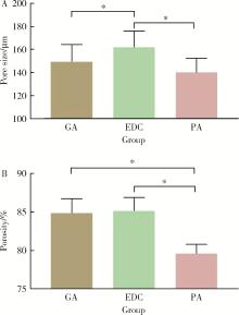
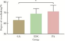
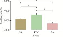
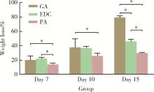
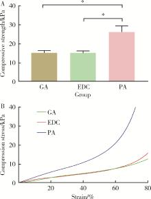
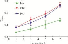
Objective: To compare the effects of three different crosslinkers on the biocompatibility, physical and chemical properties of decellularized small intestinal submucosa (SIS) porous scaffolds. Methods: The SIS porous scaffolds were prepared by freeze-drying method and randomly divided into three groups, then crosslinked by glutaraldehyde (GA), 1-ethyl-3-(3-dimethylaminopropyl) carbodi-imide (EDC) and procyanidine (PA) respectively. To evaluate the physicochemical property of each sample in different groups, the following experiments were conducted. Macroscopic morphologies were observed and recorded. Microscopic morphologies of the scaffolds were observed using field emission scanning electron microscope (FESEM) and representative images were selected. Computer software (ImageJ) was used to calculate the pore size and porosity. The degree of crosslinking was determined by ninhydrin experiment. Collagenase degradation experiment was performed to assess the resistance of SIS scaffolds to enzyme degradation. To evaluate the mechanical properties, universal mechanical testing machine was used to determine the stress-strain curve and compression strength was calculated. Human bone marrow mesenchymal cells (hBMSCs) were cultured on the scaffolds after which cytotoxicity and cell proliferation were assessed. Results: All the scaffolds remained intact after different crosslinking treatments. The FESEM images showed uniformed interconnected micro structures of scaffolds in different groups. The pore size of EDC group[(161.90±13.44) μm] was significantly higher than GA group [(149.50±14.65) μm] and PA group[(140.10±12.06) μm] (P < 0.05). The porosity of PA group (79.62%±1.14%) was significantly lower than EDC group (85.11%±1.71%) and GA group (84.83%±1.89%) (P < 0.05). PA group showed the highest degree of crosslinking whereas the lowest swelling ratio. There was a significant difference in the swelling ratio of the three groups (P < 0.05). Regarding to the collagenase degradation experiment, the scaffolds in PA group showed a significantly lower weight loss rate than the other groups after 7 days degradation. The weight loss rates of GA group were significantly higher than those of the other groups on day 15, whereas the PA group had the lowest rate after 10 days and 15 days degradation. PA group showed better mechanical properties than the other two groups. More living cells could be seen in PA and EDC groups after live/dead cell staining. Additionally, the proliferation rate of hBMCSs was faster in PA and EDC groups than in GA group. Conclusion: The scaffolds gained satisfying degree of crosslinking after three different crosslinking treatments. The samples after PA and EDC treatment had better physicochemical properties and biocompatibility compared with GA treatment. Crosslinking can be used as a promising and applicable method in the modification of SIS scaffolds.

Key words: Decellularized small intestinal submucosa, Porous scaffolds, Glutaraldehyde, 1-ethyl-3-(3-dimethylaminopropyl) carbodiimide, Procyanidine

CLC Number: 

  • R783.1

Figure 1

Macroscopic morphology of different crosslinked SIS porous scaffolds"

Figure 2

The surface and cross-section morphology of the crosslinked scaffolds by FESEM A, B and C, the top view of different SIS porous scaffolds crosslinked by GA, EDC and PA respectively; D, E and F, the side view of different SIS porous scaffolds crosslinked by GA, EDC and PA respectively.A to C, FESEM images of surface morphology of the scaffolds crosslinked by GA, EDC and PA respectively; D to F, FESEM images of cross-section morphology of the scaffolds crosslinked by GA, EDC and PA respectively (Magnification=500, scale bar=300 μm). GA, glutaraldehyde; PA, procyanidine; EDC, 1-ethyl-3-(3-dimethylaminopropyl) carbodiimide."

Figure 3

The pore size and porosity of crosslinked SIS porous scaffolds A, pore size; B, porosity; GA, glutaraldehyde; PA, procyanidine; EDC, 1-ethyl-3-(3-dimethylaminopropyl) carbodiimide. * P<0.05."

Figure 4

The crosslinking degreeof SIS porous scaffolds GA, glutaraldehyde; PA, procyanidine; EDC, 1-ethyl-3-(3-dimethylaminopropyl) carbodiimide. * P<0.05."

Figure 5

The swelling ratio of crosslinked SIS porous scaffolds GA, glutaraldehyde; PA, procyanidine; EDC, 1-ethyl-3-(3-dimethylaminopropyl) carbodiimide. * P<0.05."

Figure 6

Weight losses rates of different crosslinked SIS porous scaffolds GA, glutaraldehyde; PA, procyanidine; EDC, 1-ethyl-3-(3-dimethylaminopropyl) carbodiimide. * P<0.05."

Figure 7

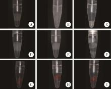
Macroscopic morphology of different crosslinked SIS porous scaffolds in digestion solution on day 7, day 10 and day 15 A, B and C, the SIS scaffold of GA group degraded in digestion solution on day 7, day 10 and day 15 respectively; D, E and F, the SIS scaffold of EDC group degraded in digestion solution on day 7, day 10 and day 15 respectively; G, H and I, the SIS scaffold of PA group degraded in digestion solution on day 7, day 10 and day 15 respectively."

Figure 8

Compressive strengthand stress-strain curves of different crosslinked SIS porous scaffolds A, compressive strength; B, stress-strain curves. GA, glutaraldehyde; PA, procyanidine; EDC, 1-ethyl-3-(3-dimethylaminopropyl) carbodiimide. * P<0.05."

Figure 9

Image of live/dead assay staining of hBMSCs indifferent crosslinked SIS scaffolds A-C, images of live/dead assay staining of hBMCSs in GA group after 24 h culture time (A, living cells, green; B, dead cells, red; C, merge of A and B); D-F, images of live/dead assay staining of hBMCSs in EDC group after 24 h culture time (D, living cells, green; E, dead cells, red; F, merge of D and E); G-I, images of live/dead assay staining of hBMCSs in PA group after 24 h culture time (G, living cells, green; H, dead cells, red; I, merge of G and H)."

Figure 10

The proliferation curves of hBMSCs cultured on different crosslinked SIS porous scaffolds GA, glutaraldehyde; PA, procyanidine; EDC, 1-ethyl-3-(3-dimethylaminopropyl) carbodiimide."

1 Sengupta D , Waldman S , Li S . From in vitro to in situ tissue engineering[J]. Ann of Biomed Eng, 2014, 42 (7): 1537- 1545.
doi: 10.1007/s10439-014-1022-8
2 Li Y , Qiao Z , Yu F , et al. Transforming growth factor-β3/chitosan sponge (TGF-β3/CS) facilitates osteogenic differentiation of human periodontal ligament stem cells[J]. Int J Mol Sci, 2019, 20 (20): 4982.
doi: 10.3390/ijms20204982
3 Veres SP , Harrison JM , Lee JM . Cross-link stabilization does not affect the response of collagen molecules, fibrils, or tendons to tensile overload[J]. J OrthopRes, 2013, 31 (12): 1907- 1913.
4 Theocharis AD, Skandalis SS, Gialeli C, et al. Extracellular matrix structure[J/OL]. Adv Drug Deliv Rev, 2016, 97: 4-27[2020-03-01]. https://www.sciencedirect.com/science/article/pii/S0169409X15002574.
5 Li M, Zhang C, Cheng M, et al. Small intestinal submucosa: A potential osteoconductive and osteoinductive biomaterial for bone tissue engineering[J/OL]. Mater Sci Eng C, 2017, 75: 149-156[2020-10-01]. https://www.sciencedirect.com/science/article/pii/S0928493116322937.
6 李博文, 吴唯伊, 唐琳, 等. 改良猪小肠黏膜下层可吸收膜在兔下颌骨缺损早期愈合中的作用[J]. 北京大学学报(医学版), 2019, 51 (5): 887- 892.
7 Sun L, Li B, Jiang D, et al. Nile tilapia skin collagen sponge modified with chemical cross-linkers as a biomedical hemostatic material[J/OL]. Colloids Surf. B, 2017, 159: 89-96[2020-03-01]. https://www.sciencedirect.com/science/article/pii/S0927776517304836.
8 Tottey S , Johnson SA , Crapo PM , et al. The effect of source animal age upon extracellular matrix scaffold properties[J]. Biomaterials, 2011, 32 (1): 128- 136.
doi: 10.1016/j.biomaterials.2010.09.006
9 Castilla B, Buttigieg J, Juan C, et al. Development and charac-terization of a novel porous small intestine submucosa-hydroxyapatite scaffold for bone regeneration[J/OL]. Mater Sci Eng C, 2017, 72: 519-525[2020-10-01]. https://www.sciencedirect.com/science/article/pii/S0928493116309675.
10 Liu X, Zheng C, Luo X, et al. Recent advances of collagen-based biomaterials: Multi-hierarchical structure, modification and biomedical applications[J/OL]. Mater Sci Eng C, 2019, 99: 1509-1522[2020-10-01]. https://www.sciencedirect.com/science/article/pii/S092849311831765X.
11 Olde D , Van L , Dijkstra P , et al. Glutaraldehyde as a cross-linking agent for collagen-based biomaterials[J]. J Mater Sci Mater Med, 1995, 6 (8): 460- 472.
doi: 10.1007/BF00123371
12 Usha R, Sreeram KJ, Rajaram A. Stabilization of collagen with EDC/NHS in the presence of l-lysine: A comprehensive study[J/OL]. Colloids Surf. B, 2012, 90: 83-90[2020-10-01]. https://www.sciencedirect.com/science/article/pii/S092777651100573X.
13 Mitra T , Sailakshmi G , Gnanamani A , et al. Preparation and characterization of a thermostable and biodegradable biopolymers using natural cross-linker[J]. Int J Biol Macromol, 2011, 48 (2): 276- 285.
doi: 10.1016/j.ijbiomac.2010.11.011
14 Sun L, Li B, Song W, et al. Comprehensive assessment of nile tilapia skin collagen sponges as hemostatic dressings[J/OL]. Mater Sci Eng C, 2020, 109: 1105-1132[2020-10-01]. https://www.sciencedirect.com/science/article/pii/S0928493118327012.
15 Ward J , Kelly J , Wang W , et al. Amine functionalization of collagen matrices with multifunctional polyethylene glycol systems[J]. Biomacromolecules, 2010, 11 (11): 3093- 3101.
doi: 10.1021/bm100898p
16 Charulatha V , Rajaram A . Influence of different crosslinking treatments on the physical properties of collagen membranes[J]. Biomaterials, 2003, 24 (5): 759- 767.
doi: 10.1016/S0142-9612(02)00412-X
17 Delgado LM , Fuller K , Zeugolis D . Collagen cross-linking: Biophysical, biochemical, and biological besponse analysis[J]. Tissue Eng Pt A, 2017, 23 (19/20): 1064- 1077.
18 Ma L , Gao C , Mao Z , et al. Collagen/chitosan porous scaffolds with improved biostability for skin tissue engineering[J]. Biomaterials, 2003, 24 (26): 4833- 4841.
doi: 10.1016/S0142-9612(03)00374-0
19 Chandika P, Ko S, Oh G, et al. Fish collagen/alginate/chitooligosaccharides integrated scaffold for skin tissue regeneration application[J/OL]. Int J Biol Macromol, 2015, 81: 504-513[2020-10-01]. https://www.sciencedirect.com/science/article/pii/S0141813015005826.
20 Shi Y , Zhang H , Zhang X , et al. A comparative study of two porous sponge scaffolds prepared by collagen derived from porcine skin and fish scales as burn wound dressings in a rabbit model[J]. Regen Biomater, 2020, 7 (1): 63- 70.
doi: 10.1093/rb/rbz036
21 Vidal C, Zhu W, Manohar S, et al. Collagen-collagen interactions mediated by plant-derived proanthocyanidins: A spectroscopic and atomic force microscopy study[J/OL]. Acta Biomater, 2016, 41: 110-118[2020-03-01]. https://www.sciencedirect.com/science/article/pii/S1742706116302446.
22 He L , Mu C , Shi J , et al. Modification of collagen with a natural cross-linker, procyanidin[J]. Int J Biol Macromol, 2011, 48 (2): 354- 359.
doi: 10.1016/j.ijbiomac.2010.12.012
23 Trifković K , Milašinović N , Djordjević V , et al. Chitosan crosslinked microparticles with encapsulated polyphenols: Water sorption and release properties[J]. J Biomater Appl, 2015, 30 (5): 618- 631.
doi: 10.1177/0885328215598940
24 Li Q, Mu L, Zhang F, et al. A novel fish collagen scaffold as dural substitute[J/OL]. Mater Sci Eng C Mater Biol Appl, 2017, 80: 346-351[2020-10-01]. https://www.sciencedirect.com/science/article/pii/S0928493117310627.
[1] LUO Hao, TIAN Fu-cong, WANG Xiao-yan. Surface roughness, gloss and sequential polishing times of various chairside computer aided design/manufacturing restorative materials [J]. Journal of Peking University (Health Sciences), 2022, 54(3): 565-571.
[2] YAN Wen-juan,ZHONG Jie,LIN Bi-chen,DING Mei-li,CHEN Xiao-xian. Application of cold flowable gutta-percha in root canal obturation after apexification [J]. Journal of Peking University (Health Sciences), 2022, 54(1): 77-82.
[3] LI Yi,WONG Lai U,LIU Xiao-qiang,ZHOU Ti,LYU Ji-zhe,TAN Jian-guo. Marginal features of CAD/CAM laminate veneers with different materials and thicknesses [J]. Journal of Peking University (Health Sciences), 2022, 54(1): 140-145.
[4] MA Xin-rong,ZHU Xiao-ming,LI Jing,LI De-li,LI He-ping,TAN Jian-guo. Effect of a novel radio-frequency atmospheric-pressure glow discharge plasma jet treatment on crosslinking of dentin collagen [J]. Journal of Peking University (Health Sciences), 2022, 54(1): 83-88.
[5] Yuan LI,Hong LIN,Tie-jun ZHANG. Comparative study on radio-opacity of dental composite resin materials’determination using film imaging and digital imaging [J]. Journal of Peking University (Health Sciences), 2021, 53(5): 995-1001.
[6] YOU Peng-yue,LIU Yu-hua,WANG Xin-zhi,WANG Si-wen,TANG Lin. Biocompatibility and effect on bone formation of a native acellular porcine pericardium: Results of in vitro and in vivo [J]. Journal of Peking University (Health Sciences), 2021, 53(4): 776-784.
[7] WANG Li-xin , XU Xiao, NI Yao-feng, SUN Hai-tao, YU Ri-yue, WEI Shi-cheng. In vivo study of liposome-modified polyetheretherketone implant on bacteriostasis and osseointegration [J]. Journal of Peking University (Health Sciences), 2021, 53(4): 758-763.
[8] WANG Jing-qi,WANG Xiao. In vivo study of strontium-doped calcium phosphate cement for biological properties [J]. Journal of Peking University (Health Sciences), 2021, 53(2): 378-383.
[9] MU Hai-li,TIAN Fu-cong,WANG Xiao-yan,GAO Xue-jun. Evaluation of wear property of Giomer and universal composite in vivo [J]. Journal of Peking University (Health Sciences), 2021, 53(1): 120-125.
[10] Wen-xin CHEN,Xu-dong BAO,Lin YUE. Curing method affecting the formation of oxygen inhibition layer on the surface of resin cement [J]. Journal of Peking University (Health Sciences), 2020, 52(6): 1117-1123.
[11] Qiu-ju LI,Wei-yu GONG,Yan-mei DONG. Effect of bioactive glass pretreatment on the durability of dentin bonding interface [J]. Journal of Peking University (Health Sciences), 2020, 52(5): 931-937.
[12] Ren-tao TANG,Xin-hai LI,Jiang-li YU,Lin FENG,Xue-jun GAO. Evaluation of microtensile bond strength between resin composite and glass ceramic [J]. Journal of Peking University (Health Sciences), 2020, 52(4): 755-761.
[13] . [J]. Journal of Peking University (Health Sciences), 2020, 52(4): 790-793.
[14] Chun-ling CAO,Cong-chong YANG,Xiao-zhong QU,Bing HAN,Xiao-yan WANG. Effects of the injectable glycol-chitosan based hydrogel on the proliferation and differentiation of human dental pulp cells [J]. Journal of Peking University(Health Sciences), 2020, 52(1): 10-17.
[15] Jing-ying HU,Li LI,Qian-mei ZHOU,Rui-yu DING,Ran SHANG,Wei BAI. Influence of different mixing pads on physical and mechanical properties of glass ionomer cement [J]. Journal of Peking University(Health Sciences), 2019, 51(5): 964-967.
Viewed
Full text


Abstract

Cited

  Shared   
  Discussed   
[1] Yun-fei LIU,Jia-jia DANG,Pan-liang ZHONG,Ning MA,Di SHI,Yi SONG. Injury mortality among Chinese aged 5 to 24 years from 1990 to 2019[J]. Journal of Peking University (Health Sciences), 2022, 54(3): 498 -504 .
[2] Wu-ping ZHOU,Shu-han YANG,Nan MU,Wei-yan JIAN. Analysis of variation trend in health workforce equity allocation in China[J]. Journal of Peking University (Health Sciences), 2022, 54(3): 477 -482 .
[3] Guang-qi LIU,Yuan-jie PANG,Jiang WU,Min LV,Meng-ke YU,Yu-tong LI,Yang-mu HUANG. Trend analysis of influenza vaccination among hospitalized elderly people in Beijing, 2013-2019[J]. Journal of Peking University (Health Sciences), 2022, 54(3): 505 -510 .
[4] Li-ye LAI,Chang-song DOU,Cui-na ZHI,Jie CHEN,Xue MA,Peng ZHAO,Bi-yun YAO. Curcumin alleviates the manganese-induced neurotoxicity by promoting autophagy in rat models of manganism[J]. Journal of Peking University (Health Sciences), 2022, 54(3): 400 -411 .
[5] Yuan MA,Yue ZHANG,Rui LI,Shu-wei DENG,Qiu-shi QIN,Liu-luan ZHU. Characteristics of amino acid metabolism in myeloid-derived suppressor cells in septic mice[J]. Journal of Peking University (Health Sciences), 2022, 54(3): 532 -540 .
[6] TIAN Jing,QIN Man,CHEN Jie,XIA Bin. Early loss of primary molar and permanent tooth germ caused by the use of devitalizer during primary molar root canal therapy: Two cases report[J]. Journal of Peking University (Health Sciences), 2022, 54(2): 381 -385 .
[7] He-wei MIN,Yi-bo WU,Xin-ying SUN. Relation of smoking status to family health and personality traits in residents aged over 18 years in China[J]. Journal of Peking University (Health Sciences), 2022, 54(3): 483 -489 .
[8] Yu-han DENG,Yong JIANG,Zi-yao WANG,Shuang LIU,Yu-xin WANG,Bao-hua LIU. Long short-term memory and Logistic regression for mortality risk prediction of intensive care unit patients with stroke[J]. Journal of Peking University (Health Sciences), 2022, 54(3): 458 -467 .
[9] Meng-ying WANG,Wen-yong LI,Ren ZHOU,Si-yue WANG,Dong-jing LIU,Hong-chen ZHENG,Zhi-bo ZHOU,Hong-ping ZHU,Tao WU,Yong-hua HU. Association study between haplotypes of WNT signaling pathway genes and nonsyndromic oral clefts among Chinese Han populations[J]. Journal of Peking University (Health Sciences), 2022, 54(3): 394 -399 .
[10] Ming-long CHEN,Xiao-han LIU,jing GUO. Relationship between social support and parental burnout in COVID-19 among Chinese young parents[J]. Journal of Peking University (Health Sciences), 2022, 54(3): 520 -525 .