Journal of Peking University (Health Sciences) ›› 2023, Vol. 55 ›› Issue (1): 88-93. doi: 10.19723/j.issn.1671-167X.2023.01.013
Previous Articles Next Articles
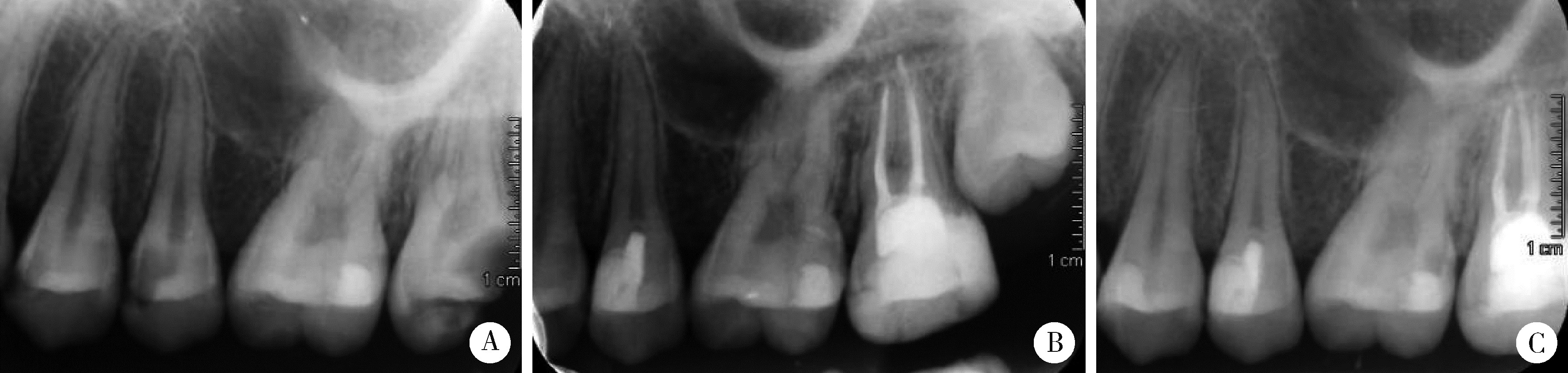
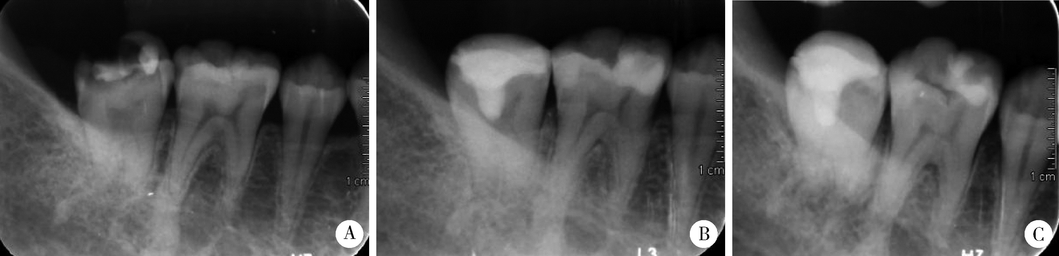
X-ray evaluation of pulp calcification in adult permanent teeth after pulpotomy
Wei YONG*( ),Kun QIAN,Wen-hao ZHU,Xiao-yi ZHAO,Chang LIU,Jie PAN
- Department of General Dentistry, Peking University School and Hospital of Stomatology & National Center of Stomatology & National Clinical Research Center for Oral Diseases & National Engineering Research Center of Oral Biomaterials and Digital Medical Devices & Beijing Key Laboratory of Digital Stomatology & NHC Research Center of Engineering and Technology for Computerized Dentistry & NMPA Key Laboratory for Dental Materials, Beijing 100081, China
CLC Number:
- R781.05


| 1 | American Association of Endodontists. Endodontic Diagnosis. Endodontics. Colleagues for Excellence. American Association of Endodontists. Clinical considerations for a regenerative procedure. 2013[EB/OL]. [2022-09-01]. https://www.aae.org/uploadedfiles/publications_and_research/research/currentregenerative-endodonticconsiderations.pdf. |
| 2 | American Academy of Pediatric Dentistry . Guideline on pulp therapy for primary and immature permanent teeth[J]. Pediatr Dent, 2016, 38 (6): 280- 288. |
| 3 |
Duncan HF , Galler KM , Tomson PL , et al. European Society of Endodontology position statement: Management of deep caries and the exposed pulp[J]. Int Endod J, 2019, 52 (7): 923- 934.
doi: 10.1111/iej.13080 |
| 4 | 刘思懿, 宫玮玉, 刘木清, 等. 成熟恒牙因龋露髓性生物陶瓷材料直接盖髓术的临床疗效观察[J]. 中华口腔医学杂志, 2020, 55 (12): 945- 951. |
| 5 | 陈嘉琪, 董艳梅. 龋源性露髓成熟恒牙活髓保存治疗的研究进展[J]. 中华口腔医学杂志, 2022, 57 (1): 95- 10. |
| 6 | 王爽, 刘鹤, 赵双云, 等. 两种生物陶瓷材料用于乳磨牙牙髓切断术的随机对照研究[J]. 中华口腔医学杂志, 2021, 56 (2): 145- 151. |
| 7 | 贾艳敏, 格根塔娜. iRoot BP Plus用于年轻恒前牙冠折露髓牙髓切断术的临床疗效观察[J]. 实用口腔医学杂志, 2020, 36 (6): 897- 900. |
| 8 |
Qudeimat D , Barrieshi-Nusair KM . Calcium hydroxide vs mineral trioxide aggregates for partial pulpotomy of permanent molars with deep caries[J]. Eur Arch Paediatr Dent, 2007, 8 (2): 99- 104.
doi: 10.1007/BF03262577 |
| 9 |
Tan SY , Yu V , Lim KC , et al. Long-term pulpal and restorative outcomes of pulpotomy in mature permanent teeth[J]. J Endod, 2020, 46 (1): 1- 8.
doi: 10.1016/j.joen.2019.11.008 |
| 10 |
Lin LM , Ricucci D , Saoud TM , et al. Vital pulp therapy of mature permanent teeth with irreversible pulpitis from the perspective of pulp biology[J]. Aust Endod J, 2020, 46 (1): 154- 165.
doi: 10.1111/aej.12392 |
| 11 | Silva L , Cosme-Silva L , Sakai VT , et al. Comparison between calcium hydroxide mixtures and mineral trioxide aggregate in primary teeth pulpotomy: A randomized controlled trial[J]. J Appl Oral Sci, 2019, 27 (1): e20180030. |
| 12 | 钱锟, 潘洁, 朱文昊, 等. 两种硅酸钙类材料用于成熟恒牙牙髓切断术的临床效果[J]. 北京大学学报(医学版), 2022, 54 (1): 113- 118. |
| 13 | Arandi NZ, Thabet M. Minimal intervention in dentistry: A literature review on biodentine as a bioactive pulp capping material[J/OL]. Biomed Res Int, 2021[2022-09-01]. https://doi.org/10.1155/2021/5569313. |
| 14 | Liu S , Wang S , Dong Y . Evaluation of a Bioceramic as a pulp capping agent in vitro and in vivo[J]. J Endod, 2015, 41 (5): 652- 657. |
| 15 | 邓婕, 钟晓波, 万朝霞, 等. CBCT评价下颌第一恒磨牙牙冠硬组织厚度增龄性变化[J]. 中国医学影像技术, 2016, 32 (2): 223- 226. |
| 16 | 史瑞棠, 侯本祥. 牙髓钙化的病因、诊断和治疗策略[J]. 中华口腔医学杂志, 2022, 57 (3): 220- 226. |
| 17 | Mass E , Zilberman UE , Mass U . Zilberman: Long term radiological evaluation of the pulp after partial pulpotomy in young permanent molars[J]. Quintessence Int, 2011, 42 (7): 547- 554. |
| 18 | Ricucci D , Loghin S , Niu LN , et al. Changes in the radicular pulp-dentine complex in healthy intact teeth and in response to deep caries or restorations: A histological and histobacteriological study[J]. J Dent, 2018, 73 (1): 76- 90. |
| 19 | Okamoto M, Takahashi Y, Komichi S, et al. Novel evaluation method of dentin repair by direct pulp capping using high-resolution micro-computed tomography[J/OL]. Clin Oral Invest[2022-09-01]. https://doi.org/10.1007/s00784-018-2374-5. |
| 20 | The American Association of Endodontists. AAE endodontic case difficulty assessment form and guidelines[S/OL]. [2022-09-01]. https://www.aae.org/specialty/clinicalresources/guidelines?position?statements/.Htm. |
| [1] | Jiajia ZHENG,Xue YANG,Quan WEN,Yuan FU,Xiao SHAO,Meili DING. Application of bioactive ceramics iRoot BP Plus® in pulpotomy for complicated crown fracture of immature permanent anterior teeth in children [J]. Journal of Peking University (Health Sciences), 2024, 56(1): 179-184. |
| [2] | Xiaoyi ZHAO,Chang LIU,Kun QIAN,Jie PAN. Efficacy and radiology evaluation of pulpotomy in mature permanent teeth [J]. Journal of Peking University (Health Sciences), 2024, 56(1): 138-143. |
| [3] | Shuang WANG,Chu-fang PENG,He LIU. Pulpotomy of human primary molars with novel bioceramic material [J]. Journal of Peking University (Health Sciences), 2022, 54(6): 1196-1201. |
| [4] | QIAN Kun,PAN Jie,ZHU Wen-hao,ZHAO Xiao-yi,LIU Chang,YONG Wei. Evaluation of bioceramic putty repairmen iRoot and mineral trioxide aggregate in mature permanent teeth pulpotomy [J]. Journal of Peking University (Health Sciences), 2022, 54(1): 113-118. |
| [5] | Yue LEI,Ying-ting YANG,Yuan ZHAN. Evaluation of bioceramic putty repairment in primary molars pulpotomy [J]. Journal of Peking University(Health Sciences), 2019, 51(1): 70-74. |
| [6] | DOU Gui-li, WU Nan, ZHAO Shuang-yun, XIA Bin. Two-year outcomes of pulpotomy in primary molars using mineral trioxide aggregate: a retrospective study [J]. Journal of Peking University(Health Sciences), 2018, 50(1): 170-175. |
| [7] | GUO Yi-dan, ZHANG Sun. Preliminary research of Er:YAG laser used for pulpotomy of Beagle dogs [J]. Journal of Peking University(Health Sciences), 2016, 48(4): 714-719. |
|
||