Journal of Peking University (Health Sciences) ›› 2024, Vol. 56 ›› Issue (1): 190-195. doi: 10.19723/j.issn.1671-167X.2024.01.030
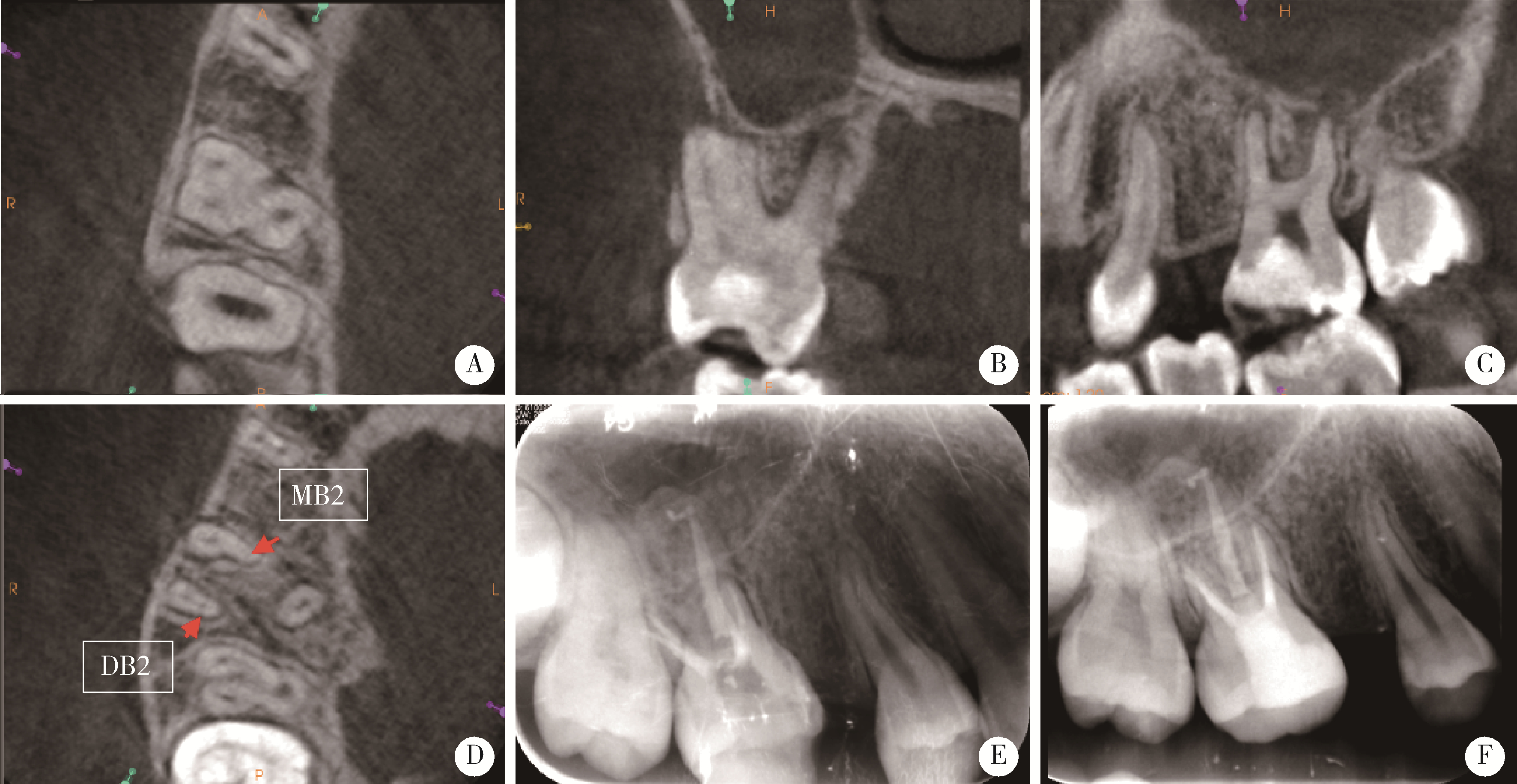
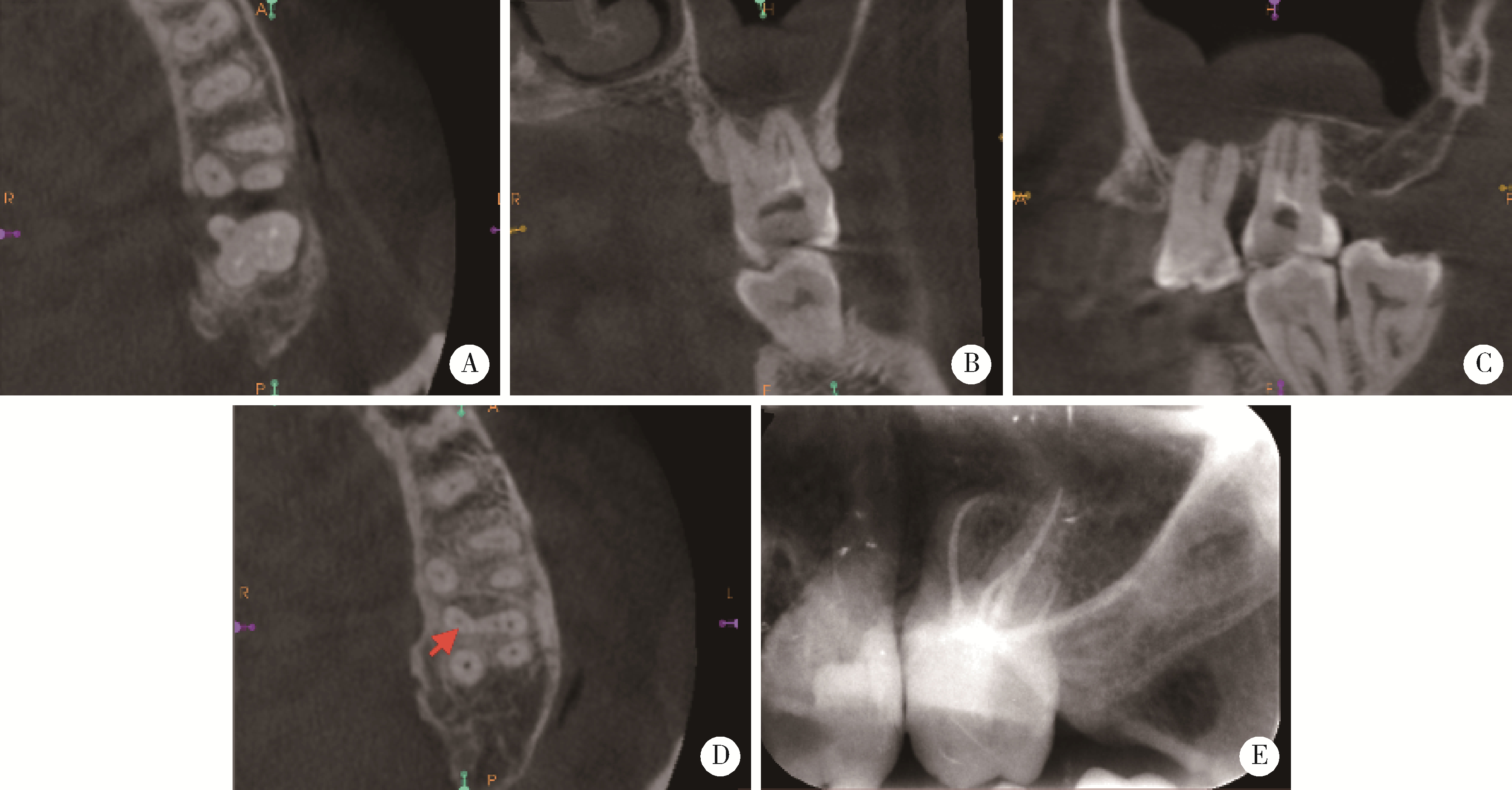
Root canal therapy of maxillary molars with atypical canals: A report of three cases
Chen CHEN1,Yuhong LIANG2,*( )
)
- 1. First Clinical Division, Peking University School and Hospital of Stomatology & National Center of Stomatology & National Clinical Research Center for Oral Diseases & National Engineering Research Center of Oral Biomaterials and Digital Medical Devices & Beijing Key Laboratory of Digital Stomatology & NHC Research Center of Engineering and Technology for Computerized Dentistry & NMPA Key Laboratory for Dental Materials, Beijing 100034, China
2. Department of Oral Emergency, Peking University School and Hospital of Stomatology, Beijing 100081, China
CLC Number:
- R781.05

| 1 |
Peters OA . Current challenges and concepts in the preparation of root canal systems: A review[J]. J Endod, 2004, 30 (8): 559- 567.
doi: 10.1097/01.DON.0000129039.59003.9D |
| 2 | 高学军, 岳林. 牙体牙髓病学[M]. 2版 北京: 北京大学医学出版社, 2013: 431. |
| 3 |
Tabassum S , Khan FR . Failure of endodontic treatment: The usual suspects[J]. Eur J Dent, 2016, 10 (1): 144- 147.
doi: 10.4103/1305-7456.175682 |
| 4 | Cohen S , Hargreaves KM . Pathways of the pulp[M]. 9th ed St. Louis, Mo: Mosby Elsevier, 2006: 230- 235. |
| 5 |
Cleghorn BM , Christie WH , Dong CC . Root and root canal morphology of the human permanent maxillary first molar: A literature review[J]. J Endod, 2006, 32 (9): 813- 821.
doi: 10.1016/j.joen.2006.04.014 |
| 6 |
Zhang R , Yang H , Yu X , et al. Use of CBCT to identify the morphology of maxillary permanent molar teeth in a Chinese subpopulation[J]. Int Endod J, 2011, 44 (2): 162- 169.
doi: 10.1111/j.1365-2591.2010.01826.x |
| 7 |
Neelakantan P , Subbarao C , Ahuja R , et al. Cone-beam computed tomography study of root and canal morphology of maxillary first and second molars in an Indian population[J]. J Endod, 2010, 36 (10): 1622- 1627.
doi: 10.1016/j.joen.2010.07.006 |
| 8 |
Kim Y , Lee SJ , Woo J . Morphology of maxillary first and second molars analyzed by cone-beam computed tomography in a korean population: variations in the number of roots and canals and the incidence of fusion[J]. J Endod, 2012, 38 (8): 1063- 1068.
doi: 10.1016/j.joen.2012.04.025 |
| 9 |
Alavi AM , Opasanon A , Ng YL , et al. Root and canal morphology of Thai maxillary molars[J]. Int Endod J, 2002, 35 (5): 478- 485.
doi: 10.1046/j.1365-2591.2002.00511.x |
| 10 |
Martins JN , Quaresma S , Quaresma MC , et al. C-shaped maxillary permanent first molar: A case report and literature review[J]. J Endod, 2013, 39 (12): 1649- 1653.
doi: 10.1016/j.joen.2013.06.032 |
| 11 |
Qian Y , Li Y , Song J , et al. Evaluation of C-shaped canals in maxillary molars in a Chinese population using CBCT[J]. BMC Med Imaging, 2022, 22 (1): 104.
doi: 10.1186/s12880-022-00831-4 |
| 12 |
Jabali AH , Chourasia HR , Wasli AS , et al. Taurodontism in maxillary and mandibular molars using cone beam computed tomography in a dental center in Saudi Arabia[J]. Ann Saudi Med, 2021, 41 (4): 232- 237.
doi: 10.5144/0256-4947.2021.232 |
| 13 |
Nair R , Khasnis S , Patil JD . Bilateral taurodontism in permanent maxillary first molar[J]. Indian J Dent Res, 2019, 30 (2): 314- 317.
doi: 10.4103/ijdr.IJDR_770_17 |
| 14 |
叶佳学, 梁宇红. 牙髓专科医师应用锥形束CT的现况调查[J]. 北京大学学报(医学版), 2023, 55 (1): 114- 119.
doi: 10.19723/j.issn.1671-167X.2023.01.017 |
| 15 | Mirza MB , Gufran K , Alhabib O , et al. CBCT based study to analyze and classify root canal morphology of maxillary molars: A retrospective study[J]. Eur Rev Med Pharmacol Sci, 2022, 26 (18): 6550- 6560. |
| 16 |
Tian XM , Yang XW , Qian L , et al. Analysis of the root and canal morphologies in maxillary first and second molars in a Chinese population using cone-beam computed tomography[J]. J Endod, 2016, 42 (5): 696- 701.
doi: 10.1016/j.joen.2016.01.017 |
| 17 |
Felsypremila G , Vinothkumar TS , Kandaswamy D . Anatomic symmetry of root and root canal morphology of posterior teeth in Indian subpopulation using cone beam computed tomography: A retrospective study[J]. Eur J Dent, 2015, 9 (4): 500- 507.
doi: 10.4103/1305-7456.172623 |
| 18 |
Tzeng LT , Chang MC , Chang SH , et al. Analysis of root canal system of maxillary first and second molars and their correlations by cone beam computed tomography[J]. J Formos Med Assoc, 2020, 119 (5): 968- 973.
doi: 10.1016/j.jfma.2019.09.012 |
| 19 |
de Moor RJ . C-shaped root canal configuration in maxillary first molars[J]. Int Endod J, 2002, 35 (2): 200- 208.
doi: 10.1046/j.1365-2591.2002.00461.x |
| 20 |
Newton CW , McDonald S . A C-shaped canal configuration in a maxillary first molar[J]. J Endod, 1984, 10 (8): 397- 399.
doi: 10.1016/S0099-2399(84)80162-4 |
| 21 |
Dankner E , Friedman S , Stabholz A . Bilateral C shape configuration in maxillary first molars[J]. J Endod, 1990, 16 (12): 601- 603.
doi: 10.1016/S0099-2399(07)80204-4 |
| 22 |
Yilmaz Z , Tuncel B , Serper A , et al. C-shaped root canal in a maxillary first molar: A case report[J]. Int Endod J, 2006, 39 (2): 162- 166.
doi: 10.1111/j.1365-2591.2006.01069.x |
| 23 |
Kottoor J , Velmurugan N , Ballal S , et al. Four-rooted maxillary first molar having C-shaped palatal root canal morphology evaluated using cone-beam computerized tomography: A case report[J]. Oral Surg Oral Med Oral Pathol Oral Radiol Endod, 2011, 111 (5): 41- 45.
doi: 10.1016/j.tripleo.2010.12.009 |
| 24 | Joshi C , Joshi S . C-shaped canal in maxillary first molars: A case report[J]. J Dent (Tehran), 2014, 11 (1): 111- 117. |
| 25 | Paksefat S , Rahimi S . Root canal treatment of a two-rooted C-shaped maxillary first molar: A case report[J]. Iran Endod J, 2014, 9 (4): 301- 303. |
| 26 | Kharouf N , Haïkel Y , Mancino D . Unusual maxillary first molars with C-shaped morphology on the same patient: Variation in root canal anatomy[J]. Case Rep Dent, 2019, 2019, 1857289. |
| 27 | Wahane KD , Bansod AV , Mattigatti S , et al. Cone-beam com-puted tomography (CBCT) analysis of an unusual configuration of the upper first molar with a C-shaped canal with apically fused roots: A case report[J]. Cureus, 2023, 15 (3): e36474. |
| 28 |
Chaintiou Piorno R , Consoli Lizzi EP , Gualtieri AF , et al. C-shaped canal system in maxillary molars evaluated by cone-beam computed tomography in an Argentine subpopulation[J]. Acta Odontol Latinoam, 2022, 35 (3): 164- 170.
doi: 10.54589/aol.35/3/164 |
| 29 |
Shifman A , Chanannel I . Prevalence of taurodontism found in radiographic dental examination of 1 200 young adult Israeli patients[J]. Community Dent Oral Epidemiol, 1978, 6 (4): 200- 203.
doi: 10.1111/j.1600-0528.1978.tb01150.x |
| 30 | Keene HJ . A morphologic and biometric study of taurodontism in a contemporary population[J]. Am J Phys Anthrop, 1966, 25, 208- 209. |
| [1] | ZHANG Ming-ming, ZHENG Ying-dong, LIANG Yu-hong. A prognostic model for assessment of outcome of root canal treatment in teeth with pulpitis or apical periodontitis#br# [J]. Journal of Peking University(Health Sciences), 2018, 50(1): 123-130. |
|
||