Journal of Peking University (Health Sciences) ›› 2023, Vol. 55 ›› Issue (2): 234-242. doi: 10.19723/j.issn.1671-167X.2023.02.006

Previous Articles     Next Articles

Evaluation of accuracy of pathological diagnosis based on thyroid core needle biopsy

Yan XIONG1,*(),Xin LI1,Li LIANG1,Dong LI1,Li-min YAN1,Xue-ying LI2,Ji-ting DI1,Ting LI1   

  1. 1. Department of Pathology, Peking University First Hospital, Beijing 100034, China
    2. Department of Biostatistics, Peking University First Hospital, Beijing 100034, China
  • Received:2022-10-11 Online:2023-04-18 Published:2023-04-12
  • Contact: Yan XIONG E-mail:yanxiong1109@163.com

RICH HTML


Abstract:

Objective: To explore the protocol for diagnosing thyroid nodules based on core needle biopsy (CNB) and study the biomarkers' application in distinguishing indeterminate samples. Methods: Patients with thyroid nodules treated at Peking University First Hospital from 2015 to 2020 were reviewed. In the study, 598 cases with CNB and matched resected specimens were retrieved. According to "diagnostic categories of thyroid CNB" proposed by the Korean Endocrine Pathology Thyroid Core Needle Biopsy Study Group, the CNB samples were diagnosed as follows: Ⅰ, unsatisfactory; Ⅱ, benign; Ⅲ, indeterminate; Ⅳ, follicular neoplasm; Ⅴ, suspicious for malignancy; and Ⅵ, malignant. The samples of CNB Ⅲ were stained by immunohistochemistry (IHC) using antibodies against CK19, Galectin-3, HBME-1, and CD56, and detected by next-generation sequencing (NGS) using an OncoAim? thyroid cancer multigene assay kit (Singlera Genomics) that detected 26 genes. Taking the resected specimens' classification as the gold standard, the predictive value of CNB for determining the malignancy of thyroid nodules and the biomarkers for distinguishing the samples of CNB Ⅲ was calculated. Results: The study included 598 patients, of which none were CNB Ⅰ, 40 cases were CNB Ⅱ, 40 cases were CNB Ⅲ, 32 cases were CNB Ⅳ, 35 cases were CNB Ⅴ, and 451 cases were CNB Ⅵ. The predictive value of CNB Ⅳ for determining follicular neoplasm was sensitivity (Sen) 100.00% and specificity (Sep) 100.00%, CNB Ⅴ-Ⅵ for determining malignancy was Sen 94.55% and Sep 100.00%, CNB Ⅱ for determining benign lesions was Sen 75.00% and Sep 99.80%. The predictive value of biomarkers for determining malignancy in cases of CNB Ⅲ was Sen 96.30% and Sep 92.31% by NGS, and Sen 81.48% and Sep 92.30% by IHC. Conclusion: The Korean "diagnostic categories of thyroid CNB", which considers the histological specificity of CNB samples and the habits of clinicians, have strong operability, high diagnosis rate, and high clinical value. Under this framework, the cases of CNB Ⅵ should be treated with surgical operation, the cases of CNB Ⅴ-Ⅵ are recommended to be treated as malignant neoplasms, and the major cases of CNB Ⅱ could be followed up without worrisome except the one considered malignant by ultrasound. The value of biomarkers in distinguishing the cases of CNB Ⅲ is significant.

Key words: Thyroid nodule, Core needle biopsy, Pathological diagnosis

CLC Number: 

  • R736.1

Table 1

Diagnostic categories of thyroid core needle biopsy proposed by the Korean Endocrine Pathology Thyroid Core Needle Biopsy Study Group[10]"

Categories Describe
Nondiagnostic or unsatisfactory
    Non-tumor adjacent thyroid tissue only
    Extrathyroid tissue only (e.g., skeletal muscle, mature adipose tissue)
    Acellular specimen (e.g., acellular fibrotic tissue, acellular hyalinized tissue, cystic fluid only)
    Blood clot only
    Other
Benign lesion
    Benign follicular nodule
    Hashimoto's thyroiditis
    Subacute granulomatous thyroiditis
    Nonthyroidal lesion (e.g., parathyroid lesions, benign neurogenic tumors, benign lymph node)
    Other
Indeterminate lesion
    Ⅲa     Indeterminate follicular lesion with nuclear atypia
    Ⅲb     Indeterminate follicular lesion with architectural atypia
    Ⅲc     Indeterminate follicular lesion with nuclear and architectural atypia
    Ⅲd     Indeterminate follicular lesion with Hürthle cell changes
    Ⅲe     Indeterminate lesion, not otherwise specified
Follicular neoplasm
    Ⅳa     Follicular neoplasm, conventional type
    Ⅳb     Follicular neoplasm with nuclear atypia
    Ⅳc     Hürthle cell neoplasm
    Ⅳd     Follicular neoplasm, not otherwise specified
Suspicious for malignancy
    Suspicious for papillary carcinoma, medullary carcinoma, poorly differentiated carcinoma, metastatic carcinoma, lymphoma, etc.
Malignant
    Papillary thyroid carcinoma, poorly differentiated carcinoma, anaplastic thyroid carcinoma, medullary thyroid carcinoma, lymphoma,
metastatic carcinoma, etc.

Table 2

Genes detected by OncoAim? thyroid cancer multigene assay kit"

Gene Transcript Variation type
Mutation Fusion
BRAF NM_004333 Exon 15 Intron 7-10
RET NM_020975 Exon 7-16 Intron 10-11
NRAS NM_002524 Exon 2-3
KRAS NM_033360 Exon 2-4
HRAS NM_176795 Exon 2-3
AKT1 NM_005163 Exon 2-7, exon 9-12
ATM NM_000051 All exon
CNNB1 NM_001904 All exon
TSHR NM_000369 All exon
APC NM_000038 All exon
TTN NM_001256850 All exon
TG NM_003235 All exon
RB1 NM_000321 All exon
MEN1 NM_000244 All exon
PDGFRA NM_006206 All exon
PIK3CA NM_006218 All exon
CDKN2A NM_000077 All exon
EIF1AX NM_001412 All exon
PTEN NM_000314 Exon 5-8
GNAS NM_000516 Exon 8-9
TP53 NM_000546 Exon 5-9
TERT NM_198253 Promoter (chr5: 1 295 183-1 295 302)
PPARG NM_005037 Intron 1
NTRK1 NM_002529 Intron 9, exon 12
NTRK3 NM_002530 Intron 13
ALK NM_004304 Intron 16, intron 19

Figure 1

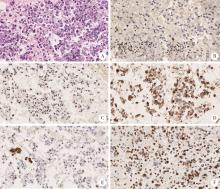
A case classified as CNB Ⅲ by thyroid core needle biopsy was diagnosed as follicular variant of papillary thyroid carcinoma in the matched resected specimen A, tumor in the CNB sample is entirely composed of follicular structures (HE ×40); B, high magnification of the lesion shows the follicular structures lined by cells with nuclei scored 1 (HE ×200); C, cytoplasm and membrane of tumor cells in the CNB sample are diffusely reactive for CK19 with strong intensity, while the normal follicular cells are reactive with weak intensity (IHC ×200); D, cytoplasm and nuclei of tumor cells in the CNB sample are diffusely reactive for Galectin-3 with strong intensity, while the normal follicular cells are nonreactive (IHC ×200); E, membrane of tumor cells in the CNB sample are partially (about 30%) reactive for HBME-1 with intermediate intensity, while the normal follicular cells are nonreactive (IHC ×200); F, tumor cells in the CNB samples are nonreactive for CD56, while membrane and cytoplasm of the normal follicular cells are diffusely reactive with strong intensity (IHC ×200); G, tumor in the matched resected specimen is infiltrative in the desmoplastic stroma and entirely composed of follicular structures (HE ×40); H, high magnification of the lesion shows the follicular structures lined by cells with nuclei scored 3 (HE ×200). CNB, core needle biopsy; HE, hematoxylin-eosin staining; IHC, immunohistochemistry."

Table 3

Predictive value of CNB Ⅴ-Ⅵ for determining malignancy"

Items Matched resected samples, n Predictive value/%
Malignant Benign Total Sen Sep PPV NPV AC
CNB Ⅴ-Ⅵ 486 0 486 94.55 100.00 100.00 65.00 95.05
Others 28 52 80
Total 514 52 566

Table 4

Predictive value of CNB Ⅱ for determining benignity"

Items Matched resected samples, n Predictive value/%
Malignant Benign Total Sen Sep PPV NPV AC
CNB Ⅱ 39 1 40 75.00 99.80 97.50 97.53 97.53
Others 13 513 526
Total 52 514 566

Table 5

Predictive value of biomarkers for determining malignancy in cases of CNB Ⅲ"

Items Matched resected samples, n Predictive value/%
Malignant Benign Total Sen Spe PPV NPV AC
CK19 Positive 25 6 31 92.60 53.85 80.65 77.78 80.00
Negative 2 7 9
Galectin-3 Positive 25 8 33 92.60 38.46 75.76 71.42 75.00
Negative 2 5 7
HBME-1 Positive 21 3 24 77.78 76.92 87.50 62.50 77.50
Negative 6 10 16
CD56 Negative 17 0 17 62.96 100.00 100.00 56.52 75.00
Positive 10 13 23
IHC-COMB1 Positive 22 1 23 81.48 92.30 95.65 70.59 85.00
Negative 5 12 17
IHC-COMB2 Positive 27 5 32 100.00 61.54 84.38 100.00 87.50
Negative 0 8 8
IHC-COMB3 Positive 27 11 38 100.00 15.38 71.05 100.00 72.50
Negative 0 2 2
OncoAim? -NGS Positive 26 1 27 96.30 92.31 96.30 92.31 95.00
Negative 1 12 13
Total 27 13 40
1 Teng WLY , Gao M , Huang G , et al. Guidelines on the diagnosis and treatment of thyroid nodules and differentiated thyroid carcinomas[J]. Chin J Endocrinol Metab, 2012, 28 (10): 779- 797.
2 Pitman MB , Abele J , Ali SZ , et al. Techniques for thyroid FNA: A synopsis of the National Cancer Institute Thyroid Fine-Needle Aspiration State of the Science Conference[J]. Diagn Cytopathol, 2008, 36 (6): 407- 424.
doi: 10.1002/dc.20829
3 Suster S . Controversies regarding the interpretation of follicular thyroid nodules[J]. Arch Pathol Lab Med, 2019, 143 (12): 1472- 1476.
doi: 10.5858/arpa.2019-0301-RA
4 Ha EJ , Baek JH , Lee JH , et al. Complications following US-guided core-needle biopsy for thyroid lesions: A retrospective study of 6 169 consecutive patients with 6 687 thyroid nodules[J]. Eur Radiol, 2017, 27 (3): 1186- 1194.
doi: 10.1007/s00330-016-4461-9
5 Jung CK , Baek JH , Na DG , et al. 2019 Practice guidelines for thyroid core needle biopsy: A report of the Clinical Practice Guidelines Development Committee of the Korean Thyroid Association[J]. J Pathol Transl Med, 2020, 54 (1): 64- 86.
doi: 10.4132/jptm.2019.12.04
6 Lloyd RV , Osamura RY , Klöppel G . WHO classification of tumours of endocrine organs[M]. 4th ed Lyon: International Agency for Research on Cancer (IARC), 2017: 65- 142.
7 Dunderovic D , Lipkovski JM , Boricic I , et al. Defining the value of CD56, CK19, Galectin 3 and HBME-1 in diagnosis of follicular cell derived lesions of thyroid with systematic review of literature[J]. Diagn Pathol, 2015, 10, 196.
doi: 10.1186/s13000-015-0428-4
8 Gharib H , Papini E , Garber JR , et al. American Association of Clinical Endocrinologists, American College of Endocrinology, and Associazione Medici Endocrinologi Medical guidelines for clinical practice for the diagnosis and management of thyroid nodules: 2016 update[J]. Endocr Pract, 2016, 22 (5): 622- 639.
9 Haugen BR , Alexander EK , Bible KC , et al. 2015 American Thyroid Association Management guidelines for adult patients with thyroid nodules and differentiated thyroid cancer: The American Thyroid Association Guidelines Task Force on thyroid nodules and differentiated thyroid cancer[J]. Thyroid, 2016, 26 (1): 1- 133.
doi: 10.1089/thy.2015.0020
10 Na DG , Baek JH , Jung SL , et al. Core needle biopsy of the thyroid: 2016 Consensus Statement and Recommendations from Korean Society of Thyroid Radiology[J]. Korean J Radiol, 2017, 18 (1): 217- 237.
doi: 10.3348/kjr.2017.18.1.217
11 Travis WDBE , Burke AP , Marx A , et al. WHO classification of tumurs of the lung, pleura, thymus and hear[M]. 4th ed Lyon: International Agency for Research on Cancer (IARC), 2015: 29- 36.
12 Jung CK , Min HS , Park HJ , et al. Pathology reporting of thyroid core needle biopsy: A proposal of the Korean Endocrine Pathology Thyroid Core Needle Biopsy Study Group[J]. J Pathol Transl Med, 2015, 49 (4): 288- 299.
doi: 10.4132/jptm.2015.06.04
13 Wolinski K , Stangierski A , Ruchala M . Comparison of diagnostic yield of core-needle and fine-needle aspiration biopsies of thyroid lesions: Systematic review and meta-analysis[J]. Eur Radiol, 2017, 27 (1): 431- 436.
doi: 10.1007/s00330-016-4356-9
14 Song S , Kim H , Ahn SH . Role of immunohistochemistry in fine needle aspiration and core needle biopsy of thyroid nodules[J]. Clin Exp Otorhinolaryngol, 2019, 12 (2): 224- 230.
doi: 10.21053/ceo.2018.01011
15 Cancer Genome Atlas Research Network . Integrated genomic characterization of papillary thyroid carcinoma[J]. Cell, 2014, 159 (3): 676- 690.
doi: 10.1016/j.cell.2014.09.050
16 D'Cruz AK , Vaish R , Vaidya A , et al. Molecular markers in well-differentiated thyroid cancer[J]. Eur Arch Otorhinolaryngol, 2018, 275 (6): 1375- 1384.
doi: 10.1007/s00405-018-4944-1
17 Landa I , Ibrahimpasic T , Boucai L , et al. Genomic and transcriptomic hallmarks of poorly differentiated and anaplastic thyroid cancers[J]. J Clin Invest, 2016, 126 (3): 1052- 1066.
doi: 10.1172/JCI85271
18 Patel KN , Angell TE , Babiarz J , et al. Performance of a genomic sequencing classifier for the preoperative diagnosis of cytologically indeterminate thyroid nodules[J]. JAMA Surg, 2018, 153 (9): 817- 824.
doi: 10.1001/jamasurg.2018.1153
19 Sadowski SM , Petrenko V , Meyer P , et al. Validation of molecular biomarkers for preoperative diagnostics of human papillary thyroid carcinoma in fine needle aspirates[J]. Gland Surg, 2019, 8 (Suppl 2): S62- S76.
20 Choi SH , Baek JH , Lee JH , et al. Evaluation of the clinical usefulness of BRAFV600E mutation analysis of core-needle biopsy specimens in thyroid nodules with previous atypia of undetermined significance or follicular lesions of undetermined significance results[J]. Thyroid, 2015, 25 (8): 897- 903.
doi: 10.1089/thy.2014.0606
21 Choi SH , Baek JH , Lee JH , et al. Initial clinical experience with BRAF(V600E) mutation analysis of core-needle biopsy specimens from thyroid nodules[J]. Clin Endocrinol, 2016, 84 (4): 607- 613.
doi: 10.1111/cen.12866
22 Crescenzi A , Trimboli P , Modica DC , et al. Preoperative assessment of TERT promoter mutation on thyroid core needle biopsies supports diagnosis of malignancy and addresses surgical strategy[J]. Horm Metab Res, 2016, 48 (3): 157- 162.
23 Jang EK , Kim WG , Kim EY , et al. Usefulness of NRAS codon 61 mutation analysis and core needle biopsy for the diagnosis of thyroid nodules previously diagnosed as atypia of undetermined significance[J]. Endocrine, 2016, 52 (2): 305- 312.
doi: 10.1007/s12020-015-0773-9
24 Marotta V , Bifulco M , Vitale M . Significance of RAS mutations in thyroid benign nodules and non-medullary thyroid cancer[J]. Cancers (Basel), 2021, 13 (15): 3785.
doi: 10.3390/cancers13153785
[1] Peng FU,Wen CHEN,Li-gang CUI,Hui-yu GE,Shu-min WANG. Applicational value of 2017 ACR TI-RADS stratification in diagnosing thyroid nodules [J]. Journal of Peking University(Health Sciences), 2019, 51(6): 1067-1070.
Viewed
Full text


Abstract

Cited

  Shared   
  Discussed   
No Suggested Reading articles found!