Journal of Peking University (Health Sciences) ›› 2023, Vol. 55 ›› Issue (2): 270-275. doi: 10.19723/j.issn.1671-167X.2023.02.010

Previous Articles     Next Articles

Activation of JAK/STAT in ovarian high-grade serous cancers and its prognostic significance

Jing YANG1,Juan DU1,2,Yu-xiang WANG1,2,Cong-rong LIU1,2,*()   

  1. 1. Department of Pathology, Peking University Third Hospital, Beijing 100191, China
    2. Department of Pathology, Peking University School of Basic Medical Sciences, Beijing 100191, China
  • Received:2022-09-19 Online:2023-04-18 Published:2023-04-12
  • Contact: Cong-rong LIU E-mail:congrong_liu@hsc.pku.edu.cn

RICH HTML


Abstract:

Objective: The activation of Janus kinase (JAK) and signal transducers and activators of transcription (STAT) plays an important role in the prognosis and targeted therapy of ovarian high-grade serous carcinoma (HGSC). Utilizing simple and practicable technique, this study aimed to evaluate the activation of JAK/STAT signaling pathway in ovarian HGSC patients, and investigated the correlation between the activation of JAK/STAT signaling pathway and the prognosis of the HGSC patients. Methods: We performed immunohistochemistry of phosphorylated STAT3 (pSTAT3) and phosphorylated STAT5 (pSTAT5) on paraffin imbedded slides of 73 ovarian HGSC patients, and evaluated the expression level and range of both markers. According to the grading score of the immunostaining of pSTAT3 and pSTAT5, we divided the 73 ovarian HGSC cases into STAT3 low/high expression and STAT5 low/high expression groups, and analyzed the prognosis of the patients in different groups, in order to explore the relationship between the expression of pSTAT3 and pSTAT5 proteins and the prognosis of the HGSC patients. Results: Some of the ovarian HGSC cases showed high expression of pSTAT3 and pSTAT5 protein level, which was related to the poorer prognosis of the HGSC patients. There was a significant difference in the expression level of pSTAT3 and pSTAT5 between the patients with better prognosis (survival time ≥3 years) and poorer prognosis (survival time < 3 years). The patients with higher protein expression of pSTAT3, pSTAT5 or both markers might have poorer prognosis, with significant shorter progression-free survival time and overall survival time (P < 0.001). Conclusion: Immunostaining of pSTAT3 and pSTAT5 proteins might be helpful to evaluate and predict the prognosis of the ovarian HGSC patients, and to identify the patients who might have higher chances to respond to the STAT inhibitors and anti-angiogenesis therapy.

Key words: Signal transducer and activator of transcription, Janus kinases, Ovarian neoplasms, High-grade serous carcinoma

CLC Number: 

  • R737.31

Table 1

Standard of grading immunohistochemical staining results of pSTAT3 and pSTAT5"

Positive cells Staining intensity Result evaluation
Percentage Score Intensity Score Group Final score*
< 10% 1 No staining 0 pSTAT3-low 0-4
10%-80% 2 Yellow 1 pSTAT3-high 5-9
>80% 3 Yellowish brown 2 pSTAT5-low 0-4
Brown 3 pSTAT5-high 5-9

Table 2

Correlation between the expression of pSTAT proteins and the clinicopathological parameters"

Parameters Counts
(n=73)
pSTAT3 pSTAT5 pSTAT3+pSTAT5
Low
expression
(n=49)
High
expression
(n=24)
P value Low
expression
(n=48)
High
expression
(n=25)
P value Low
expression
(n=53)
High
expression
(n=20)
P value
Age/years 0.13 0.52 0.19
    < 50 27 (37.0) 21 (77.8) 6 (22.2) 19 (70.4) 8 (29.6) 22 (81.5) 5 (18.5)
    ≥50 46 (63.0) 28 (60.1) 18 (39.9) 29 (59.2) 17 (40.8) 31 (67.4) 15 (32.6)
FIGO stage 0.15 0.42 0.24
    Ⅰ 9 (12.3) 4 5 5 4 6 3
    Ⅱ 14 (19.2) 10 4 9 5 11 3
    Ⅲ 38 (52.1) 29 9 28 10 30 8
    Ⅳ 12 (16.4) 6 6 6 6 6 6
PFS < 0.01 < 0.001 < 0.001
    < 3 years 36 (49.3) 15 (20.5) 21 (28.8) 13 (17.8) 23 (31.5) 17 (23.3) 19 (26.0)
    ≥3 years 37 (50.7) 34 (46.6) 3 (4.1) 35 (48.0) 2 (2.7) 36 (49.3) 1 (1.4)
OS < 0.001 < 0.001 < 0.001
    < 3 years 30 (41.1) 10 (13.7) 20 (27.4) 8 (11.0) 22 (30.1) 12 (16.4) 18 (24.7)
    ≥3 years 43 (58.9) 39 (53.4) 4 (5.5) 40 (54.8) 3 (4.1) 41 (56.2) 2 (2.7)

Figure 1

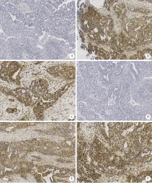
Immunohistochemistry of pSTAT3 and pSTAT5 in ovarian HGSC tissues A, pSTAT3-low (×200); B, pSTAT3-high (×200); C, pSTAT3-high (×400), pSTAT3 expressed both in the nucleus and cytoplasm; D, pSTAT5-low (×200); E, pSTAT5-high (×200); F, pSTAT5-high (×400), pSTAT5 expressed both in the nucleus and cytoplasm. HGSC, high-grade serous carcinoma; pSTAT, phosphorylated signal transducers and activators of transcription."

Figure 2

Correlation between pSTAT3 and pSTAT5 protein expression level and PFS and OS in patients with ovarian HGSC A, correlation between pSTAT3 protein expression and the PFS of patient (P < 0.001); B, correlation between pSTAT3 protein expression and the OS of patient (P < 0.001); C, correlation between pSTAT5 protein expression and the PFS of patient (P < 0.001); D, correlation between pSTAT5 protein expression and the OS of patient (P < 0.001); E, correlation between pSTAT3+pSTAT5 protein expression and the PFS of patient (P < 0.001); F, correlation between pSTAT3+pSTAT5 protein expression and the OS of patient (P < 0.001). HGSC, high-grade serous carcinoma; pSTAT, phosphorylated signal transducers and activators of transcription; PFS, progressionfree survival; OS, overall survival."

1 Roeser JC , Leach SD , McAllister F . Emerging strategies for cancer immunoprevention[J]. Oncogene, 2015, 34 (50): 6029- 6039.
doi: 10.1038/onc.2015.98
2 Frank DA . STAT3 as a central mediator of neoplastic cellular transformation[J]. Cancer Lett, 2007, 251 (2): 199- 210.
doi: 10.1016/j.canlet.2006.10.017
3 Suh YA , Jo SY , Lee HY . Inhibition of IL-6/STAT3 axis and targeting Axl and Tyro3 receptor tyrosine kinases by apigenin circumvent taxol resistance in ovarian cancer cells[J]. Int J Oncol, 2015, 46 (3): 1405- 1411.
doi: 10.3892/ijo.2014.2808
4 Chen H , Ye D , Xie X , et al. VEGF, VEGFRs expressions and activated STATs in ovarian epithelial carcinoma[J]. Gynecol Oncol, 2004, 94 (3): 630- 635.
doi: 10.1016/j.ygyno.2004.05.056
5 Saini U , Naidu S , ElNaggar AC , et al. Elevated STAT3 expression in ovarian cancer ascites promotes invasion and metastasis: A potential therapeutic target[J]. Oncogene, 2017, 36 (2): 168- 181.
doi: 10.1038/onc.2016.197
6 Perez RP , Godwin AK , Hamilton TC , et al. Ovarian cancer biology[J]. Semin Oncol, 1991, 18 (3): 186- 204.
7 Zhang H , Liu T , Zhang Z , et al. Integrated proteogenomic characterization of human high-grade serous ovarian cancer[J]. Cell, 2016, 166 (3): 755- 765.
doi: 10.1016/j.cell.2016.05.069
8 Szubert S , Moszynski R , Szpurek D , et al. The expression of platelet-derived growth factor receptors (PDGFRs) and their correlation with overall survival of patients with ovarian cancer[J]. Ginekol Pol, 2019, 90 (5): 242- 249.
doi: 10.5603/GP.a2019.0045
9 Gadducci A , Lanfredini N , Sergiampietri C . Antiangiogenic agents in gynecological cancer: State of art and perspectives of clinical research[J]. Crit Rev Oncol Hematol, 2015, 96 (1): 113- 128.
doi: 10.1016/j.critrevonc.2015.05.009
10 Aravantinos G , Pectasides D . Bevacizumab in combination with chemotherapy for the treatment of advanced ovarian cancer: A systematic review[J]. J Ovarian Res, 2014, 7, 57.
doi: 10.1186/1757-2215-7-57
[1] GE Xiao-dong, LI Mei-ling, WEN Xi-lin, LI Yi, DENG Xiao-lin, WU Xiao-feng, WEN Ming, LI Shao-lin. Optimal concentration of superparamagnetic iron oxide-short hairpin RNA dual functional molecular probe transfected into ovarian cancer cells in vitro [J]. Journal of Peking University(Health Sciences), 2015, 47(5): 754-760.
[2] MA Rui-qiong, CHENG Hong-yan, YE Xue, CHEN Jun, CUI Heng, WEI Li-hui, CHANG Xiao-hong. Expression and significance of tumor necrosis factor receptor associated protein 1 in epithelial ovarian cancer [J]. Journal of Peking University(Health Sciences), 2014, 46(1): 120-124.
Viewed
Full text


Abstract

Cited

  Shared   
  Discussed   
No Suggested Reading articles found!