Clinicopathological features and survival analysis of TFE3-rearranged renal cell carcinoma with venous tumor thrombus

  • Zhanyi ZHANG 1 ,
  • Min LU 2, 3 ,
  • Yuehao SUN 1 ,
  • Jinghan DONG 1 ,
  • Xiaofei HOU 1 ,
  • Chunlei XIAO 1 ,
  • Guoliang WANG 1 ,
  • Xiaojun TIAN 1 ,
  • Lulin MA 1 ,
  • Hongxian ZHANG 1 ,
  • Shudong ZHANG , 1, 4, *
Expand
  • 1. Department of Urology, Peking University Third Hospital, Beijing 100191, China
  • 2. Department of Pathology, Peking University Third Hospital, Beijing 100191, China
  • 3. Department of Pathology, Peking University School of Basic Medical Sciences, Beijing 100191, China
  • 4. Cancer Center, Peking University Third Hospital, Beijing 100191, China
ZHANG Shundong, email,

Received date: 2025-02-28

  Online published: 2025-08-02

Supported by

the National Natural Science Foundation of China(82273389)

the Beijing Natural Science Foundation(7232212)

Copyright

All rights reserved. Unauthorized reproduction is prohibited.

Abstract

Objective: To review the clinicopathological features of TFE3-rearranged renal cell carcinoma (TFE3-RCC) with venous tumor thrombus (VT) (TFE3-VT), to explore treatment strategies and to prognostic characteristics, and to provide diagnostic and therapeutic references for TFE3-VT patients. Methods: Patients who underwent surgery at Department of Urology, Peking University Third Hospital from January 2013 to January 2024 were enrolled, including three cohorts: Pathologically confirmed TFE3-VT patients, TFE3-RCC patients without VT (TFE3-non-VT), and non-TFE3-rearranged renal cell carcinoma patients with VT (non-TFE3-VT). Clinical history, imaging data, pathological data, and follow-up records were collected. Primary and secondary endpoints were progression-free survival (PFS) and overall survival (OS), respectively. (1) Baseline characteristics were compared between the TFE3-VT and TFE3-non-VT patients. Normally distributed continuous variables were expressed as mean±SD and compared using Student's t-test; non-normally distributed variables were expressed as M (P25, P75) and analyzed with Mann-Whitney U test; categorical variables were described as frequency and percentage [n (%)] and compared by χ2 test or Fisher's exact test. (2) Clinical history, radiological presentations, surgical data, and histopathological features of the TFE3-VT patients were comprehensively characterized. (3) Survival analysis was performed for the TFE3-VT patients. Follow-up data of the TFE3-VT patients were described in detail, and their survival outcomes were compared with the TFE3-non-VT and non-TFE3-VT patients. When compared with the TFE3-non-VT counterparts, Kaplan-Meier method was used to generate PFS and OS curves among: (1) the TFE3-RCC patients across clinical stages Ⅰ-Ⅳ; (2) TFE3-VT versus TFE3-non-VT cohorts; (3) stage Ⅲ subgroups of the TFE3-VT and TFE3-non-VT patients. Intergroup survival differences were statistically evaluated using Log-rank tests. For comparisons with the non-TFE3-VT patients, a 1 : 1 propensity score matching (PSM) was implemented to balance baseline characteristics between the two cohorts. Post-matching Kaplan-Meier curves were generated to compare PFS and OS between the matched groups, with Log-rank tests employed to determine statistical significance of survival disparities. All statistical analyses were conducted with R software (v 4.2.3), and two-tailed P < 0.05 was considered statistically significant. Results: The study included 45 TFE3-RCC patients: 13 TFE3-VT and 32 TFE3-non-VT cases. Additionally, 523 non-TFE3-VT patients were enrolled. Among the 13 TFE3-VT patients, 9 were female (69.2%) and 4 male (30.8%), with a mean age of (37.9±14.4) years, mean BMI of (22.2 ± 3.5) kg/m2, median age-adjusted Charlson comorbidity index (aCCI) of 1.0 (0.0, 1.0), and preoperative creatinine level of (75.3±15.9) μmol/L; tumors were located in the left kidney in 7 patients (53.8%) and right kidney in 6 (46.2%); preoperative distant metastasis (M1 stage) was present in 6 patients (46.2%), while 7 (53.8%) showed no metastasis; VT distribution by Mayo level comprised 7 cases (53.8%) at level 0, 1 case each at levels Ⅰ and Ⅳ (7.7% respectively), and 2 cases each at levels Ⅱ and Ⅲ (15.4% respectively); surgical approaches comprised open surgery (n=2, 15.4%), laparoscopic surgery (n=6, 46.1%), and robot-assisted laparoscopic surgery (n=5, 38.5%); mean operative time was (273±79) min, and intraoperative blood loss was (722±570) mL; mean maximum tumor diameter was (10.8±2.4) cm. All the 13 patients underwent TFE3 protein immunohistochemistry (IHC) staining, with 7 confirmed by fluorescence in situ hybridization (FISH). Tumor recurrence or metastasis occurred in 11 patients (84.6%), and 9 (69.2%) patients died during follow-up. Median PFS was 4 months (1 year PFS rate: 31%), and median OS was 13 months (1 year OS rate: 54%). Survival analysis of 45 TFE3-RCC patients revealed statistically significant differences in PFS and OS across all the clinical stages (P < 0.001). The TFE3-VT patients exhibited significantly worse PFS and OS than the TFE3-non-VT patients (P < 0.001), with persistent significance in stage Ⅲ subgroup analysis (P < 0.05). After PSM, TFE3-VT patients showed significantly inferior PFS compared with non-TFE3-VT (P=0.01), though no significant difference was shown between the OS curves (P=0.11). Conclusion: TFE3-VT predominantly occurs in young females with frequent preoperative metastases. Strongly-positive staining of TFE3 protein in IHC staining and red-green split signals in FISH tests are reliable diagnostic markers. TFE3-VT patients exhibit inferior survival compared with TFE3-non-VT patients and earlier progression than non-TFE3-VT patients.

Cite this article

Zhanyi ZHANG , Min LU , Yuehao SUN , Jinghan DONG , Xiaofei HOU , Chunlei XIAO , Guoliang WANG , Xiaojun TIAN , Lulin MA , Hongxian ZHANG , Shudong ZHANG . Clinicopathological features and survival analysis of TFE3-rearranged renal cell carcinoma with venous tumor thrombus[J]. Journal of Peking University(Health Sciences), 2025 , 57(4) : 650 -661 . DOI: 10.19723/j.issn.1671-167X.2025.04.004

TFE3重排肾细胞癌(TFE3 -rearranged renal cell carcinoma, TFE3 -RCC)是一种罕见的肾癌亚型, 因患者X染色体短臂上的TFE3基因(Xp11.2)发生易位及融合突变[1], 在既往又被称为Xp11.2易位/ TFE3基因融合相关性肾癌(Xp11.2 translocation renal cell carcinoma, Xp11.2 tRCC)。TFE3 -RCC好发于儿童及40岁以下年轻成人, 约占儿童肾细胞癌的三分之一[2], 在成年肾细胞癌中占比较低, 约1.6%~5.0%[3-4], 且在已报道的病例中女性占比更高[5]。该亚型肿瘤常伴有独特的形态学特征, 并可表现出较强的侵袭性。少数患者在确诊时已伴发静脉癌栓(venous tumor thrombus, VT)形成, VT可能累及肾静脉、下腔静脉, 甚至进入右心房, 显著增加了疾病的复杂性和治疗难度。然而, 由于TFE3 -RCC的罕见性及其基因学特征的异质性, 鲜有针对TFE3 -RCC合并VT(TFE3 -VT)患者临床、病理及预后特征的研究报道。本研究旨在通过回顾性分析TFE3 -VT患者的临床病理特征, 并与不合并VT的TFE3 -RCC(TFE3 -non-VT)患者以及非TFE3突变的肾细胞癌合并VT(non- TFE3 -VT)患者进行生存情况比较, 探索其治疗策略及预后特点, 为TFE3 -VT患者的诊断和治疗提供参考。

1 资料与方法

1.1 研究对象

本研究为单中心、回顾性队列研究, 入组范围为2013年1月至2024年1月在北京大学第三医院泌尿外科因临床诊断肾细胞癌接受手术的患者(n=3 708)。TFE3 -RCC患者的纳入标准包括: (1)经免疫组织化学(immunohistochemistry, IHC)染色或荧光原位杂交(fluorescence in situ hybridization, FISH) 检测, 术后病理诊断为TFE3 -RCC;(2)合并VT患者需同期行肾切除及VT切除术。non- TFE3 -VT患者的纳入标准为: (1)经形态学及IHC染色, 病理诊断为non- TFE3 -VT;(2)同期行肾切除及VT切除术。排除标准包括: (1)围术期(术后30 d内)死亡;(2)未切除或仅姑息性切除VT。最终, 共纳入45例TFE3 RCC患者, 其中TFE3 -VT患者13例, TFE3 -non-VT患者32例;同时纳入523例non- TFE3 -VT患者。本研究已获得北京大学第三医院医学科学研究伦理委员会批准(批准号: M2024907), 豁免签署知情同意书。

1.2 资料收集

收集全部患者的临床病史资料、影像学资料、手术资料、病理资料及生存随访资料。临床病史资料包括患者性别, 年龄, 体重指数(body mass index, BMI), 年龄校正Charlson合并症指数(age-adjusted Charlson comorbidity index, aCCI), 美国麻醉医师协会(American Society of Anesthesiologists, ASA)分级, 就诊时有无血尿、腰痛、腹部包块及发热等症状, 是否接受过新辅助治疗, 术前血肌酐水平以及围术期是否发生肺栓塞。肿瘤的影像学特征包括肿瘤侧别、VT的Mayo分级、有无远处转移。手术资料包括手术方式、手术时间、失血量、输血量(悬浮红细胞及血浆)、术后并发症的Clavien-Dindo分级、术后住院天数。病理资料包括肿瘤最大直径、免疫组织化学标记物、是否行FISH检测、pT分期、pN分期、阳性淋巴结数量及分区、有无肾周脂肪受累、瘤栓有无静脉壁侵犯、有无肉瘤样分化、有无瘤内出血坏死、2016版世界卫生组织/国际泌尿病理学会(World Health Organization/International Society of Urological Pathology, WHO/ISUP)核分级。肿瘤的TNM分期及临床分期标准依据2017第8版美国癌症联合委员会(American Joint Committee on Cancer, AJCC)肾脏肿瘤临床分期系统。随访资料包括术后有无靶向治疗、免疫治疗或放疗、有无影像学证实的复发及转移、是否死亡以及死亡原因。研究的主要结局为无进展生存期(progression free survival, PFS), 定义为手术日期与肿瘤复发、出现新发转移灶(或原先已存在的转移灶发生进展)或肿瘤特异性死亡日期之间的时间间隔; 次要结局为总生存期(overall survival, OS), 定义为手术日期与死亡日期之间的时间间隔。患者出院时建议在术后2年内每3个月进行一次全面检查, 术后3~5年每6个月、术后5年起每12个月进行一次检查, 如患者未复查则每6~12个月进行一次电话随访, 以获取生存信息。基线特征和随访数据均从北京大学第三医院泌尿外科肾癌数据库中获取, 并由两名独立的数据分析人员进行检查及补充随访;1名高年资病理医师对患者的病理报告进行核验, 以确认是否为TFE3 -RCC。

1.3 统计学分析

利用Shapiro-Wilk检验对基线资料中的连续变量进行正态性检验, 服从正态分布的连续变量以均数±标准差表示, 组间比较采用Student’ s t检验;不服从正态分布的连续变量以中位数(P25, P75)[M (P25, P75)]表示, 组间比较采用Mann-WhitneyU检验;分类变量以样本数及百分比[n (%)]表示, 组间比较采用χ2检验或Fisher’ s精确检验。利用Kaplan-Meier法绘制PFS及OS生存曲线, 并通过Log-rank检验比较生存曲线的组间差异是否有统计学意义。在TFE3 -RCC患者中, 首先比较不同临床分期(Ⅰ~Ⅳ期)患者的生存情况, 并对TFE3 -VT与TFE3-non-VT患者的生存情况进行比较。由于TFE3 -VT患者在分期中只包含临床Ⅲ~Ⅳ期, 而TFE3 -non-VT患者中仅有6例为Ⅲ期, 其余均为Ⅰ~Ⅱ期, 样本例数较少, 不适合通过倾向性评分匹配(propensity score matching, PSM)及多因素COX回归分析控制混杂因素, 因而进一步对临床Ⅲ期TFE3 -VT及TFE3 -non-VT患者进行亚组分析, 比较相同分期下两组患者生存情况的差异。随后, 采用1 ∶ 1 PSM法对TFE3 -VT与non- TFE3 -VT患者进行配对, 卡钳值设置为0.02, 用于计算倾向性评分的变量为性别(男, 女)、年龄组别(< 45岁, 45~65岁, >65岁)、是否接受过新辅助治疗以及术前有无远处转移, 匹配后标准化均值差(standard mean deviation, SMD)小于0.1视为该变量在组间分布均衡, 比较匹配后TFE3 -VT与non- TFE3 -VT患者生存情况的差异。所有统计分析均使用R语言(v 4.2.3, 美国)进行。双侧检验, P < 0.05为差异有统计学意义。

2 结果

2.1 45例TFE3-RCC患者基线特征总体比较

TFE3 -VT患者与TFE3 -non-VT患者的基线资料分布情况详见表 1TFE3 -VT患者在BMI值、手术入路、手术时间、术中失血量、术中输悬浮红细胞量、术后并发症Clavien-Dindo分级、pT分期、M分期、临床分期及核分级方面与TFE3 -non-VT患者相比差异具有统计学意义, 其中TFE3 -VT患者的平均BMI小于TFE3 -non-VT患者, 中位手术时间、失血量、输悬浮红细胞量以及肿瘤直径均大于TFE3 -non-VT患者, 接受开放手术、临床分期为Ⅲ~Ⅳ期以及核分级为高级别(Ⅲ~Ⅳ级)的比例均高于TFE3 -non-VT患者。
表1 45例TFE3重排肾细胞癌患者(合并或不合并静脉癌栓)的基线特征

Table 1 Baseline characteristics of 45 TFE3 -RCC patients with or without VT

Items TFE3-VT (n=13) TFE3-non-VT (n=32) P value
Age/years, ${\bar x}$±s 37.9 ±14.4 36.0 ±13.1 0.662
Gender, n (%) 0.638
  Male 4 (30.8) 14 (43.8)
  Female 9 (69.2) 18 (56.2)
BMI/(kg/m2), ${\bar x}$±s 22.2 ±3.5 24.8 ±3.4 0.023*
aCCI, M (P25, P75) 1.0 (0.0, 1.0) 0.0 (0.0, 1.0) 0.376
ASA level, n (%) 0.840
  1 7 (53.8) 20 (62.5)
  2 6 (46.2) 12 (37.5)
Cr/(μmoL/L), ${\bar x}$±s 75.3 ±15.9 75.6±16.6 0.953
Laterality, n (%) 0.924
  Left 7 (53.8) 15 (46.9)
  Right 6 (46.2) 17 (53.1)
Surgical approach, n (%) < 0.001*
  Laparoscopic 2 (15.4) 23 (71.9)
  Open 6 (46.1) 0 (0.0)
  Robotic 5 (38.5) 9 (28.1)
Operative time/min, M (P25, P75) 292.0 (194.0, 320.0) 138.0 (109.8, 167.8) < 0.001*
Blood loss/mL, M (P25, P75) 600.0 (300.0, 1 100.0) 40.0 (17.5, 100.0) < 0.001*
RBC transfusion/mL, M (P25, P75) 0.0 (0.0, 600.0) 0.0 (0.0, 0.0) < 0.001*
Clavien-Dindo level, n (%) 0.031*
  0-Ⅰ 9 (69.2) 31 (96.9)
  Ⅱ 4 (30.8) 1 (3.1)
Postoperative hospital stays/d, M (P25, P75) 9.0 (6.0, 14.0) 6.0 (5.0, 6.0) 0.009*
Tumor diameter/cm, M (P25, P75) 10.3 (9.6, 12.0) 4.4 (3.2, 6.5) < 0.001*
pT stage, n (%) < 0.001*
  pT1 0 (0.0) 23 (71.9)
  pT2 0 (0.0) 4 (12.5)
  pT3 11 (84.6) 5 (15.6)
  pT4 2 (15.4) 0 (0.0)
N1 stage, n (%) 5 (38.5) 3 (9.4) 0.060
M1 stage, n (%) 6 (46.2) 0 (0.0) < 0.001*
Clinical tumor stage, n (%) < 0.001*
  Stage Ⅰ 0 (0.0) 22 (68.8)
  Stage Ⅱ 0 (0.0) 4 (12.5)
  Stage Ⅲ 6 (46.2) 6 (18.8)
Stage Ⅳ 7 (53.8) 0 (0.0)
Nuclear grade, n (%) 0.003*
  Ⅰ-Ⅱ 0 (0.0) 17 (53.1)
  Ⅲ-Ⅳ 13 (100.0) 15 (46.9)
Sarcomatoid differentiation, n (%) 2 (15.4) 1 (3.1) 0.404

*P < 0.05. TFE3 -RCC, TFE3 -rearranged renal cell carcinoma; VT, venous tumor thrombus; TFE3 -VT, TFE3 -RCC patients with VT; TFE3 -non-VT, TFE3 -RCC patients without VT; BMI, body mass index; aCCI, age-adjusted Charlson comorbidity index; ASA, American Society of Anesthesiologists; Cr, serum creatinine; RBC, red blood cells.

2.2 13例TFE3 -VT患者的临床及病理特征

2.2.1 临床病史、影像学表现及手术情况

13例TFE3-VT患者的临床病史、肿瘤影像学特征及手术资料详见表 2。临床病史方面, 13例TFE3 -VT患者中, 10例(76.9%)因局部症状(如腰腹痛、血尿、包块)就诊, 2例(15.4%)因全身症状(发热、消瘦)就诊, 1例(7.7%)因体检发现。4例(30.8%)患者在术前接受了新辅助药物或放射治疗。2例(15.4%)患者在围术期出现VT引起的肺动脉栓塞, 均在抗凝治疗后好转。影像学表现方面, 13例患者在术前均进行了泌尿系统增强计算机断层扫描(computed tomography, CT)、下腔静脉增强磁共振成像、胸部平扫CT或正电子发射计算机断层显像/X线计算机体层成像(positron emission tomography/computed tomography, PET/CT)等检查。在增强CT中, 13例TFE3 -VT患者的原发肿瘤病灶均表现为软组织密度影, 增强扫描呈不均匀强化, 部分病变内部可见钙化或斑片状低密度区, 肾静脉及下腔静脉中可见明显充盈缺损(图 1)。手术方面, 13例TFE3 -VT患者均接受了肾切除联合VT切除术, 术后患者合并Clavien-Dindo Ⅰ级和Ⅱ级并发症各4例, 均经药物治疗后好转, 其余5例患者无明显并发症。
表2 13例TFE3-VT患者的临床病史、肿瘤影像学特征及手术资料

Table 2 Clinical history, radiological tumor characteristics and surgical data of 13 TFE3-VT patients

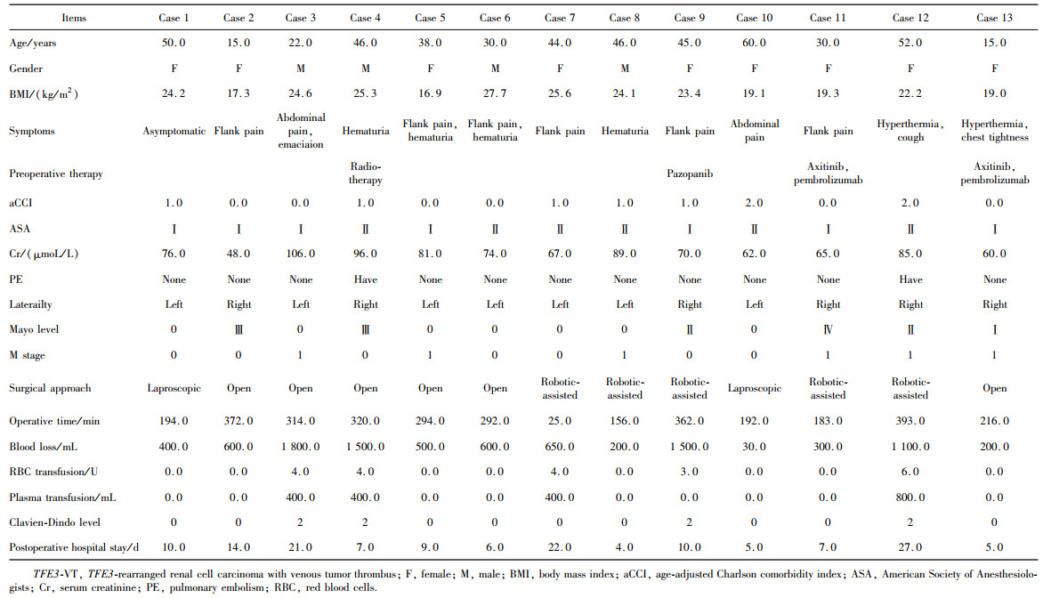
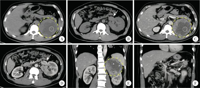
图1 1例合并Mayo 0级肾静脉癌栓的TFE3重排肾细胞癌患者(病例8)的增强CT影像

Figure 1 Contrast-enhanced CT images from a left-sided TFE3 -rearranged renal cell carcinoma patient combined with Mayo level 0 renal vein thrombus (case 8)

A, B, 9.4×9.6×9.5 cm mass was identified in the upper pole of the left kidney, the margin of the mass was well-defined; on non-contrast CT scan, the mass (yellow circle with dashed line) demonstrated heterogeneous soft tissue density (50-70 HU) with patchy areas of low density (30-40 HU); central punctate calcifications were observed within the lesion (white circle with solid line); the left renal vein was dilated (white arrow, axial plane). C, D, on contrast-enhanced CT imaging (arterial phase), the solid components of the mass showed marked heterogeneous enhancement (80-120 HU), while the cystic areas within the lesion exhibited no obvious enhancement; tumor thrombus was identified within the renal vein (white arrow), measuring approximately 3.7 cm in length (axial plane). E, F, on contrast-enhanced CT imaging (arterial phase), multiple fine septations were visible within the cystic areas and demonstrated enhancement (white arrow head); the perirenal fascia was compressed, and the lesion was in close proximity to the left adrenal gland and spleen; the left renal vein exhibited a filling defect and indicated tumor thrombus (white arrow); no significantly enlarged lymph nodes were seen (coronal plane).

2.2.2 组织病理学特征

13例TFE3 -VT患者的肿瘤最大直径、pT分期、VT静脉壁侵犯、肾周脂肪侵犯、肉瘤样分化、瘤内出血坏死、核分级、pN分期、阳性淋巴结数目及分区等病理特征详见表 3。在病理染色方面, 13例TFE3 -VT患者的苏木素-伊红(hematoxylin-eosin, H & E)染色均可见典型肾细胞癌形态, 细胞浆透明并呈片状排列(图 2)。由于各患者使用的免疫组织化学染色标志物组合存在差异, 故未对主要标志物的表达差异进行定量描述。表 3列出了几种用于鉴别TFE3 -RCC与其他RCC的免疫组织化学标志物以及每例患者的染色情况, 所有患者均进行了TFE3蛋白染色, 其中9例呈强阳性(+), 4例呈弱阳性或部分阳性, 但均在后续的FISH检测中证实为TFE3 -RCC。7例患者进行了肾谱系因子PAX8染色, 其中6例(85.7%)表现为强阳性或部分阳性。12例患者进行了肾透明细胞癌(clear cell renal cell carcinoma, ccRCC)特异性标志物CAIX染色, 其中5例(41.7%)散在阳性, 1例(8.3%)部分阳性, 余6例(50.0%)均阴性。7例患者进行了进一步的FISH检测, 均可见红-绿色分离信号, 进一步证实为TFE3 -RCC。
表3 13例TFE3-VT患者的组织病理学特征

Table 3 Histopathological features of 13 TFE3-VT patients

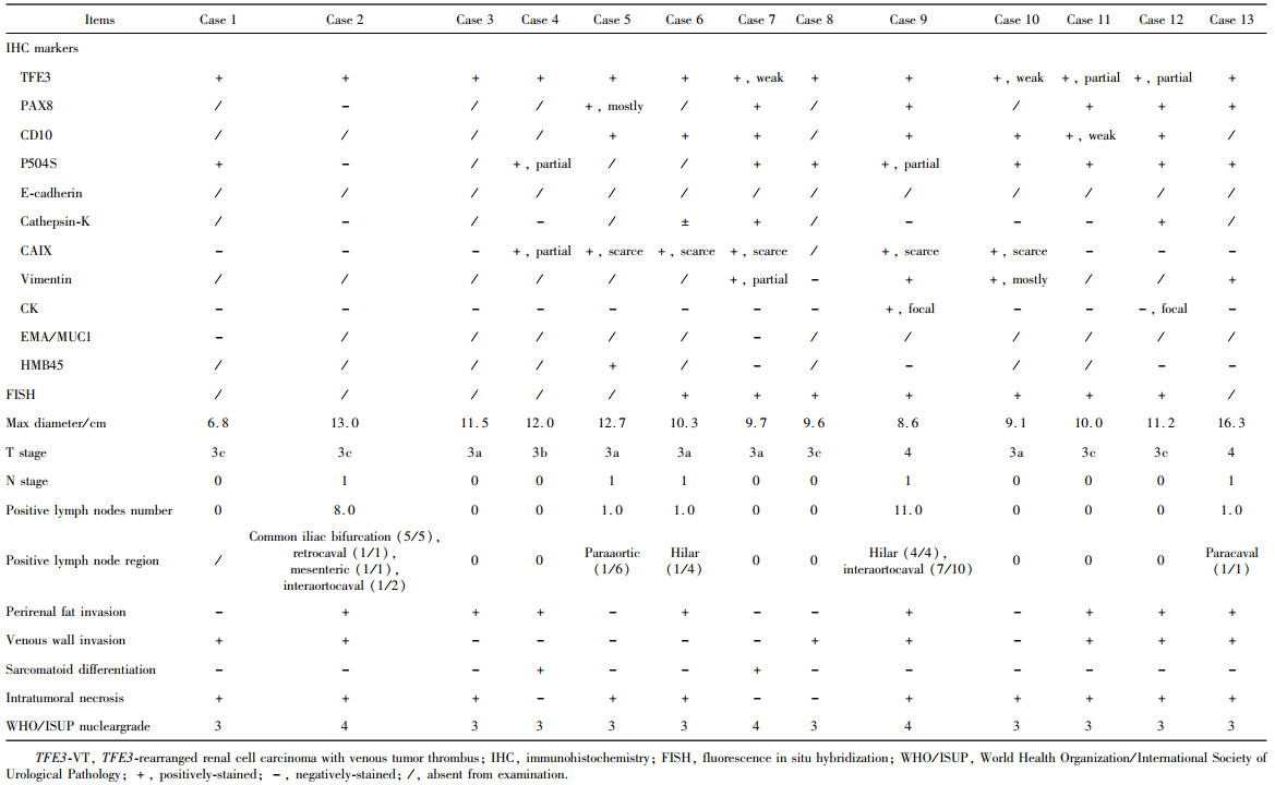
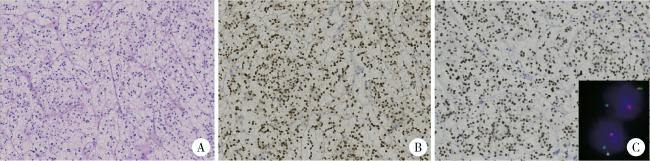
图2 1例合并Mayo Ⅱ级下腔静脉癌栓的TFE3重排肾细胞癌患者(病例9)的病理图像

Figure 2 Pathological images from a left-sided TFE3-rearranged renal cell carcinoma patient with Mayo level Ⅱ inferior vena cava thrombus (case 9)

A, Hematoxylin-Eosin staining (×200), tumor cells exhibit clear cytoplasm arranged in a sheet-like pattern; B, immunohistochemistry staining of PAX-8 (×200), staining of tumor cell nuclei was strongly positive for PAX-8; C, immunohistochemistry staining of TFE3 protein (×200), staining of tumor cell nuclei was strongly positive for TFE3, FISH-TFE3 showed red-green split signals (inset).

2.3 生存分析

2.3.1 13例TFE3 -VT患者的生存随访情况

13例TFE3 -VT患者的生存随访资料详见表 4。中位PFS为4个月, 1年PFS率为31%, 中位OS为13个月, 3年OS率为46%, 其中12例患者接受了辅助或进展后的酪氨酸激酶抑制剂(tyrosine kinase inhibitor, TKI)靶向治疗、抗PD-1免疫检查点抑制剂(immune checkpoints inhibitor, ICI)治疗或放射治疗, 11例患者(84.6%)出现肿瘤进展, 复发或转移的具体部位已在表 4列出。9例患者死亡(69.2%), 其中8例为肿瘤特异性死亡, 另1例患者不详;余4例患者中3例仍存活, 另1例失访。
表4 13例TFE3 -VT患者的术后治疗及生存结局

Table 4 Postoperative therapy and survival outcomes of 13 TFE3 -VT patients

Items Case 1 Case 2 Case 3 Case 4 Case 5 Case 6 Case 7
Postoperative therapy TKI TKI TKI None TKI TKI TKI, ICI
Type of TKI Sunitinib, axitinib Sunitinib Sunitinib None Pazopanib NA Sunitinib, axitinib, anlotinib
Type of ICI None None None None None None Triprolizumab, bevacizumab (peritoneal perfusion)
Death + + + + + - +
Death reason Cancer NA Cancer Cancer Cancer None Cancer
OS/months 56 3 9 12 8 16 13
Progression + + + + + + +
Sites of tumor relapse or metastasis Lung, liver NA Abdominal wall, surgical region Lung, liver Liver Lung, liver Liver, peritoneum, omentum, retroperitoneal region
PFS/months 13 3 4 3 1 11 6
Items Case 8 Case 9 Case 10 Case 11 Case 12 Case 13
Postoperative therapy TKI TKI TKI TKI, radiotherapy, ICI None TKI, ICI
Type of TKI Sunitinib, axitinib Pazopanib Pazopanib Sunitinib, axitinib None Axitinib
Type of ICI None None None Pembrolizumab None Pembrolizumab
Death + + - - + -
Death reason Cancer Cancer None None Cancer None
OS/months 40 1 34 31 2 16
Progression + + + - + -
Sites of tumor relapse or metastasis Lung Liver, adrenal gland, abdominal wall Surgical region, cervical and mediastinal lymph nodes, lung None NA None
PFS/months 1 0 13 31 2 16

TFE3 -VT, TFE3 -rearranged renal cell carcinoma with venous tumor thrombus; TKI, tyrosine kinase inhibitor; ICI immune checkpoints inhibitor; OS, overall survival; PFS, progression free survival; NA, not available; +, presence of survival outcome; -, censoring at the latest follow-up.

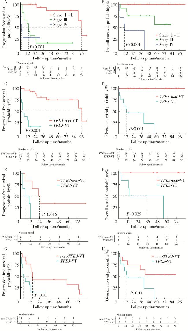
2.3.2 临床Ⅰ~Ⅳ期TFE3 -RCC患者生存情况比较

45例TFE3 -RCC患者的总体中位PFS为91个月, 5年PFS率为56%, 中位OS未达到, 5年OS率为74%, 其中临床分期Ⅰ~Ⅱ期患者的中位PFS为96个月, 5年PFS率为87%, 中位OS未达到, 5年OS率为100%;Ⅲ期患者的中位PFS为13个月, 5年PFS率为17%, 中位OS未达到, 5年OS率为56%;Ⅳ期患者的中位PFS为2个月, 1年PFS率为29%, 中位OS为9个月, 3年OS率43%。Ⅰ~Ⅱ期患者、Ⅲ期患者与Ⅳ期患者的PFS及OS曲线之间差异有统计学意义(P < 0.001)(图 3AB)。
图3 TFE3重排肾细胞癌合并静脉癌栓患者、不合并静脉癌栓的TFE3重排肾细胞癌患者以及非TFE3突变的肾细胞癌合并静脉癌栓患者的生存曲线

Figure 3 Kaplan-Meier curves of TFE3 -VT, TFE3 -non-VT, and non- TFE3 -VT

A, B, progression free survival and overall survival curves of TFE3-RCC patients with different clinical tumor stage (2017 American Joint Committee on Cancer 8th version); C, D, progression free survival and overall survival curves of TFE3-VT and TFE3-non-VT patients; E, F, progression free survi-val and overall survival curves of stage Ⅲ TFE3 -VT and TFE3 -non-VT patients.; G, H, progression free survival and overall survival curves of TFE3 -VT and non-TFE3 -VT patients after 1 ∶ 1 propensity score matching. TFE3 -RCC, TFE3 -rearranged renal cell carcinoma; TFE3 -VT, TFE3 -rearranged renal cell carcinoma with venous tumor thrombus; TFE3 -non-VT, TFE3-rearranged renal cell carcinoma patients without VT; non-TFE3 -VT, non-TFE3 -rearranged renal cell carcinoma patients with VT.

2.3.3 TFE3 -VT与TFE3 -non-VT患者的生存比较

TFE3 -non-VT患者的中位PFS为96个月, 5年PFS率为75%, 中位OS未达到, 5年OS率为100%。TFE3 -VT患者与TFE3 -non-VT患者的PFS及OS曲线之间差异有统计学意义(P < 0.001, 图 3CD)。临床Ⅲ期患者的亚组分析中, TFE3 -VT患者与TFE3 -non-VT患者的PFS(P=0.016)及OS(P=0.029)曲线之间差异仍具有统计学意义(图 3EF)。

2.3.4 TFE3 -VT与non- TFE3 -VT患者的生存比较

经PSM后, 13例TFE3 -VT患者均在non- TFE3 -VT患者中有对应的匹配病例。用于匹配的变量(性别、年龄组、新辅助治疗及术前远处转移)在匹配后的SMD值均小于0.1, 实现了组间均衡匹配(表 5)。
表5 倾向性评分匹配前后匹配因素的分布情况

Table 5 Distribution of matching factors before and after propensity score matching (PSM)

Items Before PSM SMD After PSM SMD
TFE3-VT (n=13) non-TFE3-VT (n=523) TFE3-VT (n=13) non-TFE3-VT (n=13)
Age/years, n (%) 1.432 < 0.001
  ≤45 8 (61.5) 60 (11.5) 8 (61.5) 8 (61.5)
  >45-65 5 (38.5) 312 (59.7) 5 (38.5) 5 (38.5)
  >65 0 (0.0) 151 (28.9) 0 (0.0) 0 (0.0)
Gender, n (%) 0.857 < 0.001
  Male 4 (30.8) 367 (70.2) 4 (30.8) 4 (30.8)
  Female 9 (69.2) 156 (29.8) 9 (69.2) 9 (69.2)
M1 stage, n (%) 7 (53.8) 144 (27.5) 0.556 7 (53.8) 7 (53.8) < 0.001
Neoadjuvant therapy, n (%) 4 (30.8) 66 (12.6) 0.451 4 (30.8) 4 (30.8) < 0.001

PSM, propensity score matching; TFE3 -VT, TFE3 -rearranged renal cell carcinoma with venous tumor thrombus; non-TFE3-VT, non-TFE3-rearranged renal cell carcinoma patients with venous tumor thrombus; SMD, standard mean deviation.

配对的13例non- TFE3 -VT患者中位PFS为15个月, 3年PFS率为43%, 中位OS未达到, 3年OS率为65%。TFE3 -VT患者与non- TFE3 -VT患者的PFS曲线之间差异有统计学意义(P=0.01), OS曲线之间差异无统计学意义(P=0.11, 图 3GH)。

3 讨论

本研究对13例TFE3 -VT患者的临床病理特征及生存结局进行了详细描述, 该队列中绝大多数患者为无合并症的中青年女性, 患者在就诊时伴有多种非特异性症状, 如腰痛、血尿和腹部包块等。在影像学检查中, TFE3 -VT表现为较大的肾内肿块, 泌尿系增强CT中伴有明显不均匀强化, 并可见钙化。上述特征均与既往研究[6-8]报道的TFE3 -RCC影像表现相符。从肿瘤的临床特征及围术期资料来看, VT的存在增加了围术期肺栓塞等致命性并发症的风险[9], 并显著增加了手术的困难程度和肿瘤术后复发风险。新辅助免疫治疗引起的局部粘连、较大的肿瘤体积、腔静脉切开取栓或节段性切除操作均会增加术中出血量及术后并发症的发生率。本研究中, 13例TFE3 -VT患者在术后均未出现Clavien-Dindo Ⅲ级及以上并发症, 术后均经治疗顺利出院, 反映了该术式已在本中心得到较成熟的开展, 具有良好的安全性。
病理学检查中, TFE3 -RCC肿瘤细胞通常在H&E染色中表现为透明细胞样或嗜酸性细胞样形态, 伴较大的核仁。免疫组织化学染色中, 肿瘤细胞核可见TFE3蛋白强阳性, PAX8、CD10、P504S(AMACR)及E-cadherin常阳性, 角蛋白及EMA常阴性或仅局灶阳性, Vimentin常阴性, 但可表现为弱阳性及局灶阳性[10]。ccRCC中高表达的CAIX则一般为阴性, 而在FISH检测中, 可见原癌基因TFE3断裂, 并与其他基因发生融合(具体表现为红-绿荧光信号的明显分离)[11]
本研究对TFE3 -VT的病理学检测结果与文献[10-11]报道基本相符。TFE3 -RCC在WHO泌尿系统肿瘤病理分型中经历了数次变化。2022年最新发布的《WHO泌尿和男性生殖系统肿瘤分类》(第五版)首次明确强调了基于分子特征的肾癌分型, 尤其是TFE3基因重排的分子检测在TFE3 -RCC诊断中的核心地位。相较于第四版中对该疾病以形态学为主的描述, 第五版更加强调了分子层面的鉴别诊断价值, 并指出其不同亚型(如ASPSCR1 - TFE3PRCC- TFE3等)可能具有不同的临床行为和预后, 表现出高度异质性[12-14]TFE3重排导致MiT转录因子家族的异常激活, 进而增强肿瘤细胞的增殖、迁移和血管生成能力, 是TFE3 -RCC表现为高侵袭性的重要机制[15]
进行生存分析时, 我们将TFE3 -VT与TFE3 -non-VT及non- TFE3 -VT患者进行了比较。在总体及临床Ⅲ期患者中, TFE3 -VT患者的生存预后均显著低于TFE3 -non-VT患者, 提示VT的存在可能是TFE3患者不良预后的危险因素。TFE3 -VT与匹配的non- TFE3 -VT患者相比, PFS曲线的差异存在统计学意义, 而OS曲线的差异无统计学意义, 提示TFE3突变在肾细胞癌合并VT患者中可能是发生早期进展的危险因素。
目前, 针对TFE3 -VT患者的标准治疗方式仍缺乏指南的明确推荐。根治性肾切除联合VT切除术是常见的非转移性肾细胞癌患者的标准治疗手段。然而, 对于就诊时即伴有远处转移的TFE3 -VT患者, 是否应当进行减瘤性肾切除联合VT切除术以及手术的时机, 尚缺乏充分的临床研究证据。根据Ⅲ期非劣效性随机对照试验CARMENA研究(NCT00930033)的最新数据, 在转移性ccRCC中舒尼替尼单药靶向治疗组的中位OS(19.8个月)与减瘤性肾切除术后辅以舒尼替尼组的中位OS(15.6个月)相比差异无统计学意义[HR(95%CI)= 0.97(0.79~1.19), P=0.8], 提示常规行减瘤手术可能缺乏生存获益[16]。另一项随机对照试验SURTIME研究(NCT01099423)对减瘤性肾切除术的不同时机进行探索, 结果显示, 初始舒尼替尼序贯减瘤性肾切除术(延迟手术组)与即刻减瘤性肾切除术序贯舒尼替尼(即刻手术组)的中位OS分别为32.4个月和15.0个月[HR(95% CI)=0.57(0.34~ 0.95), P=0.03], 提示系统治疗应答良好的患者可能从延迟手术中获益[17]。上述两个研究认为, 在中高风险转移性肾细胞癌患者中, 先进行手术治疗并不优于单药治疗, 但伴有症状患者, 例如有严重血尿或顽固性疼痛患者, 仍可考虑手术治疗。本研究纳入的13例TFE3 -VT患者中有6例在就诊时即合并远处转移, 6例M1期TFE3 -VT患者的中位OS为24.5个月, 7例M0期患者的中位OS则为13.0个月, M1期患者的中位OS反而较M0期患者更长, 这可能与TFE3 -VT患者的罕见性所致样本例数及结局数量少有关。虽然单纯进行减瘤性手术干预可能难以改变肿瘤的侵袭性病程, 但对原发灶及VT进行减瘤性切除是出于对如下因素的考量: (1)局部症状控制需求(6例患者均表现为顽固性血尿或持续的腰背部疼痛);(2)降低肿瘤负荷是否有潜在获益, 这也与CARMENA研究及SURTIME研究的引申结论相符。此外, 尽管TKI及ICI在常见RCC亚型中显示出良好的疗效, 其在TFE3 -VT中的作用尚需进一步研究, 能否有效用于肿瘤的系统治疗、转化治疗、新辅助治疗及辅助治疗仍无大规模临床研究数据支持。
Sun等[18]通过分析63例TFE3 -RCC患者的肿瘤微环境特征, 揭示多数病例呈现程序性细胞死亡配体1(programmed cell death-ligand 1, PD-L1)低表达及CD8+T细胞浸润不足状态, 但MED15 - TFE3融合亚型则表现出较高的PD-L1阳性率, 提示该亚型可能对ICI治疗敏感, 值得注意的是, 具有侵袭性特征的ASPL- TFE3融合亚型显示出较高的血管密度, 该特点则为抗血管生成TKI类靶向药物的选择提供了理论依据。TKI及ICI的选择方面, Bakouny等[19]基于多中心数据的回顾性队列研究发现, 接受ICI的TFE3 -RCC患者中位OS达62.4个月, 优于TKI组的10.3个月(P=0.267), 研究数据显示ICI组25%的客观缓解率与TKI组的0%之间虽差异无统计学意义(P=0.22), 但已展现潜在临床优势。本研究中, 虽然绝大多数TFE3 -VT患者表现出较差的生存结局, 但2例(病例11, 病例13)减瘤术前即出现远处转移、先接受靶向及免疫联合系统治疗并在减瘤术后维持治疗的患者在最近1次随访(术后31个月, 术后16个月)中均未发生疾病进展, 提示靶向及免疫联合疗法的潜在疗效。
本研究具有一定的局限性, 仅半数左右的患者进行了FISH定性检测, 且未进一步行分子亚型检测, 其余患者仅通过IHC染色TFE3高表达确诊, 同时, 未对VT及原发肿瘤病灶进行TFE3蛋白表达水平的空间分布差异分析, 不能充分揭示TFE3 -VT患者的分子病理特征。由于TFE3 -RCC的罕见性及TFE3 -VT患者的低比例, 本队列患者的数量十分有限, 不能判断VT是否为TFE3 -RCC的独立危险因素。在利用PSM法比较TFE3 -VT及non- TFE3 -VT的生存结局时, 虽然匹配因素在匹配队列中实现均衡分布, 但由于继续增加匹配因素后可供匹配的TFE3 -VT患者数量减少, 为保证队列的完整性, 未能纳入更多的匹配因素, 影响了组间可比性。此外, 由于本研究的回顾性特点, 不能对患者围术期系统用药进行细致评估, 无法评估靶向及免疫治疗药物对该类患者的疗效。未来可以通过开展多中心前瞻性队列研究及针对TFE3信号通路分子的基础研究, 进一步解释不同亚型的分子及免疫图谱, 并进一步优化TFE3 -RCC及TFE3 -VT患者的治疗策略, 以期改善这一罕见肾癌病理类型患者的生存预后。

利益冲突  所有作者均声明不存在利益冲突。

作者贡献声明  张展奕: 设计研究方案, 撰写论文;陆敏: 提出研究思路, 撰写论文, 总体把关和审定论文;孙悦皓: 设计研究方案, 收集、分析、整理数据;董靖晗、侯小飞、肖春雷、王国良、田晓军: 收集、分析、整理数据;马潞林、张洪宪: 设计研究方案, 总体把关和审定论文;张树栋: 提出研究思路, 总体把关和审定论文。

1
Cheng J , Han Z , Mehra R , et al. Computational analysis of pathological images enables a better diagnosis of TFE3 Xp11.2 translocation renal cell carcinoma[J]. Nat Commun, 2020, 11 (1): 1778.

2
He J , Chen X , Gan W , et al. Renal cell carcinoma associated with Xp11.2 translocation/TFE3 gene fusions: Clinical experience and literature review[J]. Future Oncol, 2015, 11 (24): 3243- 3252.

3
Komai Y , Fujiwara M , Fujii Y , et al. Adult Xp11 translocation renal cell carcinoma diagnosed by cytogenetics and immunohistochemistry[J]. Clin Cancer Res, 2009, 15 (4): 1170- 1176.

4
Zhong M , de Angelo P , Osborne L , et al. Translocation renal cell carcinomas in adults: A single-institution experience[J]. Am J Surg Pathol, 2012, 36 (5): 654- 662.

5
Cheng X , Gan W , Zhang G , et al. Clinical characteristics of XP11.2 translocation/TFE3 gene fusion renal cell carcinoma: A systematic review and meta-analysis of observational studies[J]. BMC Urol, 2016, 16 (1): 40.

6
王军卫, 夏丹, 汪朔, 等. 成人Xp11.2/TFE3基因融合相关性肾细胞癌的临床病理特征及预后分析[J]. 中华泌尿外科杂志, 2022, 43 (3): 165- 170.

7
简远熙, 吴俊霖, 杨素萍. MiT家族易位性肾细胞癌的影像学表现[J]. 临床放射学杂志, 2022, 41 (7): 1379- 1383.

8
He J , Zhou K , Zhu B , et al. Dynamic contrast-enhanced CT characterization of Xp11.2 translocation/TFE3 gene fusions versus papillary renal cell carcinomas[J]. Biomed Res Int, 2015, 2015, 298679.

9
Nyhan SM , Kostibas MP . Assessing the incidence of perioperative pulmonary embolism and optimal timing of surgery based on pre-operative renal cell cancer tumor thrombus level and growth kine-tics: Avenues to explore[J]. J Cardiothorac Vasc Anesth, 2019, 33 (10): 2735- 2736.

10
Argani P . MiT family translocation renal cell carcinoma[J]. Semin Diagn Pathol, 2015, 32 (2): 103- 113.

11
黄艳, 王玉环, 丁丽红, 等. TFE3重排肾细胞癌16例临床病理特征及预后分析[J]. 诊断病理学杂志, 2024, 31 (8): 732- 736.

12
Zhao J , Tang Y , Hu X , et al. Patients with ASPSCR1-TFE3 fusion achieve better response to ICI based combination therapy among TFE3-rearranged renal cell carcinoma[J]. Mol Cancer, 2024, 23 (1): 132.

13
Cheng AV, Wu DJ, Friedman LA, et al. MAPK1IP1L: TFE3-rearranged renal cell carcinoma: A novel fusion adding to the differential diagnosis of oncocytic renal neoplasms [J]. Virchows Arch, 2025, 1(2025-01-25)[2025-01-30]. https://pubmed.ncbi.nlm.nih.gov/39862330.

14
Moch H , Amin MB , Berney DM , et al. The 2022 World Health Organization classification of tumours of the urinary system and male genital organs. Part A: Renal, penile, and testicular tumours[J]. Eur Urol, 2022, 82 (5): 458- 468.

15
Chen W , Wu M , Du L , et al. Upregulation of GSTP1 mediated by chimeric TFE3 promotes TFE3-tRCC progression by targeting JNK signaling pathway[J]. World J Surg Oncol, 2024, 22 (1): 352.

16
Méjean A , Ravaud A , Thezenas S , et al. Sunitinib alone or after nephrectomy for patients with metastatic renal cell carcinoma: Is there still a role for cytoreductive nephrectomy?[J]. Eur Urol, 2021, 80 (4): 417- 424.

17
Bex A , Mulders P , Jewett M , et al. Comparison of immediate vs deferred cytoreductive nephrectomy in patients with synchronous metastatic renal cell carcinoma receiving sunitinib: The SURTIME randomized clinical trial[J]. JAMA Oncol, 2019, 5 (2): 164- 170.

18
Sun G , Chen J , Liang J , et al. Integrated exome and RNA sequencing of TFE3-translocation renal cell carcinoma[J]. Nat Commun, 2021, 12 (1): 5262.

19
Bakouny Z , Sadagopan A , Ravi P , et al. Integrative clinical and molecular characterization of translocation renal cell carcinoma[J]. Cell Rep, 2022, 38 (1): 110190.

Outlines

/