Current situation and exploration of clinical transformation of plasmatrix in oral implantology

  • Yulan WANG ,
  • Hao ZENG ,
  • Yufeng ZHANG , *
Expand
  • Department of Implantology, School and Hospital of Stomatology, Wuhan University, Wuhan 430079, China
ZHANG Yufeng, e-mail,

Received date: 2025-07-31

  Online published: 2025-08-29

Copyright

All rights reserved. Unauthorized reproduction is prohibited.

Abstract

With the rapid development of implant dentistry, increasing attention has been paid to the long-term stability and aesthetic outcomes of dental implants, among which sufficient volume and quality of soft and hard tissues are considered crucial contributing factors for successful treatment outcomes. Among the various available tissue regeneration strategies, plasmatrix, an autologous biomaterial derived from the patient ' s own peripheral blood, has demonstrated unique and significant clinical value in the regeneration and augmentation of both soft and hard tissues associated with dental implant therapy in recent years. This notable potential is primarily attributed to its rich content of multiple growth factors, viable cells, and a supportive fibrin scaffold, along with its excellent biocompatibility, tunable biodegradation profile, and a relatively simple and rapid preparation process that does not require complex laboratory equipment. As a result, its clinical applications have been continuously expanding across a wide range of indications. Based on a comprehensive review of the existing literature and current research evidence, this article provides an in-depth summary of the advancements in both basic science and clinical applications of plasmatrix in the context of implant dentistry. Particular attention is given to its classification from a materials science perspective, underlying molecular mechanisms, biological effects in promoting tissue regeneration, and its implementation under different clinical scenarios. Furthermore, the article discusses unresolved technical challenges and existing controversies, and outlines potential future directions for research and technological innovation, aiming to provide robust evidence-based guidance for clinical practice as well as a theoretical and methodological reference for future scientific investigations.

Cite this article

Yulan WANG , Hao ZENG , Yufeng ZHANG . Current situation and exploration of clinical transformation of plasmatrix in oral implantology[J]. Journal of Peking University(Health Sciences), 2025 , 57(5) : 836 -840 . DOI: 10.19723/j.issn.1671-167X.2025.05.004

口腔种植目前已成为牙列缺损及缺失修复的重要手段,具有良好的功能性与稳定性[1]。然而,种植牙的长期成功不仅依赖于种植体本身的设计,种植牙周围软硬组织的质量和稳定性也对其长期成功率和美观性有着重要影响[2]。种植体周围充足的骨组织可为种植体提供良好的初期稳定与长期负重支持,而健康的软组织则有助于形成生物封闭、抵御菌斑入侵并维持美学效果[3-5]
由于长期缺牙或其他疾病的影响,需要种植修复的患者常存在软硬组织缺乏的情况,给种植治疗带来了挑战。随着生物材料和组织工程的发展,各类骨替代材料(如自体骨、异体骨、合成骨等)和膜材料(如胶原膜、不可吸收膜、钛网等)在骨增量临床操作中获得了广泛应用;在软组织再生方面,自体游离龈移植与结缔组织移植仍是金标准,同时也出现了一些胶原基质、脱细胞真皮基质等生物材料的临床应用[6]
在现有的软硬组织再生材料中,血浆基质作为来源于自体血液的生物材料,具有富含血小板等活细胞与生长因子的三维纤维蛋白网络,兼具优异的组织相容性与血管生成能力,同时可协同调节炎症微环境,促进成骨细胞与成纤维细胞的增殖与分化[7],因此,它可替代或部分增强传统生物材料的效果,特别是在牙槽嵴保存术、即刻种植、骨增量、软组织增量等领域展现出了较高的临床价值。本文以血浆基质研究与口腔种植临床应用为核心,结合国内外研究进展,系统梳理血浆基质在口腔种植软硬组织再生中的基础机制、应用策略与未来的优化和发展方向,以期为其规范化应用和深入研究提供参考。

1 血浆基质的分类及作用机制

1.1 血浆基质的分类

血浆基质是通过离心分离自体血液获得的一类富含血小板、白细胞与生长因子的三维纤维蛋白支架材料,按制备方法与产物形态的不同,经历了由富血小板血浆(platelet-rich plasma, PRP)、富血小板纤维蛋白(platelet-rich fibrin, PRF)、浓缩生长因子(concentrated growth factor, CGF)到近年发展形成的新一代血浆基质——水平富血小板纤维蛋白(horizontal platelet-rich fibrin, H-PRF)的技术演进[8]。第一代血浆基质PRP需添加抗凝剂与激活剂,操作繁琐且成分不稳定;第二代血浆基质PRF取消抗凝剂,形成固体凝胶,具备缓释生长因子的能力;第三代血浆基质CGF采用多段程序离心,试图进一步提高生长因子浓度和稳定性;第四代血浆基质H-PRF将传统的角度离心改变为水平离心方式,在细胞分布均匀性、生物成分保留、细胞损伤控制等方面具有显著优势,已成为血浆基质标准化制备的新方向[7]

1.2 血浆基质促进再生的机制

血浆基质主要通过其三维纤维蛋白结构,实现包括血小板源性生长因子(platelet-derived growth factor, PDGF)、转化生长因子β (transforming growth factor-β, TGF-β)、血管内皮生长因子(vascular endothelial growth factor, VEGF)等生长因子的缓慢、持续释放,维持局部高浓度生长因子的修复微环境[9-10],因此,其可以调控多种修复相关细胞(成纤维细胞、成骨细胞、内皮细胞等)的增殖、迁移和分化,协同促进软硬组织再生[11-13]。血浆基质中的白细胞和细胞因子可调节局部免疫反应,实现部分抗菌效果,并且减少炎症,促进组织修复[7, 14]。此外,血浆基质的纤维蛋白网状结构可为细胞黏附、迁移和新生血管生成提供支架,同时作为生长因子的缓释库,促进组织修复。

1.3 血浆基质在口腔种植中的应用形式

血浆基质产物在口腔种植的临床应用上可以分为三种形式。
液态血浆基质:经手肘抽取患者静脉血于专用的液态血浆基质离心管,使用特定的离心参数制备而成,离心完成后通常可以在室温下保持十数分钟的液体状态[15],可用注射器抽取,在口腔种植中可用于冲洗拔牙窝、软组织注射、润湿种植体和其他生物材料等。
固态血浆基质:经手肘抽取患者静脉血于专用的固态血浆基质离心管,使用特定的离心参数制备而成,离心完成后可用器械夹取黄色凝胶部分,钝性分离红细胞后,根据需求将其压制成栓状或膜状,可单独或联合生物材料用于骨增量、软组织增量等[16]
血浆基质骨块:血浆基质骨块是固态和液态血浆基质与颗粒状骨替代材料按照不同比例混合后获得的混合骨移植材料,它作为一个整体,具有一定的机械强度,便于操作,并且可以根据缺损区情况进行塑形,主要用于骨增量手术中[17]

2 血浆基质在口腔种植相关领域的临床应用

在口腔种植领域,血浆基质广泛应用于引导骨组织再生术、牙槽嵴保存术、即刻种植、上颌窦底提升术及软组织增量等临床场景,以提升骨和软组织的再生效果。

2.1 引导骨组织再生术

在引导骨组织再生术中,血浆基质主要是通过混合自体骨屑或骨替代材料,形成具有机械强度的血浆基质骨块,用于水平或垂直骨增量时的骨缺损充填[18-19]。对于水平骨增量,根据术后是否可在理想位置植入理想尺寸的种植体及骨壁完整程度,将水平骨缺损分为三种类型:(1)种植体周围骨量足,但唇侧轮廓凹陷,可采用颗粒骨粉填充并覆盖固态血浆基质膜,以提升骨量与软组织质量;(2)种植体三面骨壁完整但颊侧存在缺损,适合采用血浆基质骨块填充,外覆胶原膜及固态血浆基质膜增强封闭效果与愈合速度;(3)骨量不足无法同步植入种植体时,需先期植骨,采用血浆基质骨块替代自体骨块,结合钛钉固定、胶原膜与固态血浆基质膜进行空间维持[19]
垂直骨增量术常面临空间维持困难、自体骨供区创伤大、术后愈合慢等挑战,血浆基质骨块则可作为活性因子的协同载体,混合骨形成蛋白2(bone morphogenetic protein, BMP-2)等,填入缺损区,后通过钛钉、钛网等方式固定,并表面叠加胶原膜与固态血浆基质膜封闭软组织[18]。有研究表明,血浆基质能促进新生骨形成,加快软组织愈合,降低术后感染与钛网暴露风险[16, 20-21]

2.2 牙槽嵴保存术

血浆基质在牙槽嵴保存术中可用于拔牙窝冲洗、软硬组织缺损再生。根据拔牙窝的骨壁缺损程度可进行不同操作:(1)无骨缺损(伴或不伴软组织缺损),适合采用固态血浆基质膜/栓封闭拔牙窝;(2)双侧骨壁缺损小于50%,可使用液态血浆基质冲洗,结合血浆基质骨块进行填充和塑形;(3)至少一侧骨壁缺损超过50%,需联合帐篷钉、血浆基质骨块、胶原膜及双层血浆基质膜进行复杂重建[22]。已有临床研究表明,血浆基质能加快软组织愈合,减轻拔牙后的疼痛,减少牙槽嵴吸收,增加拔牙窝内骨填充[23-24]

2.3 上颌窦底提升术

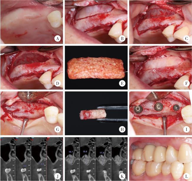
在经牙槽嵴顶上颌窦底提升术中,血浆基质可用于提升过程中分层支撑窦底黏膜,减少颗粒状骨替代材料外溢及膜穿孔风险。术中可将固态血浆基质膜折叠置入窦底下方,形成柔性缓冲层,再以血浆基质骨块逐层填入,适度敲击提升,每次提升幅度约1.5~2.0 mm。液态血浆基质可用于血浆基质骨块间隙塑形,增强成分整合[25]。在侧壁开窗上颌窦底提升术中,血浆基质膜可用于支撑上颌窦底黏膜,或是用于修补小的上颌窦底黏膜穿孔,血浆基质骨块则可以用于窦腔充填,从而发挥空间支撑和成骨引导的作用[26]。血浆基质膜可以在小范围上颌窦底提升中单独使用,在大范围上颌窦提升中血浆基质能够起到促进血管化和骨改建的作用[27-29], 如图 1所示。
图1 血浆基质骨块在上颌窦底提升术中的临床应用示例

Figure 1 Clinical application of a representative plasmatrix bone block in maxillary sinus floor elevation

A, intraoral view before surgery; B, preparation of the lateral window; C, exposure of the Schneiderian membrane; D, elevation of the Schneiderian membrane; E, plasmatrix bone block; F, repositioning of the lateral window after grafting with the bone block; G, alveolar bone condition six months after sinus floor elevation; H, intact bone column at the implant site; I, implant placement; J, preoperative CBCT showing insufficient alveolar bone height; K, CBCT at six months postoperatively showing adequate bone height; L, occlusion after prosthetic restoration. CBCT, cone beam CT.

2.4 即刻种植

在即刻种植中,血浆基质适用于多种复杂情况,包括唇侧骨壁薄或部分缺损、拔牙后软组织封闭困难、生物型薄弱或希望缩短整体治疗周期的患者,可用于拔牙窝充填、唇侧骨缺损修复、软组织封闭等。临床操作中,血浆基质骨块可用于填充种植体周围骨缺损[30];同时,其血浆基质膜状可用于封闭拔牙窝创口或增厚颊侧软组织,作为生物屏障促进血凝稳定与软组织快速愈合。血浆基质富含生长因子和白细胞,具有抗菌、抗炎和促血管生成作用,有助于减少感染风险,提高慢性感染拔牙窝的即刻种植成功率[31]。还有研究发现,采用液态血浆基质润湿种植体,能够提高其早期稳定性[32]

2.5 种植体周围软组织增量

血浆基质在种植体周围软组织管理中,可用于角化龈宽度不足、黏膜厚度较薄、牙龈生物型不理想等情况。对于种植体周围角化龈增宽,可结合根向复位瓣或条带技术,将血浆基质膜用于覆盖受植区暴露的创面[33];对于种植体周围软组织增厚,可将血浆基质膜置入隧道瓣或半厚瓣下,实现增厚软组织的效果[34];对于薄的种植体周围牙龈表型,可以通过注射液态血浆基质增厚其表型,改善软组织质量和美学效果。相关动物实验与临床研究表明,血浆基质可显著改善种植体周围软组织的生物型与稳定性[12]。相比传统自体移植,血浆基质更具操作简便、患者舒适度高等优势,成为软组织再建中有益的补充与替代方案。

2.6 种植体周围炎的治疗

血浆基质也可用于种植体周围炎的治疗中,可用于骨缺损的充填和作为屏障膜覆盖骨替代材料。在严格进行菌斑控制和肉芽组织清理后,对于骨缺损较小的情况,可以直接使用血浆基质膜充填缺损区域;对于骨缺损较大的情况,可以使用血浆基质骨块充填缺损区,并用血浆基质膜覆盖充填物,从而达到减少探诊深度和探诊出血,提高缺损内骨充填的效果。比起单纯使用骨替代材料而言,加用血浆基质后能够改善患者术后疼痛;比起单纯的翻瓣清创而言,使用血浆基质能够有效提高种植体周围的组织再生[35-36],这主要得益于血浆基质的抗菌、抗炎和再生效果。

3 血浆基质复合生物材料的研发

尽管血浆基质具有缓释多种生长因子、安全性高、生物相容性优良、操作简便等优势,但在口腔种植组织再生的应用中仍存在力学强度与体积有限、降解较快、生物活性有限等缺点,同时,因其成型性好、黏附性能佳,容易与其他材料结合,因此,可将血浆基质用作缓释载体或粘合剂成分,与其他生物材料复合,制备兼具机械支撑、生物活性与可控生长因子释放功能的三维复合支架,以满足口腔种植中对大体积骨缺损修复和长期组织再生的需求。目前的相关研究包括:血浆基质结合其他支架材料,例如混合透明质酸凝胶,使其兼具血浆基质的生长因子缓释与透明质酸的支架作用,显著提升牙龈成纤维细胞增殖、迁移及软组织再生效果[37];或是血浆基质混合壳聚糖-羟基磷灰石凝胶等,兼具机械支撑与生物活性[38];将血浆基质作为抗生素(如阿莫西林、甲硝唑、多西环素等)载体,使其具有持续抑菌能力,可显著减少术后的感染风险或用于感染性疾病的治疗[39];血浆基质负载外源活性成分或生长因子(如负载外泌体、维生素C、BMP-2等),可进一步提高其生物学活性[40];还可以结合数字化手段,基于影像学数据和口腔扫描数据设计个性化3D打印钛网或植骨导板,塑形血浆基质骨块,使其与骨缺损区紧密贴合,并且符合未来所需的骨再生形态,提高组织再生的精准性[41]

4 总结与展望

血浆基质作为一种来源于自体血液的再生材料,凭借其富含生长因子、纤维蛋白与免疫细胞等多重活性成分,在口腔种植的多个环节中展现出了良好的生物学效应和广泛的适应性。无论是在拔牙后的拔牙窝保存,即刻种植术中的骨缺损填充和软组织封闭,还是在复杂的水平和垂直骨增量术、上颌窦底提升术以及软组织表型优化中,血浆基质均可通过多种形式(液态、膜状或骨块)参与组织修复与再建。然而,目前血浆基质的应用仍面临一些挑战与限制,其成分受个体因素影响较大,生物活性与治疗效果缺乏一致性;制备过程尚未完全标准化,不同离心参数和设备可能导致质量波动;在力学性能方面,尚难完全替代传统膜材料或自体组织移植,特别是在大体积骨缺损重建中。为进一步发挥其临床潜力,未来研究应致力于构建统一的制备标准与质量评价体系,发展其与其他骨再生或屏障材料的复合策略,并结合数字化技术与个性化医学理念优化其使用路径。此外,仍需更多前瞻性临床试验与长期随访数据支持其在不同适应证中的效果与安全性验证。

国家专利  银纳米颗粒改良的血浆基质及血浆基质膜制备方法、应用,发明专利(CN115887787A),2024;一种金纳米颗粒改良的血浆基质及血浆基质膜的制备方法,发明专利(CN115887788B),2024

利益冲突  所有作者均声明不存在利益冲突。

作者贡献声明  王宇蓝:论文撰写;曾浩:论文修改;张玉峰:研究设计,论文审阅与修订;所有作者均对最终文稿进行审读并确认。

1
Srinivasan M , Kamnoedboon P , Angst L , et al. Oral function in completely edentulous patients rehabilitated with implant-supported dental prostheses: A systematic review and meta-analysis[J]. Clin Oral Implants Res, 2023, 34 (Suppl 26): 196- 239.

2
Kwok V , Caton JG , Hart ID , et al. Dental implant prognostication: A commentary[J]. J Periodontol, 2023, 94 (6): 713- 721.

DOI

3
Thoma DS , Naenni N , Figuero E , et al. Effects of soft tissue augmentation procedures on peri-implant health or disease: A syste-matic review and meta-analysis[J]. Clin Oral Implants Res, 2018, 29 (Suppl 15): 32- 49.

4
Monje A , Roccuzzo A , Buser D , et al. Influence of buccal bone wall thickness on the peri-implant hard and soft tissue dimensional changes: A systematic review[J]. Clin Oral Implants Res, 2023, 34 (Suppl 26): 8- 27.

5
Wang Y , Zhang Y , Miron RJ . Health, maintenance, and recovery of soft tissues around implants[J]. Clin Implant Dent Relat Res, 2016, 18 (3): 618- 634.

DOI

6
Sanz M , Giannobile WV . Soft and hard tissue augmentation procedures for promotion of peri-implant health and aesthetics[J]. Clin Oral Implants Res, 2018, 29 (Suppl 15): 4- 6.

7
Farshidfar N, Apaza Alccayhuaman KA, Estrin NE, et al. Advantages of horizontal centrifugation of platelet-rich fibrin in regenerative medicine and dentistry[J/OL]. Periodontol 2000, 2025. [2025-03-25]. http://doi.org/10.1111/prd.12625.

8
张玉峰. 血浆基质制品的前世今生[J]. 中华口腔医学杂志, 2021, 56 (8): 740- 746.

9
Miron RJ , Fujioka-Kobayashi M , Bishara M , et al. Platelet-rich fibrin and soft tissue wound healing: A systematic review[J]. Tissue Eng Part B Rev, 2017, 23 (1): 83- 99.

DOI

10
Farshidfar N, Amiri MA, Estrin NE, et al. Platelet-rich plasma (PRP) versus injectable platelet-rich fibrin (i-PRF): A syste-matic review across all fields of medicine[J/OL]. Periodontol 2000, 2025. [2025-03-24]. http://doi.org/10.1111/prd.12626.

11
Liu M , Liu Y , Luo F . The role and mechanism of platelet-rich fibrin in alveolar bone regeneration[J]. Biomed Pharmacother, 2023, 168, 115795.

DOI

12
Chai J , Chen Y , Shi S , et al. Efficacy of horizontal platelet-rich fibrin on gingival tissue regeneration: Cellular and histological analysis[J]. J Periodontol, 2025,

DOI

13
Ma Z , Ding J , Wang Y , et al. Study of platelet-rich fibrin promoting endothelial cell differentiation and angiogenesis induced by transplantation of adipose-derived stem cells[J]. Acta Histochem, 2023, 125 (6): 152059.

DOI

14
Zhang J , Yin C , Zhao Q , et al. Anti-inflammation effects of injectable platelet-rich fibrin via macrophages and dendritic cells[J]. J Biomed Mater Res A, 2020, 108 (1): 61- 68.

DOI

15
Miron RJ , Gruber R , Farshidfar N , et al. Ten years of injectable platelet-rich fibrin[J]. Periodontol 2000, 2024, 94 (1): 92- 113.

DOI

16
Qiu Y , Bao S , Wei H , et al. Bacterial exclusion and wound healing potential of horizontal platelet-rich fibrin (H-PRF) membranes when compared to 2 commercially available collagen membranes[J]. Clin Oral Investig, 2023, 27 (8): 4795- 4802.

DOI

17
Feng M , Wang Y , Wei Y , et al. Preparation, characterization and biological properties of a novel bone block composed of platelet rich fibrin and a deproteinized bovine bone mineral[J]. Fundam Res, 2021, 2 (2): 321- 328.

18
张玉峰, 王宇蓝. 血浆基质在口腔种植垂直骨增量中的应用[J]. 口腔疾病防治, 2022, 30 (12): 837- 843.

19
张玉峰, 王宇蓝. 血浆基质在口腔种植水平骨增量中的应用[J]. 口腔疾病防治, 2022, 30 (3): 153- 159.

20
Agrawal AA . Platelet rich fibrin is not a barrier membrane! Or is it?[J]. World J Clin Cases, 2023, 11 (11): 2396- 2404.

DOI

21
Hartmann A , Seiler M . Minimizing risk of customized titanium mesh exposures: A retrospective analysis[J]. BMC Oral Health, 2020, 20 (1): 36.

DOI

22
张玉峰, 张晓欣. 血浆基质在牙槽嵴保存术中的应用[J]. 口腔疾病防治, 2024, 32 (3): 161- 168.

23
Miron RJ , Fujioka-Kobayashi M , Moraschini V , et al. Efficacy of platelet-rich fibrin on bone formation, part 1: Alveolar ridge preservation[J]. Int J Oral Implantol (Berl), 2021, 14 (2): 181- 194.

24
Siawasch SAM , Yu J , Castro AB , et al. Autologous platelet concentrates in alveolar ridge preservation: A systematic review with meta-analyses[J]. Periodontol 2000, 2025, 97 (1): 104- 130.

DOI

25
王宇蓝, 曾浩, 夏婷, 等. 骨致密化技术联合血浆基质在穿牙槽嵴上颌窦底提升中的应用[J]. 中国口腔种植学杂志, 2025, 30 (2): 106- 110.

26
张晓欣, 张玉峰. 上颌窦底骨量不足新分类及血浆基质治疗策略[J]. 口腔生物医学, 2021, 12 (4): 215- 218.

27
Xie S , Zhang Y , Wen G , et al. The effect of autologous platelet concentrates as solely grafting material or with bone graft materials in maxillary sinus augmentation: A meta-analysis of randomized controlled trials[J]. Clin Oral Investig, 2025, 29 (2): 120.

DOI

28
Yu S , Tian Y , Wei Y , et al. Comparative microcomputed tomography and histological analysis of the effects of a horizontal platelet-rich fibrin bone block on maxillary sinus augmentation: A preclinical in vivo study[J]. Clin Oral Implants Res, 2023, 34 (6): 555- 564.

DOI

29
Yu S , Bd YT , Bd YW , et al. Early tissue and healing responses after maxillary sinus augmentation using horizontal platelet rich fibrin bone blocks[J]. BMC Oral Health, 2023, 23 (1): 589.

DOI

30
Cirmeni M , Fedele O , Giammarinaro E , et al. Immediate implant and socket preservation using sticky bone and leukocyte-platelet-rich fibrin in the anterior maxilla: A 3-year case report[J]. Clin Adv Periodontics, 2023, 13 (3): 144- 148.

DOI

31
Fang J , Xin XR , Li W , et al. Immediate implant placement in combination with platelet rich-fibrin into extraction sites with periapical infection in the esthetic zone: A case report and review of literature[J]. World J Clin Cases, 2021, 9 (4): 960- 969.

DOI

32
Öncü E , Erbeyoğlu AA . Enhancement of immediate implant stability and recovery using platelet-rich fibrin[J]. Int J Periodontics Restorative Dent, 2019, 39 (2): e58- e63.

DOI

33
Temmerman A , Cleeren GJ , Castro AB , et al. L-PRF for increa-sing the width of keratinized mucosa around implants: A split-mouth, randomized, controlled pilot clinical trial[J]. J Periodontal Res, 2018, 53 (5): 793- 800.

DOI

34
Ustaoğlu G , Paksoy T , Gümüş . Titanium-prepared platelet-rich fibrin versus connective tissue graft on peri-implant soft tissue thickening and keratinized mucosa width: A randomized, controlled trial[J]. J Oral Maxillofac Surg, 2020, 78 (7): 1112- 1123.

DOI

35
Isler SC , Soysal F , Ceyhanli T , et al. Regenerative surgical treatment of peri-implantitis using either a collagen membrane or concentrated growth factor: A 12-month randomized clinical trial[J]. Clin Implant Dent Relat Res, 2018, 20 (5): 703- 712.

DOI

36
Sun G , Cao L , Li H . Effects of platelet-rich fibrin combined with guided bone regeneration in the reconstruction of peri-implantitis bone defect[J]. Am J Transl Res, 2021, 13 (7): 8397- 8402.

37
Qiu Y , Shen K , Wei H , et al. Novel approach to soft tissue regeneration: In vitro study of compound hyaluronic acid and horizontal platelet-rich fibrin combination[J]. J Appl Oral Sci, 2024, 32, e20230294.

DOI

38
Sui X , Zhang H , Yao J , et al. 3D printing of 'green' thermo-sensitive chitosan-hydroxyapatite bone scaffold based on lyophilized platelet-rich fibrin[J]. Biomed Mater, 2023, 18 (2): 025022.

DOI

39
Ercan E , Suner SS , Silan C , et al. Titanium platelet-rich fibrin (T-PRF) as high-capacity doxycycline delivery system[J]. Clin Oral Investig, 2022, 26 (8): 5429- 5438.

DOI

40
Estrin N , Farshidfar N , Ahmad P , et al. Exosome-mediated alveolar ridge augmentation: A first human case report with histology[J]. Int J Periodontics Restorative Dent, 2025, 0 (0): 1- 20.

41
张玉峰. 数字化骨块在骨增量中的应用[J]. 中华口腔医学杂志, 2023, 58 (4): 312- 317.

Outlines

/