Expression of lumican protein in serum of patients with rheumatoid arthritis and its correlation with disease and immune activities

  • Ju YANG 1, 2 ,
  • Jing XU 3 ,
  • Juhua DAI 4 ,
  • Lianjie SHI , 5, *
Expand
  • 1. Department of Geriatrics, Suining Central Hospital, Suining 629000, Sichuan, China
  • 2. Department of Clinical Medicine, North Sichuan Medical College, Nanchong 637000, Sichuan, China
  • 3. Department of Rheumatology and Immunology, Peking University International Hospital, Beijing 102206, China
  • 4. Department of Clinical Laboratory, Peking University International Hospital, Beijing 102206, China
  • 5. Department of Rheumatology and Immunology, Peking University Shougang Hospital, Beijing 100144, China
SHI Lianjie, e-mail,

Received date: 2023-01-28

  Online published: 2025-01-06

Supported by

the Peking University International Hospital Research Funds(YN2020ZD01)

Copyright

All rights reserved. Unauthorized reproduction is prohibited.

Abstract

Objective: To investigate the serum level of lumican (LUM) and its clinical correlation with disease and immune activities in patients with rheumatoid arthritis (RA). Methods: The serum LUM levels in both RA patients and health controls (HCs) were detected by enzyme-linked immunosorbent assay (ELISA). The clinical and laboratory data of the patients were collected. The LUM levels in the patients with different clinical features were analyzed. The correlation between the clinical data, laboratory parameters, and serum LUM levels were also analyzed. Independent samples t test, Spearman correlation were used for statistical analysis. Analysis of variance and Kruskal-Wallis test, the least significant difference (LSD)-t test and Bonferroni correction were used for statistical analysis. The Pearson Chi-square test was used for comparison of the rates between the groups. Statistical significance was set at P < 0.05. Results: The levels of LUM were elevated in the RA patients than in the HCs (P < 0.000 1). Serum LUM levels were correlated with the erythrocyte sedimentation rate (ESR), C-reactive protein (CRP), rheumatoid factor (RF), immunoglobulin A (IgA), titers of platelet (PLT) and 28-joint disease activity score (DAS28, all P < 0.05). Next, we compared the serum LUM levels in the RA patients with different characteristics, and no difference was found in serum LUM levels between early-RA and RA, the same to RA with different gender (P>0.05). The levels of serum LUM were elevated in the RF positive patients (P < 0.000 1), and in the RF and anti-CCP positive patients (P < 0.05) than in the RA patients with negative RF whether the anti-CCP was positive. In addition, no differences were found between the RA patients with negative RF whether the anti-CCP was positive (P>0.05). All the levels of serum LUM were elevated in the RA patients with different CRP or ESR than in the HCs (P < 0.05), and the serum LUM levels in the RA patients with elevated ESR and CRP were significantly elevated in those with normal ESR and CRP (P < 0.05). Additionally, the results demonstrated that serum LUM levels were positively associated with RA disease activity, and they were declined in RA sustained remission than those in middle or high disease activity (P < 0.05). Furthermore, no difference was found between the RA patients in remission and HCs (P>0.05). No differences were found in the RA patients with and without complications including interstitial pneumonia disease, Sjögren's syndrome, thyroid gland diseases and osteoporosis (P>0.05). The LUM positivity rates were significantly elevated in the RF positive patients than the RF negative patients in RA (P < 0.05). Conclusion: LUM, a cyclocitrullinated protein, might be a promising biomarker which could reflect both disease activity and immune activity in RA.

Cite this article

Ju YANG , Jing XU , Juhua DAI , Lianjie SHI . Expression of lumican protein in serum of patients with rheumatoid arthritis and its correlation with disease and immune activities[J]. Journal of Peking University(Health Sciences), 2025 , 57(5) : 911 -918 . DOI: 10.19723/j.issn.1671-167X.2025.05.015

类风湿关节炎(rheumatoid arthritis, RA)是一种以不可逆软骨和骨破坏为主要特征的慢性自身免疫性疾病[1],其详细发病机制目前尚不明确,早期研究表明,适应性免疫在RA中起主导作用,近20年来的研究表明,固有免疫在RA炎症反应及驱动适应性免疫过程中也起着重要作用[2]。Toll样受体(Toll like receptor, TLR)在固有免疫调节中起着核心作用,其在RA的发病机制中扮演着重要角色[3]。RA软骨及骨组织在病变滑膜细胞的侵蚀作用下发生受损及降解,细胞外基质蛋白可能被释放,其分子片段能与TLR相互作用以触发固有免疫的信号分子[4-5]。富含亮氨酸的小蛋白聚糖(small leucine-rich proteoglycans, SLRPs)是所有组织中的一组重要生物活性细胞外基质成分,可从细胞外基质中水解释放并以其可溶形式与TLR相互作用从而激活免疫系统[6-7]
基膜聚糖(lumican, LUM)是一种瓜氨酸化蛋白,属于SLRPs家族成员之一,其基因位于染色体12q21.3-q22[8]。蛋白瓜氨酸化可诱发免疫反应,抗瓜氨酸蛋白抗体(anti-citrullinated protein antibodies, ACPAs)已成为RA中的重要血清标志物[9]。作为细胞外基质成分的LUM,具有调节细胞增殖和迁移的能力,且在不同疾病中有着不同的生物学功能。炎症性肠病小鼠模型中,LUM可通过核因子κB(nuclear factor kappa-B,NF-κB)进而调节其炎症状态[10]。此外,LUM与肿瘤的发生发展密切相关,其在肝癌、胃癌等肿瘤中已被广泛研究,在诊断及预后方面有重要临床意义[11-12]。Chang等[13]通过对RA患者的滑膜进行二维蛋白质印迹分析在RA患者的滑膜中首次发现了LUM。然而,LUM在RA患者血清中的表达及其与临床相关性尚未见相关文献报道。因此,本研究拟明确RA患者血清中LUM的表达及其临床相关性,以探讨LUM在RA中的潜在临床价值。

1 资料与方法

1.1 研究对象

选取门诊及住院的RA患者共133例,以及64名年龄与性别匹配的健康对照者,收集所有研究对象的静脉血2 mL,3 000 r/min离心10 min(离心半径为8 cm),留取上清液存于-20 ℃。RA诊断符合2010年美国风湿病学会(American College of Rheumatology, ACR)修订的RA分类标准[14]

1.2 主要试剂及仪器

LUM酶联免疫吸附试验(enzyme-linked immunosorbent assay,ELISA)试剂盒购自中国武汉Fine test公司,酶标仪购自美国Perkin Elmer公司。

1.3 血清LUM的测定

将收集的血清以体积比1 ∶ 10进行稀释,按照试剂盒说明书采用ELISA方法检测血清中LUM的含量。

1.4 临床数据的采集

收集所用患者就诊时的临床资料,包括年龄、性别和病程,记录患者留取血样时的实验室相关检查结果,包括血小板(platelet, PLT)、红细胞沉降率(erythrocyte sedimentation rate, ESR)、C反应蛋白(C-reactive protein, CRP)、抗环瓜氨酸肽(anti-cyclic citrullinated peptide, anti-CCP)抗体滴度、类风湿因子(rheumatoid factor, RF)滴度、免疫球蛋白A(immunoglobulin A, IgA)、免疫球蛋白M(immunoglobulin M, IgM)、免疫球蛋白G(immunoglobulin G, IgG)、补体C3及补体C4等。

1.5 疾病活动度评估

评估RA患者的关节肿痛数:共计28个关节,包括双手近端指间关节,双手掌指关节,双腕、双肘、双肩及双膝关节。利用28个关节的疾病活动度评分(28-joint disease activity score,DAS28)公式计算RA患者的疾病活动度:DAS28-CRP=0.56×SQRT (TJC)+0.28× SQRT (SJC)+0.36×ln(CRP+1)+ 0.14×PGA+0.96。式中,SQRT为平方根(square root),TJC为压痛关节数(tender joint count),SJC为肿胀关节数(swollen joint count),PGA为患者总体评估(patient global assessment)。DAS28 > 5.1定义为高疾病活动度(high disease activity, HDA),3.2<DAS28 ≤ 5.1定义为中疾病活动度(middle disease activity, MDA),2.6<DAS28≤3.2定义为低疾病活动度(low disease activity, LDA),DAS28 ≤ 2.6定义为缓解(remission)。

1.6 统计学分析

使用SPSS 23.0软件进行数据的统计分析处理。正态分布的数据以均数±标准差表示,两组数据间的比较采用独立样本t检验,非正态分布的数据以M(P25P75)表示,多组数据间的比较分别采用方差分析及LSD-t检验或K-W检验及Bonferroni检验,率的比较采用卡方检验,相关性分析采用Spearman相关性分析,P<0.05为差异有统计学意义。

2 结果

2.1 研究对象基本资料

共有133例诊断为RA的患者纳入本研究,包含疾病处于活动期及缓解期的患者,其中女性101例,男性32例,平均年龄58.26岁;选取年龄及性别匹配的健康对照者64例(表 1)。
表1 RA患者一般人口学、临床特征和血清LUM水平

Table 1 General demography, clinical characteristics and serum LUM level in patients with RA

Characteristics Data
RA (n=133)
  Seropositive 115 (86.5)
  Seronegative 18 (13.5)
Gender (n=133)
  Male 32 (24.1)
  Female 101 (75.9)
  Age/years 58.26±14.31
Laboratory features
  ESR/(mm/h) (n=124) 36.00 (13.25, 73.00)
  CRP/(mg/L) (n=131) 12.52 (2.80, 50.29)
  RF/(IU/mL) (n=133) 86.60 (23.05, 280.00)
  Anti-CCP/(U/mL) (n=126) 234.95 (73.58, 444.73)
  IgA/(g/L) (n=105) 2.83 (1.97, 3.90)
  IgM/(g/L) (n=105) 1.26 (0.91, 1.59)
  IgG/(g/L) (n=105) 13.73 (11.36, 17.00)
  C3/(g/L) (n=104) 1.22±0.31
  C4/(g/L) (n=104) 0.20 (0.16, 0.26)
  PLT/(×109/L) (n=125) 268 (203, 339)
RA activity (score according to DAS28-CRP) (n=133)*
  HDA (n=64) 23.66±5.13
  MDA (n=35) 22.37±5.13
  LDA (n=8) 21.92±6.15
  Remission (n=20) 18.88±5.19
Complications (n=133)
  ILD 16 (12.0)
  SS 10 (7.5)
  TGD 38 (28.6)
  OP 50 (37.6)
LUM/(μg/L) (n=133) 22.17±5.47

Data are presented as $\bar x \pm s$, n (%) or M(P25, P75). *, the amount of missing data was 6. RA, rheumatoid arthritis; LUM, lumican; ESR, erythrocyte sedimentation rate; CRP, C-reactive protein; RF, rheumatoid factor; Anti-CCP, anti-cyclic citrullinated peptide; IgA, immunoglobulin A; IgG, immunoglobulin G; IgM, immunoglobulin M; C3, complement 3; C4, complement 4; PLT, platelet counts; DAS28, 28-joint disease activity score; HDA, high disease activity; MDA, middle disease activity; LDA, low disease activity; ILD, interstitial lung disease; SS, Sjögren syndrome; TGD, thyroid gland diseases; OP, osteoporosis.

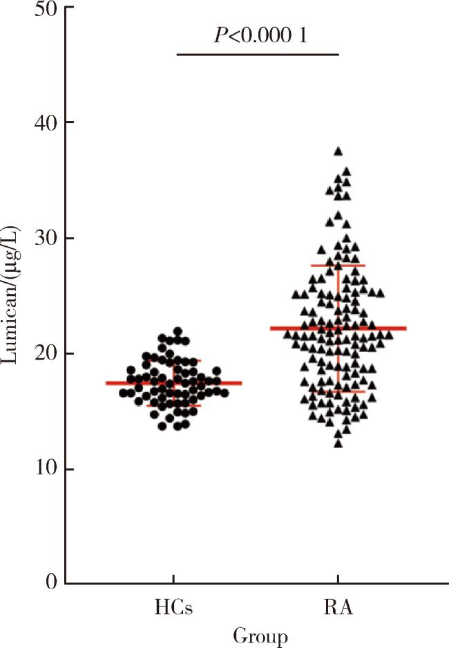
2.2 RA患者和健康对照组血清中LUM表达水平的比较

采用ELISA检测64名健康对照者和133例RA患者的血清LUM水平,分别为(17.47±1.94) μg/L和(22.17±5.47) μg/L。RA患者血清LUM水平显著高于健康对照者(P<0.01,图 1)。
图1 RA患者(n=133)和健康对照者(n=64)血清LUM的表达水平

Figure 1 Serum LUM levels in patients with RA (n=133) and HCs (n=64)

HCs, healthy controls; RA, rheumatoid arthritis; LUM, lumican.

2.3 LUM与临床指标的相关性分析

相关性分析显示,血清LUM水平与RF、IgA、PLT、ESR、CRP、DAS28-CRP等具有相关性(表 2),而与IgM、IgG、C3、C4、抗CCP抗体、病程、年龄等均无明显相关性(P>0.05)。
表2 RA患者血清LUM水平与临床指标的相关性

Table 2 Correlation between serum LUM level and clinical characteristics in patients with RA

Characteristics Serum LUM levels
R P
Age 0.132 0.130
Disease duration 0.032 0.715
ESR 0.295 0.001**
CRP 0.229 0.008**
RF 0.366 <0.001***
Anti-CCP 0.129 0.150
IgA 0.227 0.020**
IgM 0.163 0.097
IgG 0.170 0.082
C3 0.128 0.195
C4 -0.147 0.136
PLT 0.299 0.001**
DAS28-CRP 0.274 0.002**

**P<0.01, ***P<0.001, with exact P values shown in the table (two-tailed Spearman’s rank correlation test). Abbreviations as in Table 1.

2.4 不同特征RA患者血清LUM表达的差异
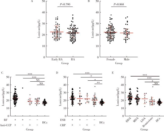
为了分析LUM在不同特征RA患者中的分布特点,本研究进一步对比了早期RA组与RA组患者的血清LUM水平,分别为(22.47±5.55) μg/L和(21.93±5.43) μg/L,两组间差异无统计学意义(P=0.790,图 2A)。LUM在女性和男性RA患者血清中的表达水平分别为(22.16±5.36) μg/L和(22.20±5.88) μg/L,两组间差异无统计学意义(P=0.868,图 2B)。
图2 不同临床及实验室特征的RA患者血清LUM水平

Figure 2 Serum LUM levels in RA patients with different clinical and laboratory characteristics

*P < 0.05, **P < 0.01, ***P < 0.001. A, serum LUM levels in RA patients with different disease durations (early RA: ≤ 2 years; RA: > 2 years); B, serum LUM levels in RA patients with different sexes; C, serum LUM levels in RA patients with different levels of RF and anti-CCP; D, serum LUM levels in RA patients with different levels of CRP and ESR; E, RA patients with different disease activity (Remission: DAS28 ≤ 2.6; LDA: 2.6 < DAS28 ≤ 3.2; MDA: 3.2 < DAS28 ≤ 5.1; HDA: DAS28 > 5.1). HCs, healthy controls; NS, no significant; Other abbreviations as in Table 1.

按照患者血清RF是否阳性以及抗CCP抗体是否阳性将患者分为四组,并对比分析各组患者的血清LUM水平:RF和抗CCP抗体均阳性组LUM为(23.31±5.40) μg/L、RF阳性而抗CCP抗体阴性组为(35.83±0.00) μg/L、RF阴性而抗CCP抗体阳性组为(18.93±3.68) μg/L、RF和抗CCP抗体均阴性组为(19.02±3.01) μg/L。以上结果表明RF阳性组患者血清LUM水平显著高于健康对照组[(17.47±1.94) μg/L],RF阴性的RA患者不论抗CCP抗体阳性还是阴性,其LUM水平与健康对照组相比差异均无统计学意义,RF和抗CCP抗体均阳性组的LUM水平显著高于RF阴性而抗CCP抗体阳性组,以及RF和抗CCP抗体均阴性组;由于RF阴性而抗CCP抗体阳性组仅1例患者,因样本量不足未对该亚组进行进一步分析,其余RA患者组间对比差异无统计学意义(图 2C)。
评估RA患者血清LUM水平与疾病活动度参数间的相关性后,依据不同水平的CRP及ESR水平将RA患者分为四组并对各组患者的血清LUM水平进行了对比:CRP和ESR均升高组的LUM水平为(23.42±5.31) μg/L、CRP升高而ESR正常组为(22.25±7.09) μg/L、CRP正常而ESR升高组为(22.00±4.59) μg/L、CRP和ESR均正常组为(20.30±5.17) μg/L。以上结果表明各组RA患者LUM水平均显著高于健康对照组[(17.47±1.94) μg/L],进一步对比不同CRP与ESR水平的RA患者血清LUM水平发现,CRP和ESR均升高组患者的血清LUM水平显著高于CRP和ESR均正常组,其余患者组间相比差异无统计学意义(图 2D)。
根据不同疾病活动度将RA患者分为四组并对各组患者的血清LUM水平进行了对比:病情处于缓解期的RA患者血清LUM水平为(18.88±5.19) μg/L、低疾病活动度的RA患者为(21.92±6.15) μg/L、中疾病活动度的RA患者为(22.37±5.13) μg/L、高疾病活动度的RA患者为(23.66±5.13) μg/L。以上结果提示RA患者血清LUM水平与疾病活动情况相关,处于缓解期的RA患者血清LUM水平与健康对照组相比差异无统计学意义,缓解期的RA患者血清LUM水平显著低于疾病处于中、高度活动期的RA组患者,其余RA组间对比差异无统计学意义(图 2E)。

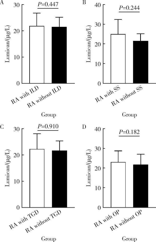
2.5 不同合并症RA患者血清LUM的表达差异

合并与未合并间质性肺疾病(interstitial lung disease, ILD)的RA患者血清LUM水平分别为(23.17±5.28) μg/L和(22.03±5.50) μg/L,差异无统计学意义(P=0.447,图 3A)。合并与未合并干燥综合征(Sjögren syndrome, SS)的RA患者血清LUM水平分别为(24.98±7.51) μg/L和(21.94± 5.24) μg/L,差异无统计学意义(P=0.244,图 3B)。合并与未合并甲状腺疾病(thyroid gland diseases, TGD)的RA患者血清LUM水平分别为(22.26± 5.90) μg/L和(22.16±5.34) μg/L,差异无统计学意义(P=0.910, 图 3C)。合并与未合并骨质疏松症(osteoporosis, OP)的RA患者血清LUM水平分别为(22.98±5.69) μg/L和(21.67±5.30) μg/L,差异无统计学意义(P=0.182, 图 3D)。
图3 合并或不合并各种并发症的RA患者血清LUM水平

Figure 3 Serum LUM levels in RA patients with or without different complications or comorbidities

A, RA patients with or without interstitial lung disease (ILD); B, RA patients with or without Sjögren syndrome (SS); C, RA patients with or without thyroid gland diseases (TGD); D, RA patients with or without osteoporosis (OP). RA, rheumatoid arthritis; LUM, lumican.

2.6 不同血清RF、抗CCP抗体水平的RA患者血清LUM阳性率

本研究中血清LUM的临界值设定为高于健康对照组平均值的2倍标准差。以21.35 μg/L为临界点,将RA患者根据血清RF是否阳性分为RF阳性组和RF阴性组,对比两组RA患者血清LUM阳性率发现,R F阳性组的血清LUM阳性率为60.0% (63/107例),显著高于RF阴性组的28.6% (8/28例,χ2=8.774,P=0.003,表 3)。将RA患者根据血清抗CCP抗体是否阳性分为抗CCP抗体阳性组和抗CCP抗体阴性组,对比两组RA患者,血清LUM阳性率分别为59.8% (64/107例)和36.8% (7/19例),两组差异无统计学意义(χ2=3.461,P=0.063,表 3)。
表3 不同RF和抗CCP抗体水平的RA患者血清LUM阳性率

Table 3 The positive rate of LUM in RA patients with different levels of serum RF or anti-CCP

Group RA with LUM, n (%) RA without LUM, n (%) χ2 value P value
RF+ 63 (60.0) 42 (40.0) 8.774 0.003
RF- 8 (28.6) 20 (71.4)
Anti-CCP+ 64 (59.8) 43 (40.2) 3.461 0.063
Anti-CCP- 7 (36.8) 12 (63.2)

Abbreviations as in Table 1.

3 讨论

本研究发现RA患者血清LUM有表达且相较于健康对照显著升高,LUM水平与CRP、ESR、DAS28呈正相关,提示LUM可能与RA疾病活动性指标存在生物学意义上的关联。此外,LUM水平还与血清IgA、RF水平呈正相关,提示其可以反映RA的免疫活动度。我们通过对比不同特征RA患者的血清LUM水平发现,不同病程及性别的RA患者,血清LUM水平间差异无统计学意义;不同CRP、ESR水平的RA患者血清LUM水平均显著高于健康对照组;此外,RA患者血清LUM水平随疾病活动度增加呈递增趋势,处于缓解期的RA患者血清LUM水平与健康对照组相比差异无统计学意义。合并与不合并并发症(包括ILD、SS、TGD和OP)的RA患者血清LUM水平差异没有统计学意义。对比不同RF和抗CCP抗体水平的RA患者血清LUM阳性率发现,RF和抗CCP抗体阴性的RA患者中,RF阳性组的血清LUM阳性率更高。
至今少有文献报道LUM在RA中的临床意义,最近有研究表明,骨关节炎(osteoarthritis,OA)及RA患者的关节滑液中LUM水平均明显升高,在OA中,LUM可通过诱导TLR4激活提供促炎刺激,但在RA中尚无进一步研究[15]。本研究中发现,RA患者血清LUM水平显著高于健康对照组,增高的血清LUM水平与CRP、ESR、DAS28等疾病活动指标呈正相关,提示RA患者血清LUM的表达可能与疾病活动度有关,这与炎症性肠病中LUM参与炎症反应相符[10]。此外,本研究发现增高的血清LUM与IgA、RF水平呈正相关,提示血清LUM与RA自身抗体的产生有关,可能成为反映免疫活动情况的指标。
流行病学提示RA患者中女性患者的数量显著多于男性,且目前一致认为早期诊断、及时治疗可以显著改善疾病预后[16-17]。目前尚未见研究提示血清LUM与病程及性别有关,本研究通过对比不同病程和性别的RA患者血清LUM水平发现组间差异无统计学意义,提示病程、性别均不是血清LUM表达的影响因素。RA中抗体为B淋巴细胞分化为浆细胞后所产生的球蛋白,其中RF与抗CCP抗体是目前RA临床诊断中的重要血清学标志物[14]。为进一步分析血清LUM的影响因素,本研究对RA患者进行了RF、抗CCP抗体双因素分组,发现RF阳性的RA患者血清LUM水平显著高于健康对照组,且RF和抗CCP抗体均阳性的患者组血清LUM水平显著高于RF阴性组,而RF阴性的RA患者组无论抗CCP抗体阳性还是阴性,其血清LUM水平与健康对照组相比差异均无统计学意义,这与相关性分析中LUM与RF呈正相关而与抗CCP抗体无相关性相符,这可能是由于RA患者体内,RF与抗CCP抗体产生的机制不同所导致的,RF是抗原刺激后B淋巴细胞所产生的自身抗体,其中B淋巴细胞共刺激可能由慢性感染启动并由固有免疫组成部分TLR介导[18-20],而抗CCP抗体则是RA患者B淋巴细胞自发分泌的一种免疫球蛋白[21]。此外,本研究中RF阳性且抗CCP抗体阴性的患者仅1例,故未进一步参与统计分析。
ESR与CRP均为急性期反应物,在RA疾病活动情况的评估中起着重要作用[1]。本研究对RA患者进行了ESR、CRP双因素分组,发现各组RA患者血清LUM水平均显著高于健康对照组,且ESR和CRP均升高的RA患者组血清LUM水平显著高于ESR和CRP均正常的患者组。ESR除受机体炎症活动影响外,还受血液多种成分影响,而CRP则在反映机体炎症活动时受其他因素影响较少[22-23],然而本研究进一步的亚组分析并未发现两者差异有统计学意义,可能是本研究样本量受限所致。ESR和CRP均升高组患者血清LUM水平显著高于ESR与CRP均正常的患者和健康对照组,这与相关性分析中LUM与炎症指标呈正相关,以及前期研究中LUM可反映炎症性肠病的炎症状态相符,提示LUM可反映机体的炎症状态[10]。为进一步分析LUM水平与RA疾病活动情况的关系,我们按疾病活动度分组发现,RA患者的血清LUM水平随疾病活动度增加呈递增趋势,这与相关性分析中血清LUM与疾病活动度指标DAS28-CRP呈正相关相符。此外,病情处于缓解期的RA患者血清LUM水平显著低于疾病处于中、高活动度的RA患者,且与健康对照组相比差异无统计学意义,提示LUM可能成为评估RA病情控制情况的血清学指标。
ILD是RA最常见也是最严重的脏器受累表现,Pilling等[24]的研究发现LUM在肺纤维化的小鼠模型中表达增多。本研究根据是否合并ILD将RA患者分为合并ILD组和未合并ILD组,对比发现两组间LUM水平差异并无统计学意义。此外,最近有研究表明LUM可以通过增加骨形成和减少骨吸收发挥骨保护作用,进而降低骨折风险[25-26]。然而本研究根据是否合并OP将RA患者分为合并OP组和未合并OP组,对比发现两组间LUM水平差异并无统计学意义。本研究也分别就合并与未合并TGD、SS等疾病进行了血清LUM水平对比,同样发现各组间差异无统计学意义,提示LUM异常并不是RA患者肺间质病变或骨质疏松发生发展中的主要致病因素。
RF与抗CCP抗体是目前RA临床诊断中的重要血清学标志物,但二者并不随患者疾病活动度变化有明显改变[14]。本研究发现RF阳性组RA患者血清LUM水平显著高于RF阴性的RA患者,结合血清LUM与疾病活动度指标的相关性,本研究进一步分析了血清RF、抗CCP抗体阴性的RA患者血清LUM阳性率,发现RF阳性的RA患者血清LUM阳性率显著高于血清RF阴性组,提示在RF阳性组RA患者中,血清LUM水平也许能更灵敏地反映其疾病活动情况。
本研究存在一定局限性,本研究为单中心研究,样本量相对较小,现有结果仍需多中心、大样本量的研究进行验证;此外,本研究为横断面研究,未完善治疗前后RA患者血清LUM水平的对比,未能进一步论证其作为评估RA病情控制情况的指标的可靠性。
综上所述,RA患者血清中高表达的LUM与CRP、ESR、DAS28等疾病活动度指标及IgA、RF等免疫活动度指标呈正相关,可能成为同时预判RA疾病活动度及免疫活动度的血清学指标,从而指导临床医师调整RA的治疗方案。

利益冲突  所有作者均声明不存在利益冲突

作者贡献声明  杨菊:搜集、分析、整理数据,完成实验,撰写论文;徐婧、戴菊华:协助搜集、分析、整理数据;石连杰:提出研究思路,设计研究方案,指导撰写论文,总体把关和审定论文。所有作者均对最终文稿进行审读并确认。

1
Smolen JS, Aletaha D, Barton A, et al. Rheumatoid arthritis[J]. Nat Rev Dis Primers, 2018, 4, 18001.

DOI

2
Edilova MI, Akram A, Abdul-Sater AA. Innate immunity drives pathogenesis of rheumatoid arthritis[J]. Biomed J, 2021, 44(2): 172- 182.

DOI

3
Elshabrawy HA, Essani AE, Szekanecz Z, et al. TLRs, future potential therapeutic targets for RA[J]. Autoimmun Rev, 2017, 16(2): 103- 113.

DOI

4
Hurysz B, Bottini N. Emerging proteoglycans and proteoglycan-targeted therapies in rheumatoid arthritis[J]. Am J Physiol Cell Physiol, 2022, 322(6): C1061- C1067.

DOI

5
Garantziotis S, Savani RC. Proteoglycans in Toll-like receptor responses and innate immunity[J]. Am J Physiol Cell Physiol, 2022, 323(1): C202- C214.

DOI

6
Matsushima N, Miyashita H, Kretsinger RH. Sequence features, structure, ligand interaction, and diseases in small leucine rich repeat proteoglycans[J]. J Cell Commun Signal, 2021, 15(4): 519- 531.

DOI

7
Nastase MV, Janicova A, Roedig H, et al. Small leucine-rich proteoglycans in renal inflammation: Two sides of the coin[J]. J Histochem Cytochem, 2018, 66(4): 261- 272.

DOI

8
Grover J, Chen XN, Korenberg JR, et al. The human lumican gene. Organization, chromosomal location, and expression in articular cartilage[J]. J Biol Chem, 1995, 270(37): 21942- 21949.

DOI

9
Wegner N, Lundberg K, Kinloch A, et al. Autoimmunity to specific citrullinated proteins gives the first clues to the etiology of rheumatoid arthritis[J]. Immunol Rev, 2010, 233(1): 34- 54.

DOI

10
Lohr K, Sardana H, Lee S, et al. Extracellular matrix protein lumican regulates inflammation in a mouse model of colitis[J]. Inflamm Bowel Dis, 2012, 18(1): 143- 151.

DOI

11
徐迈宇, 陈钢, 陈雷, 等. 肝细胞癌中Lumican的表达及意义[J]. 肝胆胰外科杂志, 2011, 23(5): 398- 399.

12
Chen X, Li X, Hu X, et al. LUM expression and its prognostic significance in gastric cancer[J]. Front Oncol, 2020, 10, 605.

DOI

13
Chang X, Zhao Y, Wang Y, et al. Screening citrullinated proteins in synovial tissues of rheumatoid arthritis using 2-dimensional Wes-tern blotting[J]. J Rheumatol, 2013, 40(3): 219- 227.

DOI

14
Aletaha D, Neogi T, Silman AJ, et al. 2010 rheumatoid arthritis classification criteria: An American College of Rheumatology/European League Against Rheumatism collaborative initiative[J]. Ann Rheum Dis, 2010, 69(9): 1580- 1588.

DOI

15
Barreto G, Senturk B, Colombo L, et al. Lumican is upregulated in osteoarthritis and contributes to TLR4-induced pro-inflammatory activation of cartilage degradation and macrophage polarization[J]. Osteoarthritis Cartilage, 2020, 28(1): 92- 101.

DOI

16
田新平, 李梦涛, 曾小峰. 我国类风湿关节炎诊治现状与挑战: 来自中国类风湿关节炎2019年年度报告[J]. 中华内科杂志, 2021, 60(7): 593- 598.

17
曾小峰, 朱松林, 谭爱春, 等. 我国类风湿关节炎疾病负担和生存质量研究的系统评价[J]. 中国循证医学杂志, 2013, 13(3): 300- 307.

18
Tan EM, Smolen JS. Historical observations contributing insights on etiopathogenesis of rheumatoid arthritis and role of rheumatoid factor[J]. J Exp Med, 2016, 213(10): 1937- 1950.

DOI

19
Shlomchik MJ, Zharhary D, Saunders T, et al. A rheumatoid factor transgenic mouse model of autoantibody regulation[J]. Int Immunol, 1993, 5(10): 1329- 1341.

DOI

20
Rifkin IR, Leadbetter EA, Busconi L, et al. Toll-like receptors, endogenous ligands, and systemic autoimmune disease[J]. Immunol Rev, 2005, 204, 27- 42.

DOI

21
Reparon-Schuijt CC, van Esch WJ, van Kooten C, et al. Secretion of anti-citrulline-containing peptide antibody by B lymphocytes in rheumatoid arthritis[J]. Arthritis Rheum, 2001, 44(1): 41- 47.

DOI

22
Bedell SE, Bush BT. Erythrocyte sedimentation rate. From folklore to facts[J]. Am J Med, 1985, 78(6 Pt 1): 1001- 1009.

23
McWilliam S, Riordan A. How to use: C-reactive protein[J]. Arch Dis Child Educ Pract Ed, 2010, 95(2): 55- 58.

DOI

24
Pilling D, Vakil V, Cox N, et al. TNF-α-stimulated fibroblasts secrete lumican to promote fibrocyte differentiation[J]. Proc Natl Acad Sci USA, 2015, 112(38): 11929- 11934.

DOI

25
Lee JY, Kim DA, Kim EY, et al. Lumican inhibits osteoclastogenesis and bone resorption by suppressing Akt activity[J]. Int J Mol Sci, 2021, 22(9): 4717.

DOI

26
Lee YS, Park SJ, Lee JY, et al. Benefits of lumican on human bone health: Clinical evidence using bone marrow aspirates[J]. Korean J Intern Med, 2022, 37(4): 821- 829.

DOI

Outlines

/