Role of the SII and SIRI in risk prediction, disease activity assessment, and prog-nostic evaluation of Behçet disease uveitis

  • Yajing GAO 1, 2, 3 ,
  • Zhengfang LI 1, 3 ,
  • Mengsi MA 1, 2, 3 ,
  • Lijun WU , 1, 3, *
Expand
  • 1. Department of Rheumatology and Immunology, People's Hospital of Xinjiang Uygur Autonomous Region, Uygur 830000, China
  • 2. Xinjiang Medical University, Uygur 830000, China
  • 3. Xinjiang Clinical Research Center for Rheumatoid Arthritis, Uygur 830000, China
WU Lijun, e-mail,

Received date: 2025-08-12

  Online published: 2025-10-30

Supported by

the Key Research and Development Project of Xinjiang Uygur Autonomous Region(2022B03002-1)

Copyright

All rights reserved. Unauthorized reproduction is prohibited.

Abstract

Objective: To evaluate the association of systemic immune-inflammation index (SII) and systemic inflammatory response index (SIRI) with Behçet disease uveitis (BU), and to assess their predictive value for inflammatory activity and clinical prognosis in BU patients. Methods: There were 194 patients diagnosed with Behçet disease (BD) and 122 healthy controls. The BD patients were classified into two subgroups based on disease activity: An active phase cohort (n=90) and a stable phase cohort (n=104). Furthermore, the patients were categorized according to the presence or absence of uveitis into two cohorts: BU (n=49) and non-BU (n=145). Among the BU cohort, 26 patients were in the active inflammatory stage, while 23 patients were in the quiescent inflammatory stage. SII and SIRI were calculated using routine blood parameters, including platelet, neutrophil, lymphocyte, and monocyte counts. Spearman correlation analysis was performed to assess the associations of SII and SIRI with BU onset, inflammatory activity, and inflammatory markers. Receiver operating characteristic (ROC) curve analysis was conducted to determine the optimal thresholds and predictive accuracy of SII and SIRI for BU onset and inflammatory activity. Results: SII and SIRI levels were significantly elevated in BD patients with ocular and vascular manifestations compared to those with stable disease (P < 0.05). No significant differences were observed in SII or SIRI levels among the patients with other clinical manifestations of BD. In the patients with BU, both SII and SIRI were significantly higher than in the non-BU and healthy control the groups (P < 0.001). Moreover, SII and SIRI levels were higher during the active inflamma-tory stage than in the inactive stage of BU (P=0.004). Spearman correlation analysis revealed that SII was positively associated with BD disease activity (ρ=0.303, P < 0.001), BU onset (ρ=0.442, P < 0.001), inflammatory activity (ρ=0.392, P=0.005), C-reactive protein (CRP, ρ=0.272, P < 0.001), and erythrocyte sedimentation rate (ESR, ρ=0.285, P < 0.001). SIRI was only positively correlated with BU onset (ρ=0.301, P=0.006). Logistic regression analysis demonstrated that eleva-ted SII was an independent risk factor for BU onset (OR=1.003, 95% CI: 1.001-1.004, P < 0.001). ROC curve analysis indicated that the optimal thresholds for SII were 711.800 [area under curve (AUC)=0.752] for predicting BU onset, 1 622.300 (AUC=0.741) for predicting inflammatory activity, and 1 634.200 (AUC=0.726) for predicting poor prognosis. The corresponding thresholds for SIRI were 1.260 (AUC=0.709), 1.390 (AUC=0.704), and 2.790 (AUC=0.678), respectively. Kaplan-Meier analysis indicated that elevated SII independently predicted adverse prognostic events (HR=3.440, 95%CI: 1.040-11.410, P=0.043). Conclusion: SII and SIRI may serve as potential clinical indicators for predicting inflammatory activity and prognosis in BD patients with uveitis. SII, in particular, demonstrates superior predictive performance for BU onset and disease activity, providing a basis for early identification of high-risk patients and clinical decision-making.

Cite this article

Yajing GAO , Zhengfang LI , Mengsi MA , Lijun WU . Role of the SII and SIRI in risk prediction, disease activity assessment, and prog-nostic evaluation of Behçet disease uveitis[J]. Journal of Peking University(Health Sciences), 2025 , 57(6) : 1067 -1073 . DOI: 10.19723/j.issn.1671-167X.2025.06.008

白塞病(Behçet disease, BD)是一种以血管炎为病理核心的多系统自身免疫性疾病[1],其眼部表现葡萄膜炎是导致不可逆视力损害的主要原因。流行病学数据显示,60%~80%的BD患者会进展为白塞病葡萄膜炎(Behçet disease uveitis, BU)[2],且发病后5年和10年致盲率分别高达24.5%和62.2%[3],严重影响患者生活质量。因此,早期诊断、正确治疗和疾病预后评估对保存和改善BU患者视功能至关重要。
目前临床中BU的诊断及活动度评估主要依靠荧光素眼底血管造影、光学相干断层扫描等影像学技术,但这些检查存在操作复杂、成本高及主观性强的局限性[4]。更重要的是,现有指标难以动态反映全身炎症状态与眼局部病变的交互作用,导致高危患者筛查及预后评估存在滞后性。
近年来,基于常规血液参数衍生的系统性炎症标志物为自身免疫性疾病的精准管理提供了新方向。全身免疫炎症指数(systemic immune inflammation index, SII)和全身炎症反应指数(systemic inflammatory response index, SIRI)通过整合中性粒细胞、淋巴细胞及血小板等参数,可多维度评估机体免疫炎症失衡状态[5]。研究证实,SII和SIRI与类风湿关节炎、系统性红斑狼疮、成人斯蒂尔(Still)病等多种自身免疫疾病的炎症状态相关,并具有显著的预后预测价值[6-7],但其在BD及BU中的应用尚未明确。值得注意的是,BD特有的血管炎表型可能通过血小板-中性粒细胞聚集体形成等机制影响炎症指标特征,提示传统单一参数可能低估BU的炎症负荷。
本研究拟通过探讨SII和SIRI与BU发病风险、疾病活动及预后的相关性,明确SII和SIRI在BU不同临床阶段的动态变化特征,构建基于SII和SIRI的BU风险分层模型,用于评估SII和SIRI作为无创临床指标指导临床决策的可行性,有望为BU的早期干预提供客观、经济的实验室依据,并揭示整合中性粒细胞、淋巴细胞和血小板的细胞参数在BD合并葡萄膜炎中的潜在机制。

1 资料与方法

1.1 研究设计与人群

本研究为一项单中心回顾性队列研究,连续性纳入2021年1月1日至2025年4月30日新疆维吾尔自治区人民医院风湿免疫科就诊的194例BD患者。纳入标准:(1)符合2014年国际白塞病分类标准(International Criteria for Behçet Disease,ICBD)[8];(2)病历资料完整;(3)入组患者及家属均知情同意。排除标准:(1)病历资料不完整;(2)合并其他致葡萄膜炎的自身免疫疾病、恶性肿瘤、感染性疾病;(3)近期有重大手术或创伤,既往有眼内手术史。
本研究同时招募了122例年龄和性别匹配的健康志愿者作为对照组,纳入标准:(1)年龄18~75岁;(2)无自身免疫疾病的个人史及家族史;(3)通过基线检查确认无已知的慢性炎症性疾病、活动性感染或恶性肿瘤病史;排除标准:(1)近3个月内有重大外伤或手术史;(2)近3个月内有使用免疫抑制剂或全身糖皮质激素史;(3)存在任何可能影响炎症指标的急性或慢性健康状况。
本研究获得新疆维吾尔自治区人民医院伦理委员会批准(批准号:KY2025093021),所有研究对象均签署知情同意书。

1.2 分组与临床分期

BD患者194例,健康对照组122例;将BD近期活动性量表评分(Behçet disease current activity form, BDCAF)[9]≥1分定义为BD疾病活动,依据BD是否活动分为BD疾病活动期(90例)和BD疾病稳定期(104例);依据是否发生葡萄膜炎分为BU组(49例)和非BU组(145例);BU组依据BD相关眼病疾病活动度评分[10]分为炎症活动期BU组(26例)和炎症稳定期BU组(23例)。

1.3 临床资料收集

收集患者的临床资料,包括基本信息(年龄、性别、病程等)和实验室指标[白细胞计数、红细胞计数、血小板计数、中性粒细胞计数、淋巴细胞计数、单核细胞计数、C反应蛋白(C-reactive protein,CRP)、红细胞沉降率(erythrocyte sedimentation rate,ESR)],计算SII、SIRI值:SII =(中性粒细胞计数×血小板计数)/淋巴细胞计数,SIRI =(中性粒细胞计数×单核细胞计数)/淋巴细胞计数。

1.4 随访与终点事件定义

基线数据定义为首次住院当天,BU随访终点时间为2025年5月1日或首次出现以下任一不良事件:黄斑水肿、视网膜血管闭塞、视网膜萎缩、视神经萎缩或失明[4]。生存时间的估计方法是从基线日期到随访结束的日期。

1.5 统计学分析

采用SPSS 26.0软件进行分析。对所有计量资料均进行正态性检验,符合正态分布以均数±标准差表示,两组间比较采用独立样本t检验;非正态分布的计量资料以M(P25P75)表示,两组间比较采用Mann-Whitney U检验;计数资料两组间比较采用χ2检验;多组间比较满足正态分布且方差齐的采用单因素方差分析(One-way ANOVA),满足正态分布不满足方差齐性的采用Welch one-way ANOVA,不满足正态分布的采用Kruskal-Wallis检验;采用Spearman相关分析评估SII、SIRI与其他变量之间的相关性;采用二元Logistic回归分析BU发病风险的危险因素。通过受试者工作特征(receiver opera-ting characteristic, ROC)曲线分析确定SII、SIRI的最佳阈值和预测效能;采用DeLong检验比较不同模型ROC曲线下面积的差异。采用Kaplan-Meier法绘制生存曲线,计算生存率和中位生存时间,组间比较采用Log-rank检验。所有统计检验均为双侧检验,P < 0.05为差异有统计学意义。

2 结果

2.1 基线临床特征

共纳入194例BD患者,其中男性83例(占42.8%),女性111例(占57.2%),男女比为1 ∶ 1.34,发病中位年龄38岁,队列中BU的患病率为25.26%(49例),研究人群的平均SII和SIRI分别为510.55(337.16, 849.29) 和0.98(0.59, 1.79)。BD患者的基线临床特征列于表 1
表1 BD患者的基线临床特征

Table 1 Baseline clinical characteristics of patients with BD

Items BD (n=194)
Age/years 37.5 (30, 48)
BD disease course/years 5 (1, 10)
WBC/ (×109/L) 6.83 (5.05, 8.45)
RBC/ (×1012/L) 4.54 (4.16, 4.99)
PLT/ (×109/L) 245 (211.25, 308.00)
NEUT/ (×109/L) 4.05 (2.81, 5.58)
LYMPH/ (×109/L) 1.84 (1.38, 2.35)
MONO/ (×109/L) 0.46 (0.34, 0.63)
CRP/ (mg/L) 2.55 (1.20, 8.54)
ESR/ (mm/h) 17 (10, 30)
SII 510.55 (337.16, 849.29)
SIRI 0.98 (0.59, 1.79)

Data are presented as M(P25P75). BD, Behçet disease; WBC, white blood cells; RBC, red blood cells; PLT, platelet; NEUT, neutrophil count; LYMPH, lymphocyte count; MONO, monocyte count; CRP, C-reactive protein; ESR, erythrocyte sedimentation rate; SII, systemic immune inflammation index; SIRI, systemic inflammatory response index.

2.2 SII、SIRI与BD全身疾病活动度的关联

为明确SII和SIRI与BD全身疾病活动度的关联,比较了BD疾病活动期和BD疾病稳定期患者之间的SII和SIRI。BD疾病活动期患者的SII和SIRI水平均高于BD疾病稳定期患者(表 2P < 0.05)。Spearman相关性分析显示,BD疾病活动与SII(ρ=0.303,P < 0.001)呈正相关,与SIRI(ρ=0.049,P=0.500)无相关性。
表2 BD疾病活动期与稳定期患者的实验室炎症指标比较

Table 2 Comparison of inflammatory markers between active and stable BD patients

Items Active BD (n=90) Stable BD (n=104) P value
WBC/ (×109/L) 7.57 (5.54, 9.75) 6.45 (4.93, 7.76) 0.002
PLT/ (×109/L) 251.50 (213.00, 325.25) 241.00 (210.00, 293.75) 0.269
NEUT/ (×109/L) 4.98 (3.17, 6.65) 3.58 (2.66, 4.73) < 0.001
LYMPH/ (×109/L) 1.74 (1.28, 2.39) 1.92 (1.53, 2.42) 0.078
MONO/ (×109/L) 0.51 (0.37, 0.66) 0.45 (0.33, 0.62) 0.193
CRP/ (mg/L) 4.08 (1.30, 18.46) 2.50 (1.20, 5.25) 0.017
ESR/ (mm/h) 21.50 (10.75, 45.50) 15.50 (8.25, 24.00) 0.001
SII 638.36 (367.84, 1 375.35) 469.78 (227.87, 680.42) 0.001
SIRI 1.19 (0.70, 2.58) 0.82 (0.53, 1.30) 0.002

Data are presented as M(P25P75). BD, Behçet disease; WBC, white blood cells; PLT, platelet; NEUT, neutrophil count; LYMPH, lymphocyte count; MONO, monocyte count; CRP, C-reactive protein; ESR, erythrocyte sedimentation rate; SII, systemic immune inflammation index; SIRI, systemic inflammatory response index.

2.3 SII、SIRI在不同临床表现BD患者中的差异

为明确SII和SIRI是否因临床表现不同而有所差异,对不同临床表现BD患者的SII和SIRI进行比较,结果显示,有葡萄膜炎的BD患者SII和SIRI水平升高(P < 0.001),有血管表现的BD患者SII和SIRI水平升高(P < 0.05),其他临床表现患者中SII和SIRI的差异无统计学意义(表 3)。
表3 比较不同临床表现BD患者的SII和SIRI水平

Table 3 Comparative analysis of SII and SIRI in clinical manifestations BD

Manifestations Cases, n(%) SII,M(P25P75) P value SIRI, M(P25P75) P value
Oral aphthae 0.772 0.894
    Yes 192 (98.96) 510.55 (335.94, 858.82) 0.98 (0.59, 1.79)
    No 2 (1.04) 487.93 (335.75, 487.93) 1.24 (0.69, 1.24)
Genital aphthae 0.499 0.525
    Yes 153 (78.87) 508.22 (331.06, 808.88) 1.03 (0.60, 1.70)
    No 41 (21.13) 524.90 (351.78, 1 259.78)) 0.81 (0.51, 1.93)
Dermal 0.575 0.710
    Yes 81 (41.75) 504.20 (326.68, 724.67) 0.99 (0.57, 1.66)
    No 113 (58.25) 524.90 (341.59, 890.59) 0.97 (0.59, 1.81)
Uveitis < 0.001 < 0.001
    Yes 49 (25.26) 823.73 (498.34, 1 622.28) 1.61 (0.94, 2.90)
    No 145 (74.74) 453.16 (296.94, 684.78) 0.82 (0.54, 1.38)
Gastrointestinal 0.110 0.640
    Yes 25 (12.89) 704.76 (351.36, 1 441.90) 1.15 (0.57, 2.07)
    No 169 (87.11) 504.20 (332.88, 791.37) 0.97 (0.59, 1.71)
Vascular 0.040 0.038
    Yes 22 (11.34) 1 107.40 (422.73, 1 457.9) 1.56 (0.73, 1.72)
    No 172 (88.66) 671.45 (328.92, 772.03) 0.98 (0.57, 1.63)
Cardiovascular 0.363 0.340
    Yes 4 (2.06) 1 086.72 (349.43, 1 369.11) 1.93 (0.74, 2.72)
    No 190 (79.94) 506.85 (335.34, 813.91) 0.97 (0.59, 1.70)
Nervous system 0.435 0.856
    Yes 15 (7.73) 504.20 (335.75, 887.92) 1.07 (0.37, 2.55)
    No 179 (92.27) 512.89 (248.41, 707.32) 0.97 (0.60, 1.72)

BD, Behçet disease; SII, systemic immune inflammation index; SIRI, systemic inflammatory response index.

2.4 BU组与非BU组的SII、SIRI水平比较

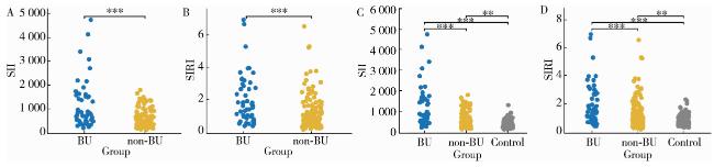
因BU患者的SII和SIRI与有其他临床表现患者相比差异有统计学意义,故具体分析BU组、非BU组及健康对照组间SII和SIRI的差异,SII和SIRI的值BU组高于非BU组及健康对照组,差异有统计学意义(图 1P < 0.001)。
图1 BU组、非BU组及健康对照组的SII和SIRI分布水平

Figure 1 SII and SIRI levels in BU, non-BU and control groups

* *P < 0.01; * * *P < 0.001. A, SII levels in BU and non-BU; B, SIRI levels in BU and non-BU; C, SII levels in BU, non-BU and control groups; D, SIRI levels in BU, non-BU and control groups. BU, Behçet disease uveitis; SII, systemic immune inflammation index; SIRI, systemic inflammatory response index.

2.5 BU发病的相关危险因素分析

以BD患者是否发生葡萄膜炎为因变量,排除既往文献[11]中有临床意义的如年龄、性别、病程、CRP、ESR等其他混杂因素后,将主要研究指标SII、SIRI纳入多因素二元Logistic回归模型,结果显示仅有SII升高是BD患者发生BU的独立危险因素(表 4P < 0.001)。
表4 BU发病风险的多因素二元Logistic回归分析

Table 4 Multivariate Logistic regression analysis of BU

Items β SE t P OR OR 95%CI
SII 0.003 0.001 19.702 < 0.001 1.003 1.001-1.004
SIRI 0.041 0.141 0.083 0.773 0.960 0.729-1.265

BU, Behçet disease uveitis; SII, systemic immune inflammation index; SIRI, systemic inflammatory response index.

2.6 活动期与稳定期BU患者的炎症指标差异

为进一步明确不同分期BU患者各炎症指标的差异,对26例炎症活动期BU组(53.06%)和23例炎症稳定期BU组(46.94%)患者的实验室炎症指标进行比较,其中白细胞计数、中性粒细胞计数、SII和SIRI在炎症活动期BU组均高于炎症稳定期BU组,差异有统计学意义(表 5P < 0.05)。
表5 活动期与稳定期BU患者的实验室炎症指标比较

Table 5 Comparative analysis of systemic inflammation in active and stable BU

Items Active BU (n=26) Stable BU (n=23) P value
WBC/ (×109/L) 9.36 (7.32, 11.49) 6.74 (5.08, 9.71) 0.002
PLT/ (×109/L) 308.50 (232.00, 366.00) 258.00 (213.00, 304.00) 0.113
NEUT/ (×109/L) 6.85 (4.81, 8.95) 4.39 (3.05, 5.74) 0.001
LYMPH/ (×109/L) 1.51 (1.06, 2.50) 1.54 (1.31, 2.09) 0.113
MONO/ (×109/L) 0.56 (0.40, 0.69) 0.51 (0.36, 0.75) 0.440
CRP/ (mg/L) 2.53 (1.18, 7.82) 2.50 (1.00, 6.56) 0.920
ESR/ (mm/h) 19.00 (11.00, 38.75) 18.00 (9.00, 33.00) 0.865
SII 1 471.74 (639.23, 1 970.36) 683.47 (347.29, 1 309.66) 0.004
SIRI 1.86 (1.36, 3.21) 1.11 (0.64, 2.26) 0.015

Data are presented as M(P25P75). BU, Behçet disease uveitis; WBC, white blood cells; PLT, platelet; NEUT, neutrophil count; LYMPH, lymphocyte count; MONO, monocyte count; CRP, C-reactive protein; ESR, erythrocyte sedimentation rate; SII, systemic immune inflammation index; SIRI, systemic inflammatory response index.

2.7 SII、SIRI与BU发病风险及炎症活动的相关性

通过Spearman相关性分析比较发现,SII与BU发病(ρ=0.442,P < 0.001)、BU炎症活动(ρ=0.392,P=0.005)、CRP(ρ=0.272,P < 0.001)、ESR(ρ=0.285,P < 0.001)呈正相关,而SIRI仅与BU发病呈正相关(ρ=0.301,P=0.006),与BU炎症活动(ρ=0.095,P=0.187)、CRP(ρ=0.071,P=0.332)、ESR(ρ=0.043,P=0.552)无相关性。

2.8 SII、SIRI对BU发病风险、炎症活动及预后的预测效能

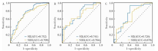
为评估SII与SIRI对BU的预测价值,进行了ROC曲线分析(图 2)。
图2 SII和SIRI预测BU的ROC曲线

Figure 2 ROC curves of SII and SIRI for BU prediction

A, onset risk prediction in BU; B, disease activity in BU; C, predicting prognosis risk in BU. SII, systemic immune inflammation index; SIRI, systemic inflammatory response index; BU, Behçet disease uveitis; ROC, Receiver operating characteristic; AUC, area under curve.

在BU发病风险预测方面,SII和SIRI的ROC曲线下的面积(area under curve, AUC) 分别为0.752(95%CI: 0.669~0.834)和0.709(95%CI: 0.624~0.794)。根据阈值(SII: 711.800; SIRI: 1.260),SII >711.800或SIRI >1.260的患者发生BU的风险增加(图 2A)。两条ROC曲线比较差异无统计学意义(DeLong检验,P=0.450)。
在区分BU疾病活动方面,SII和SIRI的AUC值分别为0.741 (95%CI: 0.602~0.879) 和0.704 (95%CI: 0.554~0.854)。SII > 1 622.300或SIRI > 1.390的患者处于炎症活动期的风险更高(图 2B)。两条ROC曲线比较差异无统计学意义(DeLong检验,P=0.180)。
在预测BU预后不良风险方面,SII (AUC=0.726, 95%CI: 0.562~0.890) 和SIRI (AUC=0.678, 95%CI: 0.512~0.845) 同样具有预测价值。SII > 1 634.200或SIRI > 2.790的患者发生预后不良风险的升高(图 2C)。两条ROC曲线比较差异无统计学意义(DeLong检验,P=0.360)。

2.9 BU预后不良的生存分析

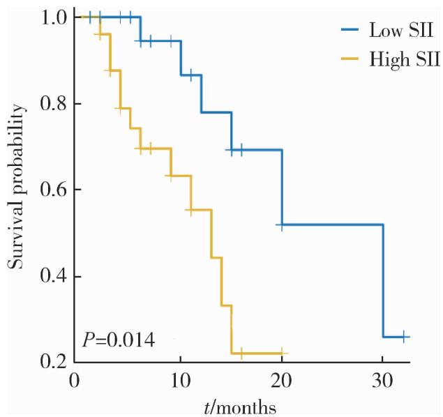
为进一步验证多因素Logistic回归分析中高SII水平是BD患者发生BU独立危险因素,依据上面ROC分析得到的最佳临界值,按照“高SII”和“低SII”将患者分组进行Kaplan-Meier分析,高SII组患者预后不良事件发生率高于低SII患者(Log-rank, P=0.009),高SII水平仍是预后不良事件的独立预测因素(HR =3.820, 95%CI: 1.310~11.100, P=0.014,图 3)。
图3 不同SII组BU患者Kaplan-Meier曲线

Figure 3 Kaplan-Meier curves of BU patients in different SII groups

SII, systemic immune inflammation index. BU, Behçet disease uveitis.

3 讨论

本研究系统评估了SII与SIRI在BU患者中的临床价值,明确了其在发病风险预测、炎症活动评估及预后判断中的潜在作用,结果提示SII不仅与BU发病风险显著相关(ρ=0.442, P < 0.001),还与疾病活动度(ρ=0.392, P=0.005)及传统炎症标志物CRP、ESR一致,提示其具备作为BU疾病活动监测与预后评估的双重潜力。
本研究中活动期BU患者的中性粒细胞计数与SII水平高于稳定期患者,提示中性粒细胞异常活化可能是BU进展的核心驱动因素。已有研究表明,BD和BU患者的中性粒细胞胞外诱捕网(neutrophil extracellular traps, NETs)释放蛋白酶3,可激活Th17细胞产生白介素17(interleukin 17, IL-17)[12-13],而IL-17通过上调P-选择素等黏附分子促进血小板、中性粒细胞聚集体形成[14],形成持续炎症的反馈环,最终导致SII在活动期升高,因此,SII有望成为评估IL-17靶向治疗效果的潜在临床指标。
本研究发现SII在以血管炎为主要表现的BD患者中显著升高,提示其在捕捉BD血管损伤与微血栓形成方面具有独特优势[7],这种差异可能源于血小板活化在BD血管损伤中的核心地位[15],以及IL-17通路对血小板活化的独特调控机制。SII整合了中性粒细胞、血小板和淋巴细胞三系参数,更能反映BD血管炎中血小板、中性粒细胞交互作用及微血栓形成过程。在以往研究中[16],炎症标志物中性粒细胞与淋巴细胞比率在BD中的诊断预测效能(AUC=0.690)低于本研究中SII的预测效能(AUC=0.752),更进一步支持了整合中性粒细胞、血小板和淋巴细胞的SII作为监测BD炎症活动的优势。
在预测效能方面,本研究确立的SII阈值体系具有良好的临床应用前景:711.8作为筛查临界值,适用于基层医疗机构在缺乏眼底造影条件下的高危患者识别, 而1 622.3和1 634.2作为临界值分别用于判断炎症活动与预后不良,提示临床需及时调整治疗方案,并在患者随访过程中早期预测BU炎症活动的可能性。鉴于SII检测成本低、可及性强,未来可探索其在基层医院、尤其在缺乏眼科专科资源的地区,作为BU筛查和随访工具的可行性。
尽管本研究在BU炎症标志物领域提供了新的视角,但仍存在以下局限性:(1)BU亚组样本量相对较小,尤其是炎症活动期BU,可能影响部分相关性分析的统计效能,未来需扩大样本量以验证本研究发现;(2)为单中心回顾性研究,研究对象主要为新疆地区患者,其民族构成与遗传背景具有一定地域性,SII与SIRI的预测阈值是否适用于其他地区人群仍需多中心、前瞻性研究加以验证;(3)未来可结合单细胞测序解析SII升高的特定中性粒细胞亚群,结合NETs标志物检测进一步验证SII与BD、BU中性粒细胞异常活化及NETs释放的关系,以期揭示中性粒细胞、血小板在BD合并葡萄膜炎进展中的潜在机制。
综上,SII和SIRI可作为BU患者风险预测、疾病活动度及预后评估的潜在临床指标,未来研究可进一步探索SII在BU疗效监测中的应用价值,并结合免疫机制研究明确其在BD炎症中的机制,从而为临床早期识别高风险BU患者及干预决策提供依据。

利益冲突  所有作者均声明不存在利益冲突。

作者贡献声明  高雅静:设计研究方案,采集、整理数据,统计学分析,论文撰写;李正芳:采集数据,统计学分析;马梦思:收集、整理数据;武丽君:总体把关和审定论文。所有作者均参与论文修改,并对最终文稿进行审读和确认。

1
Izzedine H , Jhaveri KD . Behçet's syndrome[J]. N Engl J Med, 2024, 390 (18): 1731.

2
Su G , Zhong Z , Zhou Q , et al. Identification of novel risk loci for Behçet' s disease-related uveitis in a Chinese population in a genome-wide association study[J]. Arthritis Rheumatol, 2022, 74 (4): 671- 681.

DOI

3
Li C , Li L , Wu X , et al. Clinical manifestations of Behçet' s disease in a large cohort of Chinese patients: Gender- and age-related differences[J]. Clin Rheumatol, 2020, 39 (11): 3449- 3454.

DOI

4
Zhong Z , Su G , Yang P . Risk factors, clinical features and treatment of Behçet's disease uveitis[J]. Prog Retin Eye Res, 2023, 97, 101216.

DOI

5
Islam MM , Satici MO , Eroglu SE . Unraveling the clinical significance and prognostic value of the neutrophil-to-lymphocyte ratio, platelet-to-lymphocyte ratio, systemic immune-inflammation index, systemic inflammation response index, and delta neutrophil index: An extensive literature review[J]. Turk J Emerg Med, 2024, 24 (1): 8- 19.

DOI

6
Yang CH , Wang XY , Zhang YH , et al. SIRI and SII as potential biomarkers of disease activity and lupus nephritis in systemic lupus erythematosus[J]. Front Immunol, 2025, 16, 1530534.

DOI

7
Lu Z , Xie Z , Shen K , et al. Association of dietary inflammatory index with immune-inflammatory biomarkers in rheumatoid arthritis patients: Results from NHANES 1999-2018[J]. Front Nutr, 2024, 11, 1353964.

DOI

8
Davatchi F , Assaad-Khalil S , Calamia KT , et al. The Inter-national Criteria for Behçet' s Disease (ICBD): A collaborative study of 27 countries on the sensitivity and specificity of the new criteria[J]. Acad Dermatol Venereol, 2014, 28 (3): 338- 347.

DOI

9
Bhakta BB , Brennan P , James TE , et al. Behçet' s disease: Evaluation of a new instrument to measure clinical activity[J]. Rheumatology, 1999, 38 (8): 728- 733.

DOI

10
Keino H . Evaluation of disease activity in uveoretinitis associated with Behçet's disease[J]. Immunol Med, 2021, 44 (2): 86- 97.

DOI

11
Koru L , Esen F , Turkyilmaz O , et al. Clinical characteristics of pediatric noninfectious uveitis and risk factors for severe disease: A single-center study[J]. Clin Rheumatol, 2024, 43 (9): 2933- 2942.

12
Wu Y , Ning K , Huang Z , et al. NETs-CD44-IL-17A feedback loop drives Th17-mediated inflammation in Behçet' s uveitis[J]. Adv Sci, 2025, 12 (16): 2411524.

DOI

13
Le Joncour A , Cacoub P , Boulaftali Y , et al. Neutrophil, NETs and Behçet' s disease: A review[J]. Clin Immunol, 2023, 250, 109318.

14
Dinkla S , van Cranenbroek B , van der Heijden WA , et al. Platelet microparticles inhibit IL-17 production by regulatory T cells through P-selectin[J]. Blood, 2016, 127 (16): 1976- 1986.

15
Citirik M , Ozdal PC , Keles A , et al. Platelet activation in ocular Behçet's patients with posterior segment involvement[J]. Middle East Afr J Ophthalmol, 2022, 28 (4): 203- 207.

16
Shadmanfar S , Masoumi M , Davatchi F , et al. Correlation of clinical signs and symptoms of Behçet's disease with platelet-to-lymphocyte ratio (PLR) and neutrophil-to-lymphocyte ratio (NLR)[J]. Immunol Res, 2021, 69 (4): 363- 371.

DOI

Outlines

/