Clinicopathological analysis of mesonephric-like adenocarcinoma in the corpusuteri: A report of 3 cases

  • Xiaolin WANG 1 ,
  • Luyao LI 1 ,
  • Wen ZHANG 2 ,
  • Hongyan WANG , 1, *
Expand
  • 1. Department of Pathology, the Frist Affiliated Hospital of Xi'an Jiaotong University, Xi'an 710061, China
  • 2. Department of Pathology, Northwest Women's and Children's Hospital, Xi'an 710061, China
WANG Hongyan, e-mail,

Received date: 2023-11-29

  Online published: 2025-11-07

Copyright

All rights reserved. Unauthorized reproduction is prohibited.

Abstract

Mesonephric-like adenocarcinoma (MLA) in the corpus uteri is a highly aggressive malignant tumor, which is easily confused with other types of endometrial cancer. When the tumor morphology and cellular characteristics are not consistent with the clinical biological behavior, attention should be paid to it. This study is to investigate the clinicopathologic features of MLA in the corpus uteri. Three cases of MLA in the corpus uteri diagnosed in the First Affiliated Hospital of Xi'an Jiaotong University from 2020 to 2023 were studied by clinical data, microscopic features and immunohistochemistry. The related literature was reviewed. The clinical manifestations of the three cases of MLA in the corpus uteri were nonspecific. One case was from our hospital and the other two cases were from other hospitals. The age range was 54-58 years. In the specimen description, there was a diffusely growing mass in the endometrium of the uterus, with an uneven surface and tough texture. The muscle wall was extensively invaded. At low magnification, the tumor cells were arranged in tubular, glandular, papillary, micropapillary and solid growth patterns. At high magnification, the cells lining the lumen were arranged in a single layer, cuboidal or columnar pattern, with mild to moderate atypia, with vesicular nuclei and nuclear furrows. Some of the lumens showed eosinophilic homogeneous pink staining without structural-like material. In the solid area, the cells were plat fusiform, arranged in bundles or whirlpools, with large nuclear atypia and frequent mitotic figures. A large number of intravascular cancer thrombus were observed in all the three cases. The tumor cells were positive for GATA3 and/or thyroid transcription factor-1 (TTF1), diffusely positive for pair box gene 2 (PAX2) and PAX8, and positive for CD10 in some luminal margins. Estrogen receptor (ER) was focal positive, and progesterone receptor (PR) was negative. KRAS mutation was detected in case 1. According to the 2023 updated International Federation of Gynecology and Obstetrics (FIGO) staging guidelines for endometrial cancer, all the three cases were in advanced stage. It is suggested that pathologists should make accurate diagnosis based on morphological manifestations, using a set of matched immunohistochemical markers and necessary molecular tests to avoid misdiagnosis and better guide clinical diagnosis and treatment.

Cite this article

Xiaolin WANG , Luyao LI , Wen ZHANG , Hongyan WANG . Clinicopathological analysis of mesonephric-like adenocarcinoma in the corpusuteri: A report of 3 cases[J]. Journal of Peking University(Health Sciences), 2025 , 57(6) : 1208 -1212 . DOI: 10.19723/j.issn.1671-167X.2025.06.030

子宫体中肾样腺癌(mesonephric-like adenocarcinoma,MLA)是一种罕见的发生于子宫体和卵巢的生殖系统恶性肿瘤,解剖位置及大体特征与子宫内膜癌和卵巢癌相类似,但镜下形态及免疫组织化学表型与中肾腺癌(mesonephric adenocarcinoma,MA)非常相似。子宫体MLA发病率约占所有子宫内膜癌的1%[1],其发病机制尚不清楚,组织形态学表现多样,易被误诊为低级别子宫内膜样腺癌、透明细胞癌、浆液性癌、癌肉瘤等。现回顾性分析2022—2023年于西安交通大学第一附属医院确诊的3例子宫体MLA,并复习相关文献,探讨其临床特点、组织病理学特征及免疫组织化学表型,以提高病理医师的认识和诊断水平。

1 病例资料

1.1 临床资料

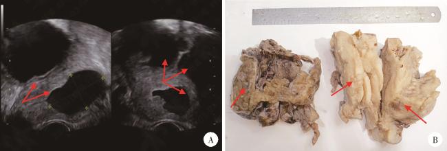
3例子宫体MLA患者均经B超检查提示宫腔占位性病变(图 1A)。其中例1为本院诊治患者,55岁,体检发现;例2为外院会诊患者,58岁,主诉下腹痛;例3为外院会诊患者,54岁,主诉绝经后阴道出血。
图1 例1患者的影像特征及肉眼改变

Figure 1 Imaging features and gross changes of case 1

A, the ultrasound image of case 1 showed mixed echogenic masses in the uterine cavity and bilateral ovaries with dark fluid areas (arrows indicate the fluid dark area of the mass); B, gross findings of case 1; The left side was the right ovary and the right side shows the whole uterus (arrows indicate the location of the mass).

1.2 大体检查

3例大体表现均为宫腔内膜面弥漫型肿物,表面凹凸不平呈颗粒状,切面灰白色,质韧,边界欠清,向下弥漫侵及肌壁,其中例1和例3累及宫颈管,表现为宫颈管内生浸润型肿物,灰白色,质硬;例1和例2累及双侧附件,附件结构不清,切面呈囊实性,实性区灰白色、质硬,囊性区内壁暗红色、粗糙,散在颗粒样凸起(图 1B)。

1.3 镜下检查

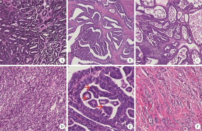
手术标本经40 g/L中性甲醛固定,常规切片,苏木精-伊红染色。镜下检查提示肿瘤细胞主要呈小管状(图 2A)、乳头状排列(图 2B),偶见微乳头状结构(例1,图 2C)及实体型区域(例3,图 2D);2例可见凝固性坏死灶。高倍镜下,管腔内衬细胞单层排列,呈立方状或柱状,核浆比增大,胞浆略嗜酸,细胞核呈卵圆形,轻到中度异型,染色质致密或呈泡状,可见核沟及核重叠(图 2E),小核仁较明显,核分裂象不易见;例3实体区细胞呈胖梭形,排列呈束状或漩涡状,核异型性较大,核分裂象易见,每10个高倍视野下约3个,类似肉瘤,但细胞核特征与小管状区域相似,且紧邻小管状区域。部分管腔内可见嗜酸性均质粉染无结构分泌物(图 2F)。
图2 3例患者的镜下特征(苏木精-伊红染色)

Figure 2 Microscopic features of the three patients (hematoxylin-eosin staining)

A, the tumor cells were tubular or glandular (×200); B, the tumor cells were papillary (×200); C, the tumor cells were micropapillary (×200); D, the tumor cells were spindle-shaped (×200); E, vesicular nuclei and nuclear furrows (red arrows) are visible at high magnification (×400); F, luminal eosinophilic homogeneous pink staining without structured secretions (×200).

1.4 免疫组织化学染色

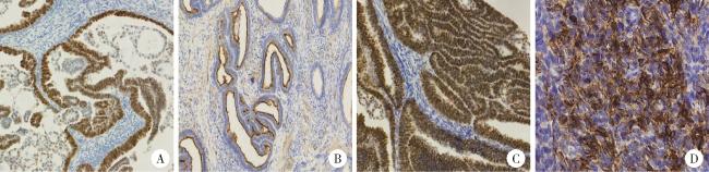
采用EnVision两步法行免疫组织化学染色,结果提示3例均配对盒基因2(pair box gene 2,PAX2)弥漫核阳性(图 3A),PAX8弥漫核阳性,CD10部分腔缘阳性(图 3B),P16局灶阳性,MSH2、MSH6、MLH1、PMS2弥漫阳性,Ki67热点区域阳性率约50%~60%;1例钙视网膜蛋白(calretinin,CR)小灶阳性,雌激素受体(estrogen receptor,ER)小灶阳性;3例孕激素受体(progesterone receptor,PR)、肾母细胞瘤(Wilm’ s tumor 1,WT-1)均阴性,p53呈野生型。GATA3和甲状腺转录因子1(thyroid transcription factor-1,TTF1)呈现不同的表达模式,例1 GATA3+/TTF1+(图 3C),例2 GATA3-/TTF1+,例3 GATA3+/TTF1-。此外,例3实体区域梭形细胞广谱CK阳性(图 3D)。
图3 3例患者的免疫组织化学染色特征(EnVision)

Figure 3 Immunohistochemical staining characteristics of the three patients (EnVision)

A, pair box gene 2 (PAX2) was diffusely positive in the nucleus (×200); B, the luminal margin was positive for CD10 (×200); C, thyroid transcription factor-1 (TTF1) was positive in the nucleus (×200); D, case 3 was positive for broad-spectrum CK in solid spindle cells (×200).

1.5 分子特征

本组例1进一步采用荧光定量聚合酶链反应技术行KRASNRASBRAF分子检测,发现KRAS基因突变。

1.6 手术-病理分期

3例均诊断为子宫体MLA,属于子宫内膜癌的侵袭性组织学类型,均侵及深肌层,具体病理分期见表 1。根据2023年国际妇产科联盟(International Federation of Gynecology and Obstetrics,FIGO)子宫内膜癌分期[2],例1为FIGO ⅢA1期,例2为FIGO ⅣB期,例3为FIGO ⅡC期。
表1 3例患者手术-病理分期

Table 1 Surgical-pathological staging of the three patients

Items Case 1 Case 2 Case 3
Depth of infiltration 2/3 myometrium Full myometrium and serosa 3/5 myometrium
Tumor emboli in blood vessels and lymphatic vessels >4 >4 >4
Limited spread Involving the cervix and both adnexa Involving both adnexa Involving the cervix
Pelvic lymph nodes Negative Negative Negative
Distant metastasis Negative Peritoneal metastasis Negative

1.7 治疗及预后

3例患者均行全子宫双附件及盆腔淋巴结清扫手术并联合同步放化疗,至2024年3月已分别随访13个月、16个月、12个月,目前均未发现复发或转移。

2 讨论

2016年McFarland等[3]首次提出MLA这一术语,用于描述一组在形态学和免疫组织化学上类似于宫颈MA,但发生在宫颈外的肿瘤,最常见于子宫内膜和卵巢,现已被纳入第5版《世界卫生组织女性生殖器官肿瘤分类》中。MLA的起源及发病机制尚不清楚,有学者发现MLA与子宫内膜异位症、子宫腺肌病及其他苗勒管(Müllerian duct)肿瘤相关,所以倾向认为MLA起源于苗勒管,随后沿着中肾方向分化[1, 4]
子宫体MLA的发病年龄范围较广,从26~91岁均有报道,中位年龄61岁[1],最常见的症状是绝经后阴道出血或异常子宫出血,约半数以上病例发现时已处于晚期。
子宫体MLA肉眼表现与其他类型的子宫内膜癌相似。低倍镜下肿瘤表现为多种生长模式,如小管状、腺管状、乳头状、微乳头状、实性、性索样、小梁状、粉刺样、网状、筛状、肾小球样、梭形细胞等,以小管状、腺管状最常见,这种混合存在的生长模式易与其他类型子宫内膜癌相混淆,如浆液性癌、透明细胞癌、子宫内膜样癌、癌肉瘤等,尤其是在子宫内膜活检诊断中;本组例1可见乳头及微乳头成分,类似浆液性癌,例3可见实体梭形细胞成束排列,类似肉瘤。管腔内嗜酸性粉染物是MLA的一个特征性表现,然而并非每个病例均有此特征。高倍镜下癌细胞呈扁平、立方或柱状,胞质轻度嗜酸,胞核中度异型,可见泡状核,核常重叠或成角,可见核沟;有的可见不规则的梭形细胞呈漩涡或束状排列,类似于子宫内膜样癌的未成熟鳞状细胞,但没有显示细胞间桥、明显的细胞边界或角化。肿瘤区域内无鳞状、纤毛及黏液化生,且周围无中肾管残迹。一组237例子宫内膜癌的回顾性研究发现,与子宫内膜样癌患者相比,MLA肿瘤体积更大,深肌层浸润、FIGO Ⅲ~Ⅳ期更常见[5]
MLA的免疫组织化学染色具有特征性,有学者通过分析694例MA、MLA、子宫内膜样癌、宫颈腺癌患者的免疫组织化学表型发现, GATA3是MA和MLA的最佳标志物,其敏感性和特异性分别为91%和94%[6]。研究还认为TTF1在绝大多数MLA病例中高表达,可与GATA3联合辅助诊断MLA,且二者有时呈现相反的染色模式,即GATA3-/TTF1+,如本组例2。MLA中CD10多为局灶表达,主要表现为腔缘染色阳性,其敏感性为73%,特异性为83%;而CR敏感性、特异性均较低,仅少数病例中见局灶阳性表达,本组例3实体区显示有灶状阳性。Euscher等[7]对23例子宫内膜MLA的免疫表型特征进行研究也发现了类似的结果,因此建议使用一组免疫组织化学试剂来辅助诊断MLA,如GATA3、TTF1、CD10、CR等。MLA中PAX8、PAX2常呈弥漫阳性表达,p53呈野生型表达,P16局灶阳性表达[4-5]。ER、PR常阴性,但也有学者发现少数病例中ER可呈弱阳性表达(如本组例3即为ER小灶阳性), 他们认为,与ER相比,PR阴性是更可靠的指标[1]。Kim等[8]发现MLA均表现为微卫星稳定性,MSH2、MSH6、MLH1、PMS2表达阳性,本组3例免疫组织化学表达均与该研究一致。
Mirkovic等[9]发现子宫体和卵巢MLA与MA的分子特征相似,均表现为KRAS突变、1q增加、10号和12号染色体增加,无PTEN突变,表明MLA与MA在生物学行为上有重叠;此外,研究还发现半数病例存在PIK3CA突变,该突变常见于子宫内膜样癌,MA中未见报道,表明MLA也与苗勒管(子宫内膜样)分化癌有重叠。本组例1检测出KRAS突变,与以往研究相符。
由于MLA生长模式的多样性,还需与其他类型肿瘤鉴别:(1)宫颈MA累及宫体:虽然二者镜下特点及分子遗传学极其相似,但根据病变解剖位置可做出诊断,且病灶周边常有中肾残余或增生;(2)子宫内膜样癌:肿瘤主要呈腺管状排列,常伴鳞状分化、纤毛及黏液化生,ER、PR弥漫阳性;(3)浆液性癌:肿瘤常呈复杂乳头状结构和/或腺样结构,形态不规则,腺腔内无嗜酸性物,细胞核异型性较大,核仁明显,呈p53突变型,P16强阳性表达;(4)透明细胞癌:肿瘤排列呈管状、囊状、乳头状和实性片状,管腔内有时可见嗜酸性物,囊管状、乳头状区衬覆靴钉样细胞,实性区由透明细胞组成,核异型性明显,肝细胞核因子1β(hepatocyte nuclear factor 1 beta,HNF1β)、Napsin A蛋白酶阳性表达;(5)子宫体MA:以往有学者认为子宫体MA肿瘤完全或主要位于子宫肌层,常伴有中肾增生或残留,而MLA主要位于子宫内膜,虽然可能会发生肌层侵犯,但肿瘤应该出现在子宫内膜,随后侵犯肌层,而不是主要位于子宫肌层内[10]
由于目前国内外子宫体MLA病例报道较少,无相关大样本前瞻性研究,因此尚无标准的治疗方案,临床上仍以经验性手术联合放疗和(或)化疗为主,具体疗效尚不确切,本组3例患者在手术治疗后的随访过程中尚未发现复发或转移。
综上,子宫体MLA具有高度侵袭性,镜下以腺管状结构常见,细胞轻到中度异型,但通常发现时已处于晚期,因此,当肿瘤的镜下特征与临床生物学行为不相符时,需考虑该疾病。此外,因子宫体MLA具有多种不同的生长模式,易与其他类型子宫内膜癌混淆,提示病理医生在诊断过程中切记不能遗漏,需基于形态学表现,并检测包括GATA3、TTF1、CD10、CR、PAX8、PAX2、p53、P16、ER、PR以及MSH2、MSH6、MLH1、PMS2等在内的一组免疫组织化学标记物,条件允许时可同时进行分子检测以做出准确的诊断。

利益冲突  所有作者均声明不存在利益冲突。

作者贡献声明  王晓林:收集、分析、整理数据,撰写论文;李璐瑶、张雯:收集、分析、整理数据;王鸿雁:提出研究思路,总体把关和审定论文。所有作者均参与论文修改,并对最终文稿进行审读和确认。

1
Deolet E , Van Dorpe J , Van de Vijver K . Mesonephric-like adenocarcinoma of the endometrium: Diagnostic advances to spot this wolf in sheep's clothing. A review of the literature[J]. J Clin Med, 2021, 10 (4): 698.

DOI

2
Berek JS , Matias-Guiu X , Creutzberg C , et al. FIGO staging of endometrial cancer: 2023[J]. Int J Gynaecol Obstet, 2023, 162 (2): 383- 394.

DOI

3
McFarland M , Quick CM , McCluggage WG . Hormone receptor-negative, thyroid transcription factor 1-positive uterine and ovarian adenocarcinomas: Report of a series of mesonephric-like adenocarcinomas[J]. Histopathology, 2016, 68 (7): 1013- 1020.

DOI

4
Pors J , Segura S , Chiu DS , et al. Clinicopathologic characteristics of mesonephric adenocarcinomas and mesonephric-like adenocarcinomas in the gynecologic tract: A multi-institutional study[J]. Am J Surg Pathol, 2021, 45 (4): 498- 506.

DOI

5
Kim HG , Kim H , Yeo MK , et al. Mesonephric-like adenocarcinoma of the uterine corpus: Comprehensive analyses of clinicopathological, molecular, and prognostic characteristics with retrospective review of 237 endometrial carcinoma cases[J]. Cancer Genomics Proteomics, 2022, 19 (4): 526- 539.

DOI

6
Pors J , Cheng A , Leo JM , et al. A comparison of GATA3, TTF1, CD10, and calretinin in identifying mesonephric and mesonephric-like carcinomas of the gynecologic tract[J]. Am J Surg Pathol, 2018, 42 (12): 1596- 1606.

DOI

7
Euscher ED , Bassett R , Duose DY , et al. Mesonephric-like carcinoma of the endometrium: A subset of endometrial carcinoma with an aggressive behavior[J]. Am J Surg Pathol, 2020, 44 (4): 429- 443.

DOI

8
Kim H , Kim HS . Mesonephric-like adenocarcinoma of the uterine corpus: Comparison between mismatch repair protein immuno-staining and microsatellite instability testing[J]. Anticancer Res, 2023, 43 (4): 1785- 1795.

DOI

9
Mirkovic J , McFarland M , Garcia E , et al. Targeted genomic profiling reveals recurrent KRAS mutations in mesonephric-like adenocarcinomas of the female genital tract[J]. Am J Surg Pathol, 2018, 42 (2): 227- 233.

DOI

10
Howitt BE , Nucci MR . Mesonephric proliferations of the female genital tract[J]. Pathology, 2018, 50 (2): 141- 150.

DOI

Outlines

/