Journal of Peking University(Health Sciences) ›› 2019, Vol. 51 ›› Issue (2): 210-220. doi: 10.19723/j.issn.1671-167X.2019.02.003

Previous Articles     Next Articles

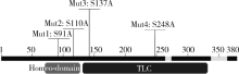
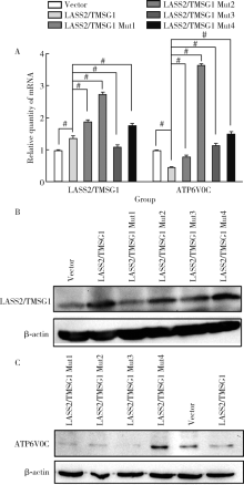
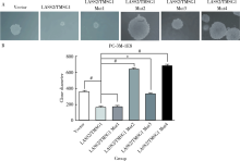
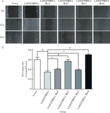
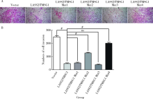
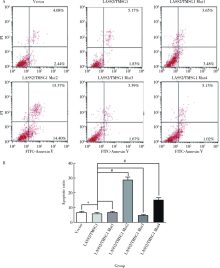
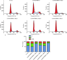
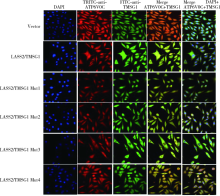
Novel tumor metastasis suppressorgene LASS2/TMSG1 S248A mutant promotes invasion of prostate cancer cells through increasing ATP6V0C expression

Kuan-gen ZHANG1,Yu-he ZHOU1,Ya-kun SHAO1,Fang MEI1,Jiang-feng YOU1,Bei-ying LIU2,Fei PEI1,3,()   

  1. 1. Department of Pathology, Peking University School of Basic Medical Sciences, Beijing 100191China
    2. School of Mechanical Engineering, University of Science & Technology Beijing, Beijing 100083 China;
    3. Department of Pathology, Peking University Third Hospital, Beijing 100191,China
  • Received:2018-08-20 Online:2019-04-18 Published:2019-04-26
  • Contact: Fei PEI E-mail:peifei@bjmu.edu.cn
  • Supported by:
    the National Sciences Foundation of China(81572533);the National Sciences Foundation of Beijing(7182078)

Abstract:

Objective: LASS2/TMSG1 gene is a novel tumor metastasis suppressor gene cloned from human prostate cancer cell line PC-3M in 1999 by Department of Pathology,Peking University of Basic Medical Sciences. It was found out that protein encoded by LASS2/TMSG1 could interact with the c subunit of vacuolar-ATPase (ATP6V0C). In this study, we explored the effect of LASS2/TMSG1 and its mutants on proliferation, migration and invasion of human prostate cancer cells and its molecular mechanism.Methods: We constructed four LASS2/TMSG1 mutants and stably transfected the variants to human prostate cancer cell line PC-3M-1E8 cell with high metastatic potential. The stable transfectants were identified by qPCR and Western blot through analyzing the expression of LASS2/TMSG1 and ATP6V0C, the cell biology functions of LASS2/TMSG1 and its four mutants were studied using growth curve,MTT assay, soft agar colony formation assay, wound migration assay, Matrigel invasion study and flow cytometry. Furthermore, immunofluorescence was used to analysis the interaction of LASS2/ TMSG1 mutants and ATP6V0C.Results: LASS2/TMSG1 mRNA and protein in LASS2/TMSG1 group and Mut1-Mut4 groups were higher than that in Vector group; Western blot showed that ATP6V0C protein in LASS2/TMSG1 wild group was lower than that in Vector group, but ATP6V0C protein in LASS2/TMSG1 S248A group was obviously higher than that in Vector group. MTT test and growth curve assay showed growth ability in LASS2/TMSG1 S248A group was increasing compared with other groups from day 5. Soft Agar colony formation experiment showed anchor independent growth ability in LASS2/TMSG1 S248A group was higher than those in the other groups (P<0.05), Cell migrations (from 35.3%±3.2% to 70.3%±3%) in LASS2/TMSG1 S248A group was increasing compared with LASS2/TMSG1 wild group (P<0.01), and more cells passed through Matrigel in LASS2/TMSG1 S248A group compared with LASS2/TMSG1 wild group (from 50±3.2 to 203±6.5, P<0.01), the apoptosis rate in LASS2/TMSG1 S248A group was obviously higher than that in LASS2/TMSG1 wild group (from 7% to 15.1%, P<0.05), and the G0/G1 ratio in LASS2/TMSG1 S248A group was obviously higher than that in LASS2/TMSG1 wild group (from 51.0% to 85.4%). Furthermore, double immunofluorescent staining observed the colocalization between ATP6V0C and LASS2/TMSG1 protein and its mutations, the expression of ATP6V0C in LASS2/TMSG1 S248A group increased significantly compared with the other groups.Conclusion: LASS2/TMSG1 S248A promotes proliferation, migration and invasion of prostate cancer cells through increasing ATP6V0C expression, suggesting that aa248-250 is an important function site for LASS2/TMSG1 in invasion suppression of prostate cancer cells.

Key words: Prostate cancer, Mutants, Vacuolar ATPase, ATP6V0C, LASS2/TMSG1

CLC Number: 

  • R737.25

Table 1

Primers of LASS2/TMSG1 and its mutants"

Gene Primer
LASS2/TMSG1 Forward: 5'-GGG ACA AGT TTG TAC AAA AAA GCA GGC TTA ATG CTC CAG ACC TTG TAT GAT TA-3'
Reverse: 5'-GGG GAC CAC TTT GTA CAA GAA AGC TGG GTA GTC ATT CTT ACG ATG GTT GT-3'
M1: S91A Forward: 5'-AAC ATT TCT ACC TGA CCG CTG GCA AGC AGC CCA AGC-3'
Reverse: 5'-GCT TGG GCT GCT TGC CAG CGG TCA GGT AGA AAT GTT-3'
M2: S110A Forward: 5'-CTG GCG GCC AGC GAG CCC GCT CT-3'
Reverse: 5'-AGA GCG GGC TCG CTG GCC GCC AG-3'
M3: S137A Forward: 5'-GGT AAA ATG TGA ATC TCC AGG CGG CTT CTC GGA ACT TCT TG-3'
Reverse: 5'-CAA GAA GTT CCG AGA AGC CGC CTG GAG ATT CAC ATT TTA CC-3'
M4: S248A Forward: 5'-CCG ATT ACC TGC TGG AGG CAG CCA AGA TGT TTA AC-3'
Reverse: 5'-GTT AAA CAT CTT GGC TGC CTC CAG CAG GTA ATC GG-3'

Figure 1

Four direct point mutations from serine to alanine of LASS2/TMSG1 LASS2/TMSG1 Mut1,S91A;LASS2/TMSG1 Mut2,S110A;LASS2/TMSG1 Mut3, S137A; LASS2/TMSG1 Mut4, S248A."

Figure 2

The influence of LASS2/TMSG1 gene and its mutations on LASS2/TMSG1 and ATP6V0C expression in prostate cancer cell lines PC-3M-1E8 A,Using semi-quantitative real-time RT-PCR, the expression of LASS2/TMSG1 mRNA in the LASS2/TMSG1 group and Mut1-Mut4 groups is significantly higher than that in the Vector group; and LASS2/TMSG1 mRNA in Mut1, Mut2 and Mut4 groups is higher than that in the LASS2/TMSG1 group; Moreover, ATP6V0C mRNA in LASS2/TMSG1 group is lower than that in Vector group, and ATP6V0C mRNA in Mut1-Mut4 groups is higher than that in LASS2/TMSG1 group (#P<0.01); B, Using Western blot, LASS2/TMSG1 protein in LASS2/TMSG1 group and Mut1-Mut4 groups is higher than that in Vector group; C, Western blot showed that ATP6V0C protein in LASS2/TMSG1 group is lower than that in Vector group, but ATP6V0C protein in Mut4 group is obviously higher than that in Vector group."

Figure 3

The influence of LASS2/TMSG1 gene and its mutations on proliferation in prostate cancer cell lines PC-3M-1E8 MTT test(A) and growth curve essay(B) showed growth ability in Mut2 group and Mut4 group is increasing compared with other groups from day 5,Mut2 group>Mut4 group>Mut3 group>Vector group>Mut1 group>LASS2/TMSG1 group in turn."

Figure 4

The influence of LASS2/TMSG1 gene and its mutations on anchor independent growth ability in prostate cancer cell lines PC-3M-1E8 Soft Agar colony formation experiment showed anchor independent growth ability in Mut2 group and Mut4 group is higher than other groups(#P<0.01), Mut4 group>Mut2 group>Mut3 group>Vector group>Mut1 group>LASS2/TMSG1 group in turn."

Figure 5

The influence of LASS2/TMSG1 gene and its mutations on cell migration in prostate cancer cell lines PC-3M-1E8 Cell migration(scratch repair rate within 24 hours) in Mut2 group and Mut4 group is increasing compared with LASS2/TMSG1 group(#P<0.01), Mut4 group> Vector group> Mut2 group>Mut1 group>Mut3 group>LASS2/TMSG1 group in turn."

Figure 6

The influence of LASS2/TMSG1 gene and its mutations on cell invasion in prostate cancer cell lines PC-3M-1E8 More cells passed through Matrigel in Vector group and Mut4 group compared with LASS2/TMSG1 group(#P<0.01), Vector group>Mut4 group>Mut2 group>Mut1 group>LASS2/TMSG1 group>Mut3 group in turn. ns, not significant."

Figure 7

The influence of LASS2/TMSG1 gene and its mutations on cell apoptosis in prostate cancer cell lines PC-3M-1E8 Apoptosis ratio in Mut2 group and Mut4 group is obviously higher than that in LASS2/TMSG1 group and Vector group(#P<0.01), Mut2 group(apoptosis rate:29.77%)>Mut4 group(15.15%)>Mut1 group(7.13%)>LASS2/TMSG1 group(7%)>Vector group(6.52%)>Mut3 group(5.06%)in turn."

Figure 8

The influence of LASS2/TMSG1 gene and its mutations on cell cycle in prostate cancer cell lines PC-3M-1E8 G0/G1 ratio in Mut1 group and Mut4 group is obviously higher than that in LASS2/TMSG1 group, Mut4 group (G0/G1 ratio: 85.46%)>Mut1 group(79.88%)>Mut2 group (65.50%)>Vector group (64.98%)>Mut3 group (60.96%)>LASS2/TMSG1 group (51.06%)in turn."

Figure 9

Double immunofluorescent staining observed the colocalization between ATP6V0C and LASS2/TMSG1 prote in and its mutations Double immunofluorescent staining observed the colocalization between ATP6V0C and LASS2/TMSG1 protein and its mutations, showing that LASS2/TMSG1 protein(green) and ATP6V0C protein(red) are mainly co-localized in plasma (yellow). The expression of ATP6V0Cin Mut4 group increased significantly compared to other groups, and the co-localization signal was increased evidently. DAPI, 4', 6-diamidino-2-phenylindole."

[1] Cronin KA, Lake AJ, Scott S , et al. Annual Report to the nation on the status of cancer, part Ⅰ: national cancer statistics[J]. Cancer, 2018,124(13):2785-2800.
doi: 10.1002/cncr.31551
[2] 刘宇欣, 郑杰, 方伟岗 , 等. 具有不同转移潜能的前列腺癌细胞亚系的分离鉴定[J]. 中华病理学杂志, 1999,28(5):361-364.
[3] 马春树, 刘宇欣, 郑杰 , 等. 应用mRNA差异显示技术克隆肿瘤转移相关基因LASS2/TMSG1[J]. 中国科学, 2002,32(3):270-275.
[4] Strausberg RL, Feingold EA, Grouse LH , et al. Generation and initial analysis of more than 15,000 full-length human and mouse cDNA sequences[J]. Proc Natl Acad Sci USA, 2002,99(26):16899-16903.
doi: 10.1073/pnas.242603899
[5] Pan H, Qin WX, Huo KK , et al. Cloning, mapping, and charaterization of a human homologue of the yeast longevity assurance gene LAG1[J]. Genomics, 2001,77(1/2):58-64.
doi: 10.1006/geno.2001.6614
[6] Pewzner-Jung Y, Ben Dor S, Futerman AH . When do Lasses (longevity assurance genes) become CerS (ceramide synthases)? Insights into the regulation of ceramide synjournal[J]. J Biol Chem, 2006,281(35):25001-25005.
doi: 10.1074/jbc.R600010200
[7] 裴裴, 由江峰, 宁钧宇 , 等. 人肿瘤转移抑制基因TMSG-1单克隆抗体的制备、鉴定及在肿瘤检测中的应用[J]. 中华病理学杂志, 2005,34(1):15-21.
[8] Yu W, Wang L, Pei F , et al. A novel tumor metastasis suppressor gene LASS2/TMSG1 interacts with vacuolar ATPase through its homeodomain[J]. J Cell Biochem, 2013,114(3):570-583.
doi: 10.1002/jcb.v114.3
[9] Ohta T, Numata M, Yagishita H , et al. Expression of 16 kDa proteolipid of vacuolar-type H(+)-ATPase in human pancreatic cancer[J]. Br J Cancer, 1996,73(12):1511-1517.
doi: 10.1038/bjc.1996.285
[10] Sennoune SR, Bakunts K, Martinez GM , et al. Vacuolar H+-ATPase in human breast cancer cells with distinct metastatic potential: distribution and functional activity[J]. Am J Physiol Cell Physiol, 2004,286(6):1443-1452.
doi: 10.1152/ajpcell.00407.2003
[11] Lee I, Skinner MA, Guo HB , et al. Expression of the vacuolar H+-ATPase 16-kDa subunit results in the Triton X-100-insoluble aggregation of beta1 integrin and reduction of its cell surface expression[J]. J Biol Chem, 2004,279(51):53007-53014.
doi: 10.1074/jbc.M405717200
[12] Vitavska O, Wieczorek H, Merzendorfer H . A novel role for subunit C in mediating binding of the H+-V-ATPase to the actin cytoskeleton[J]. J Biol Chem, 2003,278(20):18499-18505.
doi: 10.1074/jbc.M212844200
[13] Holliday LS, Lu M, Lee BS , et al. The amino-terminal domain of the B subunit of vacuolar H+-ATPase contains a filamentous actin binding site[J]. J Biol Chem, 2000,275(41):32331-32337.
doi: 10.1074/jbc.M004795200
[14] Goldstein DJ, Andresson T, Sparkowski JJ , et al. The BPV-1 E5 protein, the 16 kDa membrane pore-forming protein and the PDGF receptor exist in a complex that is dependent on hydrophobic transmembrane interactions[J]. EMBO J, 1992,11(13):4851-4859.
doi: 10.1002/embj.1992.11.issue-13
[15] Skinner MA , Wildeman AG. beta(1) integrin binds the 16-kDa subunit of vacuolar H(+)-ATPase at a site important for human papillomavirus E5 and platelet-derived growth factor signaling[J]. J Biol Chem, 1999,274(33):23119-23127.
doi: 10.1074/jbc.274.33.23119
[16] Liotta LA, Kohn EC . The microenvironment of the tumour-host interface[J]. Nature, 2001,411(6835):375-379.
doi: 10.1038/35077241
[17] Zou P, Yang Y, Pei F , et al. Silencing of vacuolar ATPase c subunit ATP6V0C inhibits invasion of prostate cancer cell through LASS2/TMSG1 independent manner[J]. Oncol Rep, 2018,39(1):298-306.
[18] Kim SS, Chae HS, Bach JH , et al. P53 mediates ceramide-induced apoptosis in SKN-SH cells[J]. Oncogene, 2002,21(13):2020-2028.
doi: 10.1038/sj.onc.1205037
[19] Kim HJ, Ghil KC, Kim MS , et al. Potentiation of ceramide-induced apoptosis by p27kip1 overexpression[J]. Arch Pharm Res, 2005,28(1):87-92.
doi: 10.1007/BF02975141
[20] Yang H, Sadda MR, Li M , et al. S-adenosylmethionine and its metabolite induce apoptosis in HepG2 cells: Role of protein phosphatase 1 and Bcl-x(S)[J]. Hepatology, 2004,40(1):221-231.
doi: 10.1002/hep.v40:1
[21] Lee JY, Bielawska AE, Obeid LM . Regulation of cyclin-depen-dent kinase 2 activity by ceramide[J]. Exp Cell Res, 2000,261(2):303-311.
doi: 10.1006/excr.2000.5028
[22] Zhu XF, Liu ZC, Xie BF , et al. Ceramide induces cell cycle arrest and upregulates p27kip in nasopharyngeal carcinoma cells[J]. Cancer Lett, 2003,193(2):149-154.
doi: 10.1016/S0304-3835(03)00050-8
[1] BAI Gao-chen,SONG Yi,JIN Jie,YU Wei,HE Zhi-song. Clinical efficacy of docetaxel combined with carboplatin in patients with metastatic castration-resistant prostate cancer [J]. Journal of Peking University (Health Sciences), 2021, 53(4): 686-691.
[2] Kui-xia SUN,Cun-ling YAN,Zhi-yan LI,Ping LIU,Wei ZHANG,Qun HE. Clinical value of serum isoform [-2] proprostate-specific antigen and its derivatives in predicting aggressive prostate cancer [J]. Journal of Peking University (Health Sciences), 2020, 52(2): 234-239.
[3] Wen-qing LI,Si-mei REN,Xing-bo LONG,Yu-qing TIAN. Palmitoylome profiling indicates that androgens promote the palmitoylation of metabolism-related proteins in prostate cancer-derived LNCaP cells [J]. Journal of Peking University (Health Sciences), 2020, 52(2): 227-233.
[4] TANG Xu, ZHAO Wei-hong, SONG Qin-qin, YIN Hua-qi, DU Yi-qing, SHENG Zheng-zuo, WANG Qiang, ZHANG Xiao-wei, LI Qing, LIU Shi-jun, XU Tao. Influence of SOX10 on the proliferation and invasion of prostate cancer cells [J]. Journal of Peking University(Health Sciences), 2018, 50(4): 602-606.
[5] ZOU Peng-cheng, YANG Yi-feng, XU Xiao-yan LIU Bei-ying, MEI Fang, YOU Jiang-feng, LIU Qi-chen, PEI Fei . Silencing of vacuolar ATPase c subunit ATP6V0C inhibits invasion of prostate cancer cells [J]. Journal of Peking University(Health Sciences), 2017, 49(6): 937-947.
[6] JI Guang-jie, HUANG Cong, SONG Gang, LI Xue-song, SONG Yi, ZHOU Li-qun. Predictive factor analysis of time to progression of castration-resistant prostate cancer after androgen deprivation therapy [J]. Journal of Peking University(Health Sciences), 2017, 49(4): 657-662.
Viewed
Full text


Abstract

Cited

  Shared   
  Discussed   
[1] . [J]. Journal of Peking University(Health Sciences), 2009, 41(2): 188 -191 .
[2] . [J]. Journal of Peking University(Health Sciences), 2009, 41(3): 376 -379 .
[3] . [J]. Journal of Peking University(Health Sciences), 2009, 41(4): 459 -462 .
[4] . [J]. Journal of Peking University(Health Sciences), 2010, 42(1): 82 -84 .
[5] . [J]. Journal of Peking University(Health Sciences), 2007, 39(3): 319 -322 .
[6] . [J]. Journal of Peking University(Health Sciences), 2007, 39(3): 333 -336 .
[7] . [J]. Journal of Peking University(Health Sciences), 2007, 39(3): 337 -340 .
[8] . [J]. Journal of Peking University(Health Sciences), 2007, 39(4): 346 -350 .
[9] . [J]. Journal of Peking University(Health Sciences), 2007, 39(4): 351 -354 .
[10] . [J]. Journal of Peking University(Health Sciences), 2007, 39(4): 361 -364 .