Journal of Peking University(Health Sciences) ›› 2019, Vol. 51 ›› Issue (5): 900-906. doi: 10.19723/j.issn.1671-167X.2019.05.018
Previous Articles Next Articles
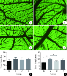
Comparative study of differentiation potential of mesenchymal stem cells derived from orofacial system into vascular endothelial cells
Jing XIE1,Yu-ming ZHAO1,Nan-quan RAO1,Xiao-tong WANG2,Teng-jiao-zi FANG1,Xiao-xia LI1,Yue ZHAI1,Jing-zhi LI1,Li-hong GE1,Yuan-yuan WANG1,△( )
)
- 1. Department of Pediatric Dentistry, Peking University School and Hospital of Stomatology & National Clinical Research Center for Oral Diseases & National Engineering Laboratory for Digital and Material Technology of Stomatology & Beijing Key Laboratory of Digital Stomatology, Beijing 100081, China
2. Department of Oral Emergency, Peking University School and Hospital of Stomatology & National Clinical Research Center for Oral Diseases & National Engineering Laboratory for Digital and Material Technology of Stomatology & Beijing Key Laboratory of Digital Stomatology, Beijing 100081, China
CLC Number:
- R78


| [1] | Sakaguchi Y, Sekiya I, Yagishita K , et al. Comparison of human stem cells derived from various mesenchymal tissues: superiority of synovium as a cell source[J]. Arthritis Rheumatol, 2005,52(8):2521. |
| [2] | Folkman J . Angiogenesis in cancer, vascular, rheumatoid and other disease[J]. Nat Med, 1995,1(1):27. |
| [3] | Carmeliet P . Angiogenesis in life, disease and medicine[J]. Nature, 2005,438(7070):932. |
| [4] | Canan A, Huseyin A, Riza KA , et al. Angiogenesis in inflammatory bowel disease[J]. In J Inflammation, 2015,2015(3):970890. |
| [5] | Tateishi-Yuyama E, Matsubara H, Murohara T . Therapeutic angiogenesis for patients with limb ischemia by autologous transplantation of bone-marrow cells: a pilot study and a randomized controlled trial[J]. Acc Current J Rev, 2002,360(9331):427. |
| [6] | Hou L, Kim JJ, Woo YJ , et al. Stem cell-based therapies to promote angiogenesis in ischemic cardiovascular disease[J]. Am J Physiol, 2016,310(4):H455. |
| [7] | Shi S, Gronthos S . Perivascular niche of postnatal mesenchymal stem cells in human bone marrow and dental pulp[J]. J Bone Miner Res, 2003,18(4):696-704. |
| [8] | Belotti A, Elli E, Speranza T , et al. Circulating endothelial cells and endothelial activation in essential thrombocythemia: results from CD146 + immunomagnetic enrichment: flow cytometry and soluble E-selectin detection [J]. Am J Hematol, 2012,87(3):319. |
| [9] | Jouve N, Despoix N, Espeli M , et al. The involvement of CD146 and its novel ligand galectin-1 in apoptotic regulation of endothelial cells[J]. J Biol Chem, 2013,288(4):2571-2579. |
| [10] | Nakamura S, Yamada Y, Katagiri W , et al. Stem cell proliferation pathways comparison between human exfoliated deciduous teeth and dental pulp stem cells by gene expression profile from promising dental pulp[J]. J Endodont, 2009,35(11):1536-1542. |
| [11] | Duttenhoefer F, Lara d FR, Meury T, et al. 3D scaffolds co-seeded with human endothelial progenitor and mesenchymal stem cells: evidence of prevascularisation within 7 days[J]. Eur Cells Mater, 2013,26(4):49. |
| [12] | Moore MC, Pandolfi V, Mcfetridge PS . Novel human-derived extracellular matrix induces in vitro, and in vivo, vascularization and inhibits fibrosis[J]. Biomaterials, 2015,49:37. |
| [13] | Nourse MB, Halpin DE, Scatena M , et al. VEGF induces differentiation of functional endothelium from human embryonic stem cells: implications for tissue engineering[J]. Arterioscler Thromb Vas Biol, 2010,30(1):80-89. |
| [14] | Oswald J, Boxberger S, Jørgensen B , et al. Mesenchymal stem cells can be differentiated into endothelial cells in vitro[J]. Stem Cells, 2004,22(3):377. |
| [15] | Sugiyama M, Iohara K, Wakita H , et al. Dental pulp-derived CD31 -/CD146 - side population stem/progenitor cells enhance recovery of focal cerebral ischemia in rats [J]. Tissue Eng Part A, 2011,17(9/10):1303-1311. |
| [16] | Park S, Sorenson CM, Sheibani N . PECAM-1 isoforms, eNOS and endoglin axis in regulation of angiogenesis[J]. Clin Sci, 2015,129(3):217. |
| [17] | Zhang Z, Neiva KG, Lingen MW , et al. VEGF-dependent tumor angiogenesis requires inverse and reciprocal regulation of VEGFR1 and VEGFR2[J]. Cell Death Differ, 2010,17(3):499. |
| [18] | Ruszkowskaciastek B, Sokup A, Socha M W , et al. A preliminary evaluation of VEGF-A, VEGFR1 and VEGFR2 in patients with well-controlled type 2 diabetes mellitus[J]. J Zhejiang Univ Sci B, 2014,15(6):575-581. |
| [19] | Rondaij MG, Bierings R, Kragt A , et al. Dynamics and plasticity of Weibel-Palade bodies in endothelial cells[J]. Arterioscler Thromb Vasc Biol, 2006,26(5):1002. |
| [20] | Kleibeuker EA, Schulkens IA, Castricum KC , et al. Examination of the role of galectins during in vivo angiogenesis using the chick chorioallantoic membrane assay[J]. Methods Mol Biol, 2015,1207:305. |
| [21] | Dehelean CA, Feflea S, Gheorgheosu D , et al. Anti-angiogenic and anti-cancer evaluation of betulin nanoemulsion in chicken chorioallantoic membrane and skin carcinoma in BALB/c mice[J]. J Biomed Nanotechnol, 2013,9(4):577-589. |
| [1] | Yao ZHANG,Jinxin GUO,Shijia ZHAN,Enyu HONG,Hui YANG,Anna JIA,Yan CHANG,Yongli GUO,Xuan ZHANG. Role and mechanism of cysteine and glycine-rich protein 2 in the malignant progression of neuroblastoma [J]. Journal of Peking University (Health Sciences), 2024, 56(3): 495-504. |
| [2] | Yu-yang YE,Lin YUE,Xiao-ying ZOU,Xiao-yan WANG. Characteristics and microRNA expression profile of exosomes derived from odontogenic dental pulp stem cells [J]. Journal of Peking University (Health Sciences), 2023, 55(4): 689-696. |
| [3] | SHUAI Ting,LIU Juan,GUO Yan-yan,JIN Chan-yuan. Knockdown of long non-coding RNA MIR4697 host gene inhibits adipogenic differentiation in bone marrow mesenchymal stem cells [J]. Journal of Peking University (Health Sciences), 2022, 54(2): 320-326. |
| [4] | Xiao-min GAO,Xiao-ying ZOU,Lin YUE. Mediated pathways of exosomes uptake by stem cells of apical papilla [J]. Journal of Peking University(Health Sciences), 2020, 52(1): 43-50. |
| [5] | Xia LIU,Ying ni LI,Xiao li SUN,Qing lin PENG,Xin LU,Guo chun WANG. Effects of integrin metalloproteinases on osteogenic differentiation [J]. Journal of Peking University(Health Sciences), 2018, 50(6): 962-967. |
| [6] | TANG Xu, ZHAO Wei-hong, SONG Qin-qin, YIN Hua-qi, DU Yi-qing, SHENG Zheng-zuo, WANG Qiang, ZHANG Xiao-wei, LI Qing, LIU Shi-jun, XU Tao. Influence of SOX10 on the proliferation and invasion of prostate cancer cells [J]. Journal of Peking University(Health Sciences), 2018, 50(4): 602-606. |
| [7] | WANG Zi-cheng, CHENG Li, LV Tong-de, SU Li, LIN Jian, ZHOU Li-qun. Inflammatory priming adipose derived stem cells significantly inhibit the proliferation of peripheral blood mononuclear cells [J]. Journal of Peking University(Health Sciences), 2018, 50(4): 590-594. |
| [8] | CHEN Wei, HU Fan-lei, LIU Hong-jiang, XU Li-ling, LI Ying-ni, LI Zhan-guo. Myeloid-derived suppressor cells promoted autologous B cell proliferation in rheumatoid arthritis [J]. Journal of Peking University(Health Sciences), 2017, 49(5): 819-823. |
| [9] | JIA Wei-qian, ZHAO Yu-ming, GE Li-hong. Recombinant human transforming growth factor β1 promotes dental pulp stem cells proliferation and mineralization [J]. Journal of Peking University(Health Sciences), 2017, 49(4): 680-681. |
| [10] | CAI Yi, GUO Hao, LI Han-zhong, WANG Wen-da, ZHANG Yu-shi. MicroRNA differential expression profile in tuberous sclerosis complex cell line TSC2-/- MEFs and normal cell line TSC2+/+ MEFs [J]. Journal of Peking University(Health Sciences), 2017, 49(4): 580-584. |
| [11] | SIMA Zi-han, HONG Ying-ying, LI Tie-jun△. Effects of PTCH1 mutations on the epithelial proliferation derived from keratocystic odontogenic tumour [J]. Journal of Peking University(Health Sciences), 2017, 49(3): 522-526. |
| [12] | GAO Xiang, CHEN Xiang-mei, ZHANG Ting, ZHANG Jing, CHEN Mo, GUO Zheng--yang, SHI Yan-yan, LU Feng-min, DING Shi-gang. Relationship between macrophage capping protein and gastric cancer cell’s proliferation and migration ability [J]. Journal of Peking University(Health Sciences), 2017, 49(3): 489-494. |
| [13] | YANG Di, XU Jun-hui, DENG Fu-rong△, GUO Xin-biao . Effects of silver nanoparticle on hemichannel activation and anti-proliferation in HaCaT cells [J]. Journal of Peking University(Health Sciences), 2017, 49(3): 371-375. |
| [14] | SUI Hua-xin, LV Pei-jun, WANG Yu-guang, WANG Yong, SUN Yu-chun. Effect of lowlevel laser irradiation on proliferation and osteogenic differentiation of human adipose-derived stromal cells [J]. Journal of Peking University(Health Sciences), 2017, 49(2): 337-343. |
| [15] | LI Jing-wen, YIN Xiao-hui, LUAN Qing-xian. Comparative study of proliferative and periodontal differentiation propensity of induced pluripotent stem cells at different passages [J]. Journal of Peking University(Health Sciences), 2017, 49(1): 16-024. |
|
||