Journal of Peking University(Health Sciences) ›› 2019, Vol. 51 ›› Issue (5): 937-943. doi: 10.19723/j.issn.1671-167X.2019.05.024

Previous Articles     Next Articles

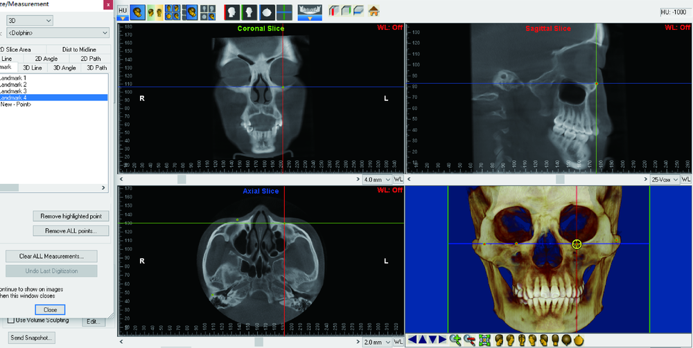
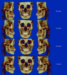
Using three-dimensional craniofacial images to construct horizontal reference plane

Min-jung KIM,Yi LIU()   

  1. Department of Orthodontics, Peking University School and Hospital of Stomatology & National Clinical Research Center for Oral Diseases & National Engineering Laboratory for Digital and Material Technology of Stomatology & Beijing Key Laboratory of Digital Stomatology, Beijing 100081, China
  • Received:2017-09-07 Online:2019-10-18 Published:2019-10-23
  • Contact: Yi LIU E-mail:lyortho@163.com

Abstract:

Objective: To compare four different three-dimensional horizontal planes and detect anatomical landmarks so as to provide theoretical reference for horizontal reference plane constructed by three-dimensional cephalometry. Methods: The subjects of this study were 32 facial symmetry patients (menton from mid-sagittal plane ≤2 mm). Cone-bead computed tomography (CBCT) was obtained before orthodontic treatment, and the data were imported into Dolphin imaging soft in DICOM format. The sagittal plane was passing through the Nasion, Sella and Dent. Four horizontal reference planes were constructed by three points of bilateral porion and bilateral orbitale. Plane 1: horizontal reference plane constructed by right porion and bilateral orbitale. Plane 2: horizontal reference plane constructed by left porion and bilateral orbitale. Plane 3: horizontal reference plane constructed by bilateral porion and right orbitale. Plane 4: horizontal reference plane constructed by bilateral porion and left orbitale. Pitch, yaw, roll for four planes were measured three dimensionally. All the samples were measured two times by one judge at an interval of two weeks. The two times measuring results were evaluated with Intraclass correlation coefficient (ICC) for verifying reliability. The multiple sets of repeated measurement analysis were used to compare the four different planes. Based on ages, the samples were divided into two groups (group 1: ages 13 to 17, group 2: over 18 years), the mean and standard deviation of landmark coordinates measured with Dent as the origin point, the circumference formula was applied to calculate the change of landmark position generated by head rotation. Results: No significant differences of pitch, yaw and roll among the four planes (P=0.196, 0.314, and 0.341). One degree of pitch rotation made changes of porion and orbitale approximate 0.5 mm, and 1.6 mm, respectively. One degree of yaw rotation made changes of porion and orbitale approximate 1.1 mm, and 1.5 mm, respectively. One degree of roll rotation made changes of porion and orbitale approximate 1.2 mm, and 0.7 mm, respectively. Conclusion: There was no significant difference among the four horizontal planes constructed by any three points of bilateral orbitales and bilateral porions. It has the highest concordance using bilateral orbitales and one porion to construct horizontal plane in this study, probably the best option in clinical practice. Different head rotation generated different distance changes of anatomical landmarks.

Key words: Head position, Frankfort plane, Cone beam computed tomography, Three-dimensional cephalometry

CLC Number: 

  • R783.5

Table 1

Define of hard and soft tissue landmarks for head orientation"

Landmark Definition
Menton The lowest point on the symphyseal shadow of the mandible
Sella The geometric center of the pituitary fossa
Dentes epistrophei The most posterior point of dentes epistrophei
Nasion The most anterior point on frontonasal suture in the midsagittal plane
Orbitale The lowest point on the inferior rim of the orbit
Porion The most superiorly positioned point of the external auditory meatus

Table 2

Define of plus and minus to confirmed direction of 3D rotation"

Items + -
Pitch Downward Upward
Yaw Right posterior Left after
Roll Lower right Left lower

Table 3

Define of 3D rotation degrees"

Rotarion Definition
Pitch Rotates around the X-axis, intersection between horizontal reference plane and rotated horizontal plane projected to MSP
Yaw Rotates around the Y-axis, intersection between MSP reference plane and rotated MSP projected to horizontal reference plane
Roll Rotates around the Z-axis, intersection between horizontal reference plane and rotated horizontal plane projected to coronal plane

Table 4

Rotation degree differences between three planes with Rp plane respectively/(°)"

Items Rp-Lp Rp-Ro Rp-Lo
Pitch -0.078±0.892 0.023±0.505 -0.121±0.850
Yaw 0.032±0.177 -0.002±0.010 0.002±0.008
Roll -0.013±0.091 -0.048±0.753 0.117±0.636

Figure 1

Tracing the bilateral orbitale and bilateral porion"

Figure 2

Define of horizontal planes:Rp plane constructed by bilateral orbitale and right porion; Lp plane constructed by bilateral orbitale and left porion; Ro plane constructed by bilateral porion and right orbitale; Lo plane constructed by bilateral porion and left orbitale"

Figure 3

Define of 3-dimensional rotation degree"

Table 5

Landmark coordinates in different age groups/mm"

Age groups Coordinate Right orbitale Left orbitale Right porion Left porion
X 31.017±2.651 31.067±2.180 59.344±3.323 61.578±5.017
13-17 years old Y 26.967±3.998 26.800±3.944 26.700±3.803 26.739±3.826
Z 80.622±3.944 80.467±4.149 4.811±3.790 4.033±3.524
X 32.364±2.623 32.507±3.546 59.064±6.088 60.964±4.532
≥18 years old Y 28.393±4.482 28.193±4.452 28.179±4.519 27.943±4.715
Z 83.300±2.986 83.179±3.417 6.207±2.806 7.421±2.252

Table 6

Based on different ages apply the circumference formula calculated change of bilateral porion and orbitale position generated by head rotation in different directions/mm"

Age groups Rotation Right orbitale Left orbitale Right porion Left porion
Pitch 1.484 1.480 0.474 0.472
13-17 years old Yaw 1.508 1.505 1.039 1.077
Roll 0.717 0.716 1.135 1.172
Pitch 1.536 1.533 0.504 0.505
≥18 years old Yaw 1.560 1.559 1.037 1.072
Roll 0.751 0.751 1.142 1.170
[1] Proffit WR . Contemporary orthodontics[M]. 5th ed. St.Louis: Elsevier, 2013: 134-137.
[2] Jacobson A . Radiographic cephalometry: from basics to 3-D imaging[M]. 2nd ed. Chicago: Quintessence Pub, 2006: 153-160.
[3] Moorrees CFA, Kean MR . Natural head position, a basic consi-deration in the interpretation of cephalometric radiographs[J]. Am J Phys Anthropol, 1958,16(2):213-234.
[4] Finlay LM . Craniometry and cephalometry: a history prior to the advent of radiography[J]. Angle Orthod, 1980,50(4):312-321.
[5] Downs WB . Analysis of the dentofacial profile[J]. Angle Orthod, 1956,26(4):191-212.
[6] Zebeib AM, Naini FB . Variability of the inclination of anatomic horizontal reference planes of the craniofacial complex in relation to the true horizontal line in orthognathic patients[J]. Am J Orthod Dentofacial Orthop, 2014,146(6):740-747.
[7] Barbera AL, Sampson WJ, Townsend GC . Variation in natural head position and establishing corrected head position[J]. Homo, 2014,65(3):187-200.
[8] Hsung T, Lo J, Li T , et al. Automatic detection and reproduction of natural head position in stereo-photogrammetry[J]. PLoS One, 2015,10(6):e130877.
[9] Kovacs L, Zimmermann A, Brockmann G , et al. Three-dimensional recording of the human face with a 3D laser scanner[J]. J Plast Reconstr Aesthet Surg, 2006,59(11):1193-1202.
[10] Xia JJ, McGrory JK, Gateno J, et al. A new method to orient 3-dimensional computed tomography models to the natural head position: a clinical feasibility study[J]. J Oral Maxillofac Surg, 2011,69(3):584-591.
[11] Tian K, Li Q, Wang X , et al. Reproducibility of natural head position in normal Chinese people[J]. Am J Orthod Dentofacial Orthop, 2015,148(3):503-510.
[12] Damstra J, Fourie Z, DeWit M, et al. A three-dimensional comparison of a morphometric and conventional cephalometric mid-sagittal planes for craniofacial asymmetry[J]. Clin Oral Investig, 2012,16(1):285-294.
[13] Lee JK, Jung PK, Moon CH . Three-dimensional cone beam computed tomographic image reorientation using soft tissues as reference for facial asymmetry diagnosis[J]. Angle Orthod, 2014,84(1):38-47.
[14] Oh S, Kim CY, Hong J . A comparative study between data obtained from conventional lateral cephalometry and reconstructed three-dimensional computed tomography images[J]. J Korean Assoc Oral Maxillofac Surg, 2014,40(3):123-129.
[15] Severt TR, Proffit WR . The prevalence of facial asymmetry in the dentofacial deformities population at the University of North Carolina[J]. Int J Adult Orthodon Orthognath Surg, 1997,12(3):171-176.
[16] Steiner C . Cephalometrics for you and me[J]. Am J Orthod, 1953,39(10):729-755.
[17] Steiner C . Cephalometrics in clinical practice[J]. Angle Orthod, 1959,29(1):8-29.
[18] Steiner C . The use of cephalometrics as an aid to planning and assessing orthodontic treatment[J]. Am J Orthod, 1960,46(10):721-735.
[19] Kim MS, Lee EJ, Song IJ , et al. The location of midfacial landmarks according to the method of establishing the midsagittal reference plane in three-dimensional computed tomography analysis of facial asymmetry[J]. Imaging Sci Dent, 2015,45(4):227.
[20] Kim HJ, Kim BC, Kim JG , et al. Construction and validation of the midsagittal reference plane based on the skull base symmetry for three-dimensional cephalometric craniofacial analysis[J]. J Craniofac Surg, 2014,25(2):338-342.
[21] Xiong Y, Zhao Y, Yang H , et al. Comparison between interactive closest point and procrustes analysis for determining the median sagittal plane of three-dimensional facial data[J]. J Craniofac Surg, 2016,27(2):441-444.
[22] 王斯维, 黎敏, 杨慧芳 , 等. 3种生成大视野锥形束CT数据正中矢状面方法的比较[J]. 北京大学学报(医学版), 2016,48(2):330-335.
doi: 10.3969/j.issn.1671-167X.2016.02.028
[23] Lim YK, Chu EH, Lee DY , et al. Three-dimensional evaluation of soft tissue change gradients after mandibular setback surgery in skeletal class Ⅲ malocclusion[J]. Angle Orthod, 2010,80(5):896-903.
[24] Kim MG, Lee JW, Cha KS , et al. Three-dimensional symmetry and parallelism of the skeletal and soft-tissue poria in patients with facial asymmetry[J]. Korean J Orthod, 2014,44(2):62-68.
[1] Chang CAO,Fei WANG,En-bo WANG,Yu LIU. Application of β-TCP for bone defect restore after the mandibular third molars extraction: A split-mouth clinical trial [J]. Journal of Peking University(Health Sciences), 2020, 52(1): 97-102.
[2] Xiao-yan XIE,Shu-mei JIA,Zhi-hui SUN,Zu-yan ZHANG. Diagnostic accuracy of cone beam computed tomography with different resolution settings for external root resorption [J]. Journal of Peking University(Health Sciences), 2019, 51(1): 75-79.
Viewed
Full text


Abstract

Cited

  Shared   
  Discussed   
[1] Author. English Title Test[J]. Journal of Peking University(Health Sciences), 2010, 42(1): 1 -10 .
[2] . [J]. Journal of Peking University(Health Sciences), 2009, 41(2): 188 -191 .
[3] . [J]. Journal of Peking University(Health Sciences), 2009, 41(3): 376 -379 .
[4] . [J]. Journal of Peking University(Health Sciences), 2009, 41(4): 459 -462 .
[5] . [J]. Journal of Peking University(Health Sciences), 2010, 42(1): 82 -84 .
[6] . [J]. Journal of Peking University(Health Sciences), 2007, 39(3): 319 -322 .
[7] . [J]. Journal of Peking University(Health Sciences), 2007, 39(3): 333 -336 .
[8] . [J]. Journal of Peking University(Health Sciences), 2007, 39(3): 337 -340 .
[9] . [J]. Journal of Peking University(Health Sciences), 2007, 39(3): 225 -328 .
[10] . [J]. Journal of Peking University(Health Sciences), 2007, 39(4): 346 -350 .