Journal of Peking University (Health Sciences) ›› 2020, Vol. 52 ›› Issue (3): 582-585. doi: 10.19723/j.issn.1671-167X.2020.03.028

Previous Articles     Next Articles

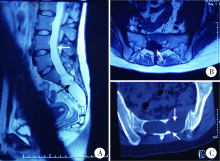
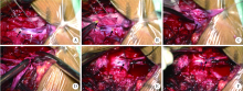
Clinical study of 21 cases of sacral cysts containing fila terminale

Guo-zhong LIN,Zhen-yu WANG(),Jing-cheng XIE,Bin LIU,Chang-cheng MA,Xiao-dong CHEN   

  1. Department of Neurosurgery, Peking University Third Hospital, Beijing 100191, China
  • Received:2018-04-04 Online:2020-06-18 Published:2020-06-30
  • Contact: Zhen-yu WANG E-mail:wzyu02@hotmail.com
  • Supported by:
    Capital Foundation for Clinical Characteristics and Application Research(Z171100001017120)

Abstract:

Objective: To summarize the characteristics of sacral cysts containing fila terminale and to explore the surgical treatment methods.Methods: The clinical features, imaging characteristics and surgical methods of 21 cases of sacral cysts containing fila terminale from July 2010 to March 2017 were reviewed and analyzed. Lumbosacral and perineal pain, weakness of the lower limbs and bladder and bowel dysfunction were the common clinical symptoms. MRI showed that the cysts located in the sacral canal. The lower T1 and higher T2 signals were found on MRI. There were fila terminale within the cysts which tethered the spinal cord. No enhancement was visible within the lesion. The key steps of operation included the resection of the cyst wall, the cutting off of the fila terminale, the release of the tethered cord and the reconstruction of the cisterna terminalis.Results: The total and subtotal resections of cyst walls were achieved in 14 and 7 cases, respectively. The fila terminales were separated and cut off in all the cases, and the tethered cords were released completely. The reconstructions of the cisterna terminalis were accomplished in all the cases. There was no new-onset dysfunction except for 7 cases of mild numbness around anus postoperatively. Pathological examinations confirmed that the cyst wall was fibrous connective tissue, and hyperplasia of fibrous tissue and/or adipose tissue was found within the thickened fila terminale. The lumbosacral and perineal pain disappeared. The weakness of the lower extremities and the bladder and bowel dysfunction gradually improved. The period of follow-up ranged from 3 months to 7 years (average: 2.25 years). The spinal function of all the patients restored to McCormick grade Ⅰ. Only 1 case encountered recurrence of cyst.Conclusion: The sacral cysts containing fila terminale are rare. The common symptoms include lumbosacral and perineal pain and symptoms of tethered cord. MRI is helpful to the diagnosis, which shows the signal of cerebrospinal fluid and the fila terminale in the cyst as well as tlow-placed conus medullaris. Microsurgery should remove the cyst wall, cut off the fila terminale, release the tethered cord and reconstruct the cisterna terminalis.

Key words: Sacral cyst, Fila terminale, Tethered cord syndrome, Microsurgery, Electromyography

CLC Number: 

  • R651.2

Figure 1

Preoperative images of a case of sacral cysts containing fila terminale A,sagittal T2 MRI revealed a cyst in the sacral canal (black arrow) with low position conus medullaris located at the lower edge of the lumbar 4 (white arrow); B, axial T1 MRI showed thickened fila terminale in the cyst which was infiltrated by fat (white arrow); C, axial CT showed that bone defect area appeared in the anterior and posterior walls of sacral canal (white arrow)."

Figure 2

Intraoperative images of a case of sacral cysts containing fila terminale A, dorsal wall of the cyst and thedural sac was opened and a thickened fila terminale infiltrated by fat was found in the cyst (black arrow), which entered the cyst through fistula (white arrow); B, the fila terminale was treated by electrocoagulation and cut off at the end of the subdural fila terminale, the broken end of the fila terminale was visible (black arrow); C, then the wall of cyst was bluntly dissected (black arrow); D, the cyst wall and caudal fila terminale were resected (black arrow); E, the end of the dural sac was dissected (black arrow); F, the dura was sutured to reconstruct the terminal cistern (black arrow)."

[1] 陈晓东, 王振宇, 谢京城, 等. 症状性骶管内囊肿的诊断与治疗[J]. 中国脊柱脊髓杂志, 2006,16(2):138-141.
[2] Sun J, Wang Z, Li Z, et al. Reconstruction of nerve root sheaths for sacralextradural spinal meningeal cysts with spinal nerve root fibers[J]. Sci China Life Sci, 2013,56(11):1007-1013.
[3] Nabors MW, Pait TG, Byrd EB, et al. Updated assessment and current classification of spinal meningeal cysts[J]. J Neurosurg, 1988,68(3):366-377.
pmid: 3343608
[4] 谢京城, 王振宇, 陈晓东. 骶管终丝脊膜囊肿合并脊髓拴系综合征的诊断和治疗[J]. 中国临床神经外科杂志, 2015,20(11):651-653.
[5] Hukuda S, Mochizuki T, Ogata M, et al. Operations for cervical spondylotic myelopathy. A comparison of the results of anterior and posterior procedures[J]. J Bone Joint Surg Br, 1985,67(4):609-615.
[6] Selcuki M, Mete M, Barutcuoglu M, et al. Tethered cord syndrome in adults: Experience of 56 patients[J]. Turk Neurosurg, 2015,25(6):922-929.
[7] 汪寒松. MRI在诊断骶管囊肿中的应用[J]. 中国当代医药, 2014,21(2):127-128.
[8] Lee GY, Paradiso G, Tator CH, et al. Surgical management of tethered cord syndrome in adults: indications, techniques, and long-term outcomes in 60 patients[J]. J Neurosurg Spine, 2006,4(2):123-131.
[9] Sun JJ, Wang ZY, Liu B, et al. Neck transfixion for sacralextradural spinal meningeal cysts without spinal nerve root fibers[J]. Eur Spine J, 2016,25(6):1945-1952.
[10] 林国中, 王振宇, 刘斌. 神经电生理监测技术在椎管内病变术中的应用[J]. 北京大学学报(医学版), 2012,44(5):776-779.
[1] Mei-ge LIU,Pu FANG,Yan WANG,Lu CONG,Yang-yi FAN,Yuan YUAN,Yan XU,Jun ZHANG,Dao-jun HONG. Clinical, pathological and genetic characteristics of 8 patients with distal hereditary motor neuropathy [J]. Journal of Peking University (Health Sciences), 2021, 53(5): 957-963.
[2] LIN Guo-zhong, MA Chang-cheng, WANG Zhen-yu, XIE Jing-cheng, LIU Bin, CHEN Xiao-dong. Minimally invasive treatment of cervical1-2 epidural neurilemmoma [J]. Journal of Peking University (Health Sciences), 2021, 53(3): 586-589.
[3] Jing WANG,Jun-peng CHEN,Yang WANG,Xiang-liang XU,Chuan-bin GUO. Application of digital mandibular movement record and masticatory muscle electromyography in the evaluation of stomatognathic function in patients with mandibular tumor [J]. Journal of Peking University(Health Sciences), 2019, 51(3): 571-578.
[4] CHEN Li-hua, YANG Yi, WEI Qun, LI Yun-jun, LI Wen-de, GAO Jin-bao,YU Bin, ZHAO Hao, XU Ru-xiang. Microsurgical management of petroclival meningiomas combined trans-subtemporal and suboccipital retrosigmoid keyhole approach [J]. Journal of Peking University(Health Sciences), 2016, 48(4): 738-742.
[5] DUAN Hong-Zhou, LI Liang, ZHANG Yang, ZHANG Jia-Yong, BAO Sheng-De. Surgical treatment of paraclinoid aneurysms [J]. Journal of Peking University(Health Sciences), 2015, 47(4): 679-684.
[6] PENG Jing, LONG Hai, YUAN Yi-Ming, CUI Wan-Shou, ZHANG Zhi-Chao, PAN Wen-Bo. Comparison of the outcomes of microscopic varicocelectomy and laparoscopic varicocelectomy [J]. Journal of Peking University(Health Sciences), 2014, 46(4): 541-543.
Viewed
Full text


Abstract

Cited

  Shared   
  Discussed   
[1] Author. English Title Test[J]. Journal of Peking University(Health Sciences), 2010, 42(1): 1 -10 .
[2] . [J]. Journal of Peking University(Health Sciences), 2009, 41(2): 188 -191 .
[3] . [J]. Journal of Peking University(Health Sciences), 2009, 41(3): 376 -379 .
[4] . [J]. Journal of Peking University(Health Sciences), 2009, 41(4): 459 -462 .
[5] . [J]. Journal of Peking University(Health Sciences), 2010, 42(1): 82 -84 .
[6] . [J]. Journal of Peking University(Health Sciences), 2007, 39(3): 319 -322 .
[7] . [J]. Journal of Peking University(Health Sciences), 2007, 39(3): 333 -336 .
[8] . [J]. Journal of Peking University(Health Sciences), 2007, 39(3): 337 -340 .
[9] . [J]. Journal of Peking University(Health Sciences), 2007, 39(3): 225 -328 .
[10] . [J]. Journal of Peking University(Health Sciences), 2007, 39(4): 346 -350 .