Journal of Peking University (Health Sciences) ›› 2023, Vol. 55 ›› Issue (5): 939-942. doi: 10.19723/j.issn.1671-167X.2023.05.025
Previous Articles Next Articles
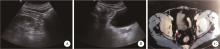
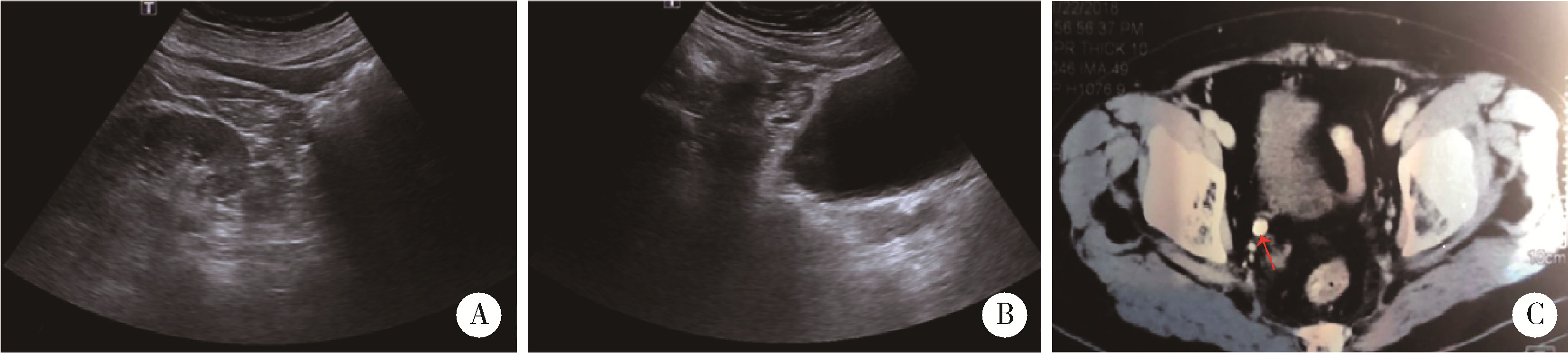
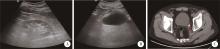
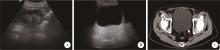
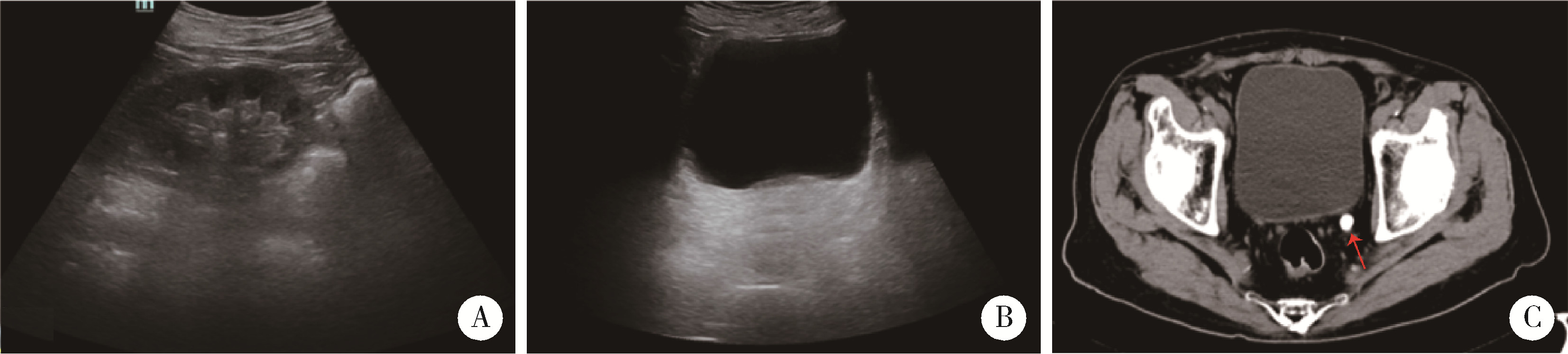
Diagnosis and treatment of four cases of asymptomatic and non-hydrous ureteral calculi
Cai-peng QIN,Fei WANG,Yi-qing DU,Xiao-wei ZHANG,Qing LI,Shi-jun LIU,Tao XU*( )
)
- Department of Urology, Peking Univesity People's Hospital, Beijing 100044, China
CLC Number:
- R693.4


| 1 | Wimpissinger F , Türk C , Kheyfets O , et al. The silence of the stones: Asymptomatic ureteral calculi[J]. J Urol, 2007, 178 (4 Pt 1): 1341- 1344. |
| 2 |
Marchini GS , Vicentini FC , Monga M , et al. Irreversible renal function impairment due to silent ureteral stones[J]. Urology, 2016, 93, 33- 39.
doi: 10.1016/j.urology.2016.02.042 |
| 3 | He L , Yang H , Fan L , et al. Comparison of symptomatic versus asymptomatic urolithiasis: Surgical outcomes and medium-term follow-up[J]. Int J Clin Exp Med, 2016, 9 (2): 4300- 4307. |
| 4 |
Wimpissinger F , Springer C , Kurtaran A , et al. Functional aspects of silent ureteral stones investigated with MAG-3 renal scintigraphy[J]. BMC Urol, 2014, 14 (1): 1- 5.
doi: 10.1186/1471-2490-14-1 |
| 5 |
Marchini GS , Vicentini FC , Mazzucchi E , et al. Silent ureteral stones: Impact on kidney function: Can treatment of silent ureteral stones preserve kidney function?[J]. Urology, 2012, 79 (2): 304- 308.
doi: 10.1016/j.urology.2011.07.1436 |
| 6 |
Weizer AZ , Auge BK , Silverstein AD , et al. Routine postoperative imaging is important after ureteroscopic stone manipulation[J]. J Urol, 2002, 168 (1): 46- 50.
doi: 10.1016/S0022-5347(05)64829-X |
| 7 |
Glowacki LS , Beecroft ML , Cook RJ , et al. The natural history of asymptomatic urolithiasis[J]. J Urol, 1992, 147 (2): 319- 321.
doi: 10.1016/S0022-5347(17)37225-7 |
| 8 |
Khaitan A , Gupta NP , Hemal AK , et al. Post-ESWL, clinically insignificant residual stones: Reality or myth?[J]. Urology, 2002, 59 (1): 20- 24.
doi: 10.1016/S0090-4295(01)01494-7 |
| 9 |
Shokeir AA . Renal colic: Pathophysiology, diagnosis and treatment[J]. Eur Urol, 2001, 39 (3): 241- 249.
doi: 10.1159/000052446 |
| 10 | Sfakianakis GN , Cohen DJ , Braunstein RH , et al. MAG3-F0 scintigraphy in decision making for emergency intervention in renal colic after helical CT positive for a urolith[J]. J Nucl Med, 2000, 41 (11): 1813- 1822. |
| 11 |
Lanzone JA , Gulmi FA , Chou SY , et al. Renal hemodynamics in acute unilateral ureteral obstruction: Contribution of endothelium-derived relaxing factor[J]. J Urol, 1995, 153 (6): 2055- 2059.
doi: 10.1016/S0022-5347(01)67401-9 |
| 12 |
Zabihi N , Teichman JM . Dealing with the pain of renal colic[J]. Lancet, 2001, 358 (9280): 437- 438.
doi: 10.1016/S0140-6736(01)05668-9 |
| 13 | Miller OF , Kane CJ . Time to stone passage for observed ureteral calculi: A guide for patient education[J]. J Urol, 1999, 162 (3 Pt 1): 688- 690. |
| 14 |
German I , Lantsberg S , Crystal P , et al. Non contrast compu-terized tomography and dynamic renal scintigraphy in the evaluation of patients with renal colic: Are both necessary?[J]. Eur Urol, 2002, 42 (2): 188- 191.
doi: 10.1016/S0302-2838(02)00271-3 |
| 15 |
Eisner BH , Pedro R , Namasivayam S , et al. Differences in stone size and ureteral dilation between obstructing proximal and distal ureteral calculi[J]. Urology, 2008, 72 (3): 517- 520.
doi: 10.1016/j.urology.2008.03.034 |
| 16 |
Kelleher JP , Plail RO , Dave SM , et al. Sequential renography in acute urinary tract obstruction due to stone disease[J]. Br J Urol, 1991, 67 (2): 125- 128.
doi: 10.1111/j.1464-410X.1991.tb15092.x |
| 17 |
Gandolpho L , Sevillano M , Barbieri A , et al. Scintigraphy and Doppler ultrasonography for the evaluation of obstructive urinary calculi[J]. Braz J Med Biol Res, 2001, 34 (6): 745- 751.
doi: 10.1590/S0100-879X2001000600007 |
| [1] | Qiang FU,Guan-ying GAO,Yan XU,Zhuo-hua LIN,You-jing SUN,Li-gang CUI. Comparative study of ultrasound and magnetic resonance imaging in the diagnosis of asymptomatic anterosuperior acetabular labrum tears [J]. Journal of Peking University (Health Sciences), 2023, 55(4): 665-669. |
| [2] | LI Jin-yong, SUN Hong-liang, YE Zhi-dong, FAN Xue-qiang, LIU Peng. Carotid plaque composition and volume evaluated by multi-detector computed tomography angiography [J]. Journal of Peking University(Health Sciences), 2018, 50(5): 833-839. |
|
||