Journal of Peking University (Health Sciences) ›› 2024, Vol. 56 ›› Issue (6): 963-971. doi: 10.19723/j.issn.1671-167X.2024.06.004

Previous Articles     Next Articles

Mechanism of melatonin regulating the expression level of rhythm genes to alleviate interstitial pulmonary fibrosis

Bingle LI1, Lingyan ZHU2, Yongfu WANG2,*(), Li BAI3,4,*()   

  1. 1. Baotou Medical College, Inner Mongolia University of Science and Technology, Baotou 014010, Inner Mongolia, China
    2. Department of Rheumatology and Immunology, the First Affiliated Hospital of Baotou Medical College, Baotou 014010, Inner Mongolia, China
    3. The Central Lab, the First Affiliated Hospital of Baotou Medical College, Baotou 014010, Inner Mongolia, China
    4. Inner Mongolia Autoimmune Key Laboratory, Baotou 014010, Inner Mongolia, China
  • Received:2024-08-02 Online:2024-12-18 Published:2024-12-18
  • Contact: Yongfu WANG, Li BAI E-mail:wyf18686198868@163.com;182020022@btmc.edu.cn
  • Supported by:
    Natural Science Foundation of Inner Mongolia Autonomous Region(2024QN08029);Natural Science Foundation of Inner Mongolia Autonomous Region(2023MS08052)

RICH HTML


Abstract:

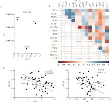
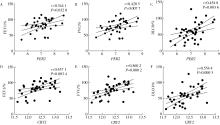
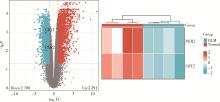
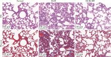
Objective: To investigate the intervention of melatonin (MT) in the expression of circadian genes in patients with pulmonary fibrosis and to analyze the mechanism by which it alleviates the progression of pulmonary fibrosis. Methods: By utilizing the Gene Expression Omnibus (GEO) database, we identified differentially expressed circadian genes between patients with pulmonary fibrosis and controls. We analyzed the correlation between circadian genes and pulmonary function as well as genes related to pulmonary fibrosis. A bleomycin-induced mouse model of pulmonary fibrosis (BLM group) was constructed to observe the expression differences of PER2 and CRY2 by sequencing and immunohistochemical staining in the BLM group and after MT intervention (BLM+MT group). Hematoxylin and eosin (HE) staining and Masson staining were used to observe the effects of MT on fibrosis. We used Western blot to detect the expression of P-smad2/3 in lung epithelial cells induced by transforming growth factor β (TGF-β). Reverse transcription quantitative real-time PCR technology was employed to investigate the rhythmic expression changes of circadian genes in the control group, TGF-β group, and TGF-β+MT group. Finally, luzindole, a MT receptor antagonist, was used to intervene in TGF-β+MT group, and Western blot was used to explore the receptor dependence of MT in alleviating TGF-β-induced epithelial-mesenchymal transition. Results: (1) Analysis of the GEO dataset (GSE) revealed a negative correlation between circadian genes PER2 and CRY2 and the expression of TGF-β, and a positive correlation with pulmonary function indicators in patients. (2) Transcriptome sequencing analysis of lung tissue in BLM group found that the expression of PER2 and CRY2 was significantly reduced compared with the normal group. Histopathological staining results showed that the lung tissue structure of the normal group was intact and clear, with thin alveolar septa; in the BLM group, there was a large increase in collagen fibers and disordered alveolar structure; compared with the BLM group, the BLM+MT group had reduced collagen fiber proliferation and inflammatory cell infiltration; the expression of PER2 and CRY2 in the BLM group was lower than in the normal group, and the expression in the BLM+MT group was increased compared with the BLM group. (3) In vitro lung epithelial cell experiments with TGF-β intervention showed that compared with the control group, the expression of P-smad2/3 increased in the TGF-β group, and MT intervention inhibited the inducing effect of TGF-β on P-smad2/3, while intervention with the MT receptor antagonist reversed this phenomenon. The results indicated that MT could inhibit the activation of the TGF-β pathway, and this process was dependent on MT receptors. (4) The 48-hour rhythm experiment in lung epithelial cells showed that the mRNA rhythm of PER2 and CRY2 in the TGF-β+MT group was close to 24 hours and showed a trend towards restoring the rhythm of the control group, while the addition of the MT receptor blocker tended to make the rhythm duration and amplitude of both groups approach that of the TGF-β group. Conclusion: MT, by binding to its receptors, can restore the periodic expression of the circadian genes PER2 and CRY2, thereby inhibiting the activation of the TGF-β classical pathway and suppressing the pathological process of epithelial-mesenchymal transition in pulmonary fibrosis. This finding provides new molecular targets and potential therapeutic strategies for the treatment of pulmonary fibrosis.

Key words: Melatonin, Rhythm genes, Pulmonary fibrosis

CLC Number: 

  • R563

Table 1

List of primers in RT-qPCR amplification"

Gene Primer sequence (5′-3′)
GAPDH Forward: GGAGCGAGATCCCTCCAAAAT
Reverse: GGCTGTTGTCATACTTCTCATGG
PER2 Forward: GACATGAGACCAACGAAAACTGC
Reverse: AGGCTAAAGGTATCTGGACTCTG
CRY2 Forward: GCTAGAGTGACGGAGATGCC
Reverse: TCAGGAGTCCTTGCTTGCTG

Figure 1

GSE47460 data set in the control group with UIP group of circadian gene expression differences and genetic correlation analysis and pulmonary fibrosis A, differential expression analysis of rhythm genes in the GSE47460 dataset between the control group and usually interstitial pneumonia (UIP) group; Padj, adjusted P value; FC, fold change. B, heat map of the association of PER2 and CRY2 with pulmonary fibrosis genes in UIP. C, correlation analysis of PER2 and CRY2 with TGF-β; The value of the axis is normalized-log2-based signal intensity. TGF-β, transforming growth factor β."

Figure 2

Correlation of PER2 (A-C) and CRY2 (D-F) with pulmonary function indexes FEV1, forced expiratory volume in one second; FVC, forced vital capacity; DLCO, diffusing capacity of the lungs for carbon monoxide. The value of the axis is normalized-log2-based signal intensity."

Figure 3

Heat map and volcano map of differential analysis of genes sequenced between normal and BLM group mice BLM, bleomycin; FC, fold change."

Figure 4

HE and Masson staining were performed in the normal group, BLM group and BLM+MT group of mouse lung tissue BLM, bleomycin; MT, melatonin."

Figure 5

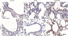
Immunohistochemical results of PER2 and CRY2 in each group of mice lung tissue BLM, bleomycin; MT, melatonin."

Figure 6

The expression differences of smad2/3 and P-smad2/3 in the control group, TGF-β group and TGF-β+MT group in lung epithelial cells TGF-β, transforming growth factor β; MT, melatonin."

Figure 7

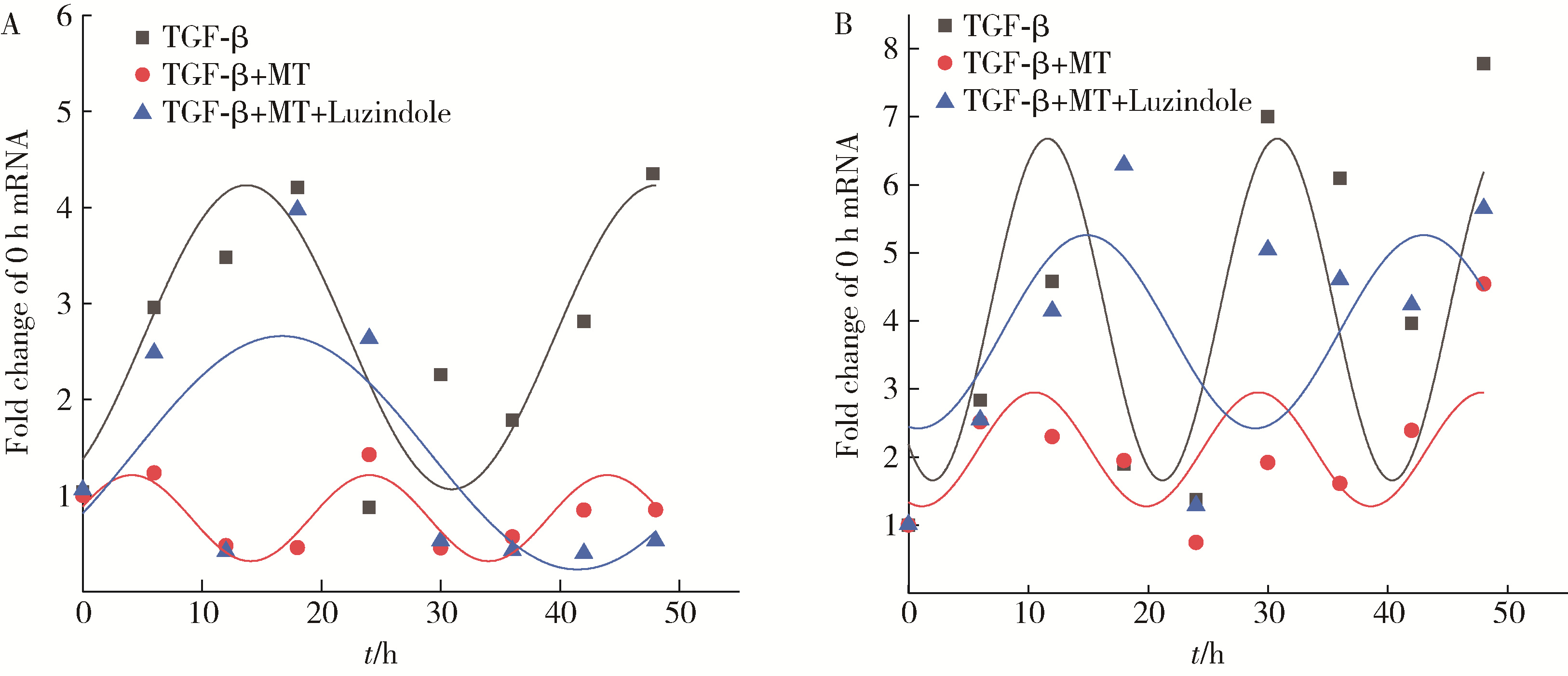
Epithelial markers decreased and mesenchymal markers increased in each group of lung epithelial cell lines (A), and the 48-hour mRNA rhythm changes of cell rhythm genes PER2 (B) and CRY2 (C) in each group TGF-β, transforming growth factor β; Col-1, collagen type 1; E-cad, E-cadherin; MT, melatonin."

Figure 8

48-hour mRNA rhythm fitting curves of PER2 and CRY2 in the TGF-β group, TGF-β+MT group, and melatonin receptor blocker group in lung epithelial cell lines TGF-β, transforming growth factor β; MT, melatonin."

Figure 9

Western blot was used to detect the protein expression of Col-1 and E-cad in the control group, TGF-β group, TGF-β+MT group and melatonin receptor blocker group TGF-β, transforming growth factor β; Col-1, collagen type 1; E-cad, E-cadherin; MT, melatonin."

1 Koike N , Yoo SH , Huang HC , et al. Transcriptional architecture and chromatin landscape of the core circadian clock in mammals[J]. Science, 2012, 338 (6105): 349- 354.
doi: 10.1126/science.1226339
2 Yamazaki S , Numano R , Abe M , et al. Resetting central and peripheral circadian oscillators in transgenic rats[J]. Science, 2000, 288 (5466): 682- 685.
doi: 10.1126/science.288.5466.682
3 Cox KH , Takahashi JS . Circadian clock genes and the transcriptional architecture of the clock mechanism[J]. J Mol Endocrinol, 2019, 63 (4): R93- R102.
doi: 10.1530/JME-19-0153
4 Fukuhara C , Tosini G . Peripheral circadian oscillators and their rhythmic regulation[J]. Front Biosci, 2003, 8, d642- d651.
doi: 10.2741/1042
5 Oishi A , Jockers R . Measuring protein-protein interactions of melatonin receptors by bioluminescence resonance energy transfer (BRET)[J]. Methods Mol Biol, 2022, 2550, 207- 218.
6 Cipolla-Neto J , Amaral FGD . Melatonin as a hormone: New phy-siological and clinical insights[J]. Endocr Rev, 2018, 39 (6): 990- 1028.
doi: 10.1210/er.2018-00084
7 Kandalepas PC , Mitchell JW , Gillette MU . Melatonin signal transduction pathways require E-box-mediated transcription of Per1 and Per2 to reset the SCN Clock at dusk[J]. PLoS One, 2016, 11 (6): e0157824.
doi: 10.1371/journal.pone.0157824
8 Zhao SQ , Gao Y , Zhang Y , et al. cAMP/PKA/CREB signaling pathway-mediated effects of melatonin receptor genes on clock gene expression in Bactrian camel ovarian granulosa cells[J]. Domest Anim Endocrinol, 2021, 76, 106609.
doi: 10.1016/j.domaniend.2021.106609
9 Sisto M , Ribatti D , Lisi S . Organ fibrosis and autoimmunity: The role of inflammation in TGFbeta-dependent EMT[J]. Biomolecules, 2021, 11 (2): 310.
doi: 10.3390/biom11020310
10 Frangogiannis N . Transforming growth factor-beta in tissue fibrosis[J]. J Exp Med, 2020, 217 (3): e20190103.
doi: 10.1084/jem.20190103
11 Andugulapati SB , Gourishetti K , Tirunavalli SK , et al. Biochanin- A ameliorates pulmonary fibrosis by suppressing the TGF-beta mediated EMT, myofibroblasts differentiation and collagen deposition in in vitro and in vivo systems[J]. Phytomedicine, 2020, 78, 153298.
doi: 10.1016/j.phymed.2020.153298
12 Cunningham PS , Meijer P , Nazgiewicz A , et al. The circadian clock protein REVERBα inhibits pulmonary fibrosis development[J]. Proc Natl Acad Sci USA, 2020, 117 (2): 1139- 1147.
doi: 10.1073/pnas.1912109117
13 Chen SJ , Yu F , Feng X , et al. DEC1 is involved in circadian rhythm disruption-exacerbated pulmonary fibrosis[J]. Cell Commun Signal, 2024, 22 (1): 245.
doi: 10.1186/s12964-024-01614-w
14 Dong C , Gongora R , Sosulski ML , et al. Regulation of transforming growth factor-beta1 (TGF-beta1)-induced pro-fibrotic activities by circadian clock gene BMAL1[J]. Respir Res, 2016, 17, 4.
doi: 10.1186/s12931-016-0320-0
15 Thannickal VJ , Toews GB , White ES , et al. Mechanisms of pulmonary fibrosis[J]. Annu Rev Med, 2004, 55, 395- 417.
doi: 10.1146/annurev.med.55.091902.103810
16 Inui N , Sakai S , Kitagawa M . Molecular pathogenesis of pulmonary fibrosis, with focus on pathways related to TGF-beta and the Ubiquitin-Proteasome pathway[J]. Int J Mol Sci, 2021, 22 (11): 6107.
doi: 10.3390/ijms22116107
17 Chanda D , Otoupalova E , Smith SR , et al. Developmental pathways in the pathogenesis of lung fibrosis[J]. Mol Aspects Med, 2019, 65, 56- 69.
doi: 10.1016/j.mam.2018.08.004
18 Rodriguez-Santana C , Lopez-Rodriguez A , Martinez-Ruiz L , et al. The relationship between Clock genes, sirtuin 1, and mitochondrial activity in head and neck squamous cell cancer: Effects of melatonin treatment[J]. Int J Mol Sci, 2023, 24 (19): 15030.
doi: 10.3390/ijms241915030
19 Shin NR , Park JW , Lee IC , et al. Melatonin suppresses fibrotic responses induced by cigarette smoke via downregulation of TGF-beta1[J]. Oncotarget, 2017, 8 (56): 95692- 95703.
doi: 10.18632/oncotarget.21680
20 Li Y , Zhou Y , Zhao C , et al. The circadian clock gene, BMAL1, promotes radiosensitization in nasopharyngeal carcinoma by inhibiting the epithelial-to-mesenchymal transition via the TGF-beta1/Smads/Snail1 axis[J]. Oral Oncol, 2024, 152, 106798.
doi: 10.1016/j.oraloncology.2024.106798
21 Guo SN , Jiang XQ , Chen N , et al. Melatonin regulates circadian clock proteins expression in allergic airway inflammation[J]. Heliyon, 2024, 10 (6): e27471.
doi: 10.1016/j.heliyon.2024.e27471
22 Wan L , Shi XY , Ge WR , et al. The Instigation of the associations between melatonin, Circadian genes, and epileptic spasms in infant rats[J]. Front Neurol, 2020, 11, 497225.
doi: 10.3389/fneur.2020.497225
23 Fernandez-Ortiz M , Sayed RKA , Roman-Montoya Y , et al. Age and chronodisruption in mouse heart: Effect of the NLRP3 Inflammasome and melatonin therapy[J]. Int J Mol Sci, 2022, 23 (12): 6846.
doi: 10.3390/ijms23126846
[1] Shan HE,Xin CHEN,Qi CHENG,Lingjiang ZHU,Peiyu ZHANG,Shuting TONG,Jing XUE,Yan DU. Tofacitinib inhibits the transformation of lung fibroblasts into myofibroblasts through JAK/STAT3 pathway [J]. Journal of Peking University (Health Sciences), 2024, 56(3): 505-511.
Viewed
Full text


Abstract

Cited

  Shared   
  Discussed   
No Suggested Reading articles found!