Journal of Peking University (Health Sciences) ›› 2026, Vol. 58 ›› Issue (1): 214-219. doi: 10.19723/j.issn.1671-167X.2026.01.029

Previous Articles     Next Articles

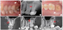
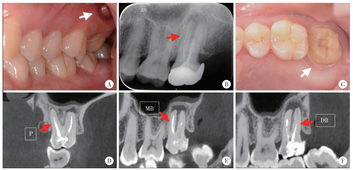
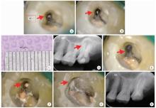
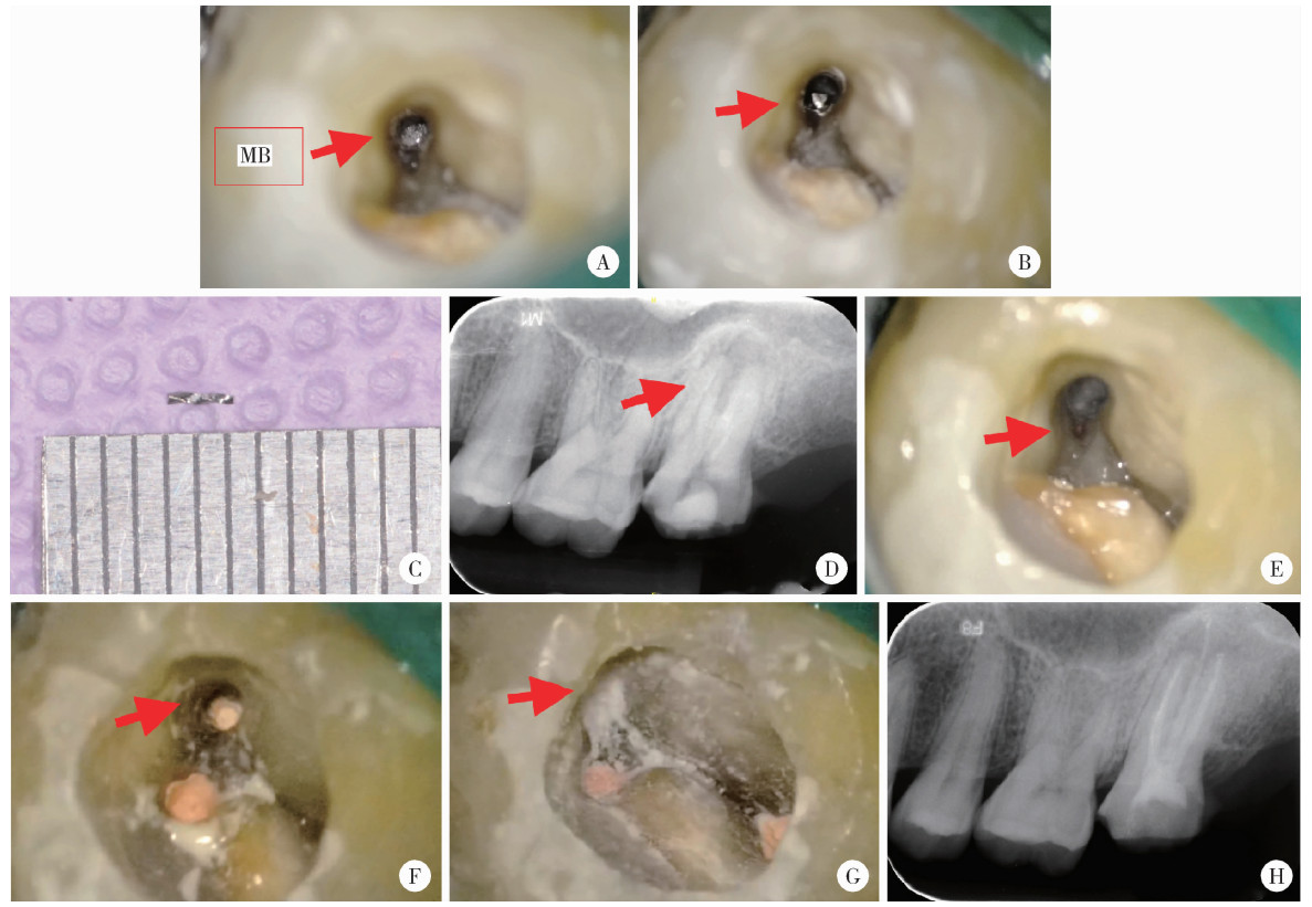
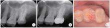
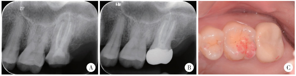
Endodontic retreatment of a maxillary second molar with chronic apical periodontitis and separated instrument: A case report

Hailing ZANG1, Yuhong LIANG2,*()   

  1. 1. Department of Stomatology, Peking University International Hospital, Beijing 102206, China
    2. Department of Oral Emergency, Peking University School and Hospital of Stomatology & National Center for Stomatology & National Clinical Research Center for Oral Diseases & National Engineering Research Center of Oral Biomaterials and Digital Medical Devices & Beijing Key Laboratory of Digital Stomatology & NHC Research Center of Engineering and Technology for Computerized Dentistry & NMPA Key Laboratory for Dental Materials, Beijing 100081, China
  • Received:2025-10-13 Online:2026-02-18 Published:2026-01-05
  • Contact: Yuhong LIANG

RICH HTML


Abstract:

This case report describes the micro-endodontic retreatment and comprehensive management of a complex left maxillary second molar presenting with multiple complications. The patient, a 41-year-old female, presented with recurrent gingival swelling associated with the tooth for more than six months. The tooth had a history of root canal treatment, fiber post placement, and full crown restoration two years earlier. Cone-beam CT (CBCT) revealed a separated metallic instrument approximately 5 mm in length in the apical region of the mesiobuccal root, a fiber post in the palatal root, underfilled root canal obturation, and periapical radiolucency involving the mesiobuccal, distobuccal and palatal roots. A diagnosis of chronic periapical periodontitis (post-endodontic treatment) was established. The primary challenges included the limited access to the left maxillary second molar, the deeply embedded apical instrument segment, and the high risk for complications, such as root fracture or secondary perforation during retrieval attempts. The treatment strategy involved removal of the separated instrument and retreatment under a dental operating microscope. With ultrasonic assistance, the palatal fiber post and the coronal segment (2.5 mm) of the separated instrument from the mesiobuccal canal were successfully removed. An iatrogenic lateral perforation occurred in the mesiobuccal canal near the furcation area during the instrument retrieval procedure. Given the depth and high risk of further damage from retrieving the remaining apical segment (2 mm), a conservative and flexible approach was adopted using the bypass technique. After successful bypassing, thorough chemo-mechanical debridement was performed, followed by obturation using the warm vertical compaction technique. The iatrogenic perforation on the distal wall of the mesiobuccal canal was immediately repaired with a bioceramic material (iRoot BP) for its superior sealing and bioactivity. The tooth was then restored with a full crown. A 10-month follow-up showed that the patient was asymptomatic with complete fistula healing and radiographic examination revealed favorable healing of the periapical lesion. This case demonstrates that, in complex root canal retreatment cases involving retained instruments and iatrogenic perforations, the integration of advanced diagnostic tools (e. g. CBCT), microsurgical techniques (e. g. microscope and ultrasound), and modern restorative materials (e. g. bioceramics), along with a dynamic strategy combining instrument retrieval and bypassing, is essential for achieving predictable long-term success.

Key words: Root canal retreatment, Instrument separation, Perforation, Bypass, Bioceramics

CLC Number: 

  • R781.3

Figure 1

Intraoral examination, preoperative radiograph and cone-beam CT images of left maxillary second molar A, preoperative intraoral view of apical swelling (white arrow); B, preoperative periapical radiograph shows a metallic high-density fractured instrument (red arrow) within the mesiobuccal canal; C, the clinical view after crown removal reveals a resin filling and a palatal fiber post (white arrow); D, the palatal post extends to the mid-root level with an apical radiolucency; E, the image confirms the separated instrument in the apical third of the mesiobuccal root, associated with an apical radiolucency communicating with the distobuccal apical lesion; F, underfilled obturation is observed in the distobuccal root canal. P, palatal; MB, mesiobuccal; DB, distobuccal."

Figure 2

Clinical images and postoperative periapical radiograph illustrating the microscopic removal of the fractured instrument, perforation repair and root canal filling for left maxillary second molar A, fractured instrument tip observed in the mesiobuccal canal (red arrow) under dental microscope; B, instrument becomes loose (red arrow) after ultrasonic vibration; C, the retrieved 2.5 mm instrument segment; D, post-retrieval PA shows 2 mm residual in the apical region (red arrow); E, a lateral perforation is visible in the mesiobuccal canal near the furcation area after instrument retrieval (red arrow); F, the apical portion of the mesio-buccal canal is filled with gutta-percha (red arrow); G, the perforation is repaired with iRoot BP bioceramic material (red arrow); H, postoperative periapical radiograph confirms flush filling and a well-sealed perforation repair. MB, mesiobuccal."

Figure 3

Follow-up periapical radiographs and intraoral photograph of left maxillary second molar A, 3-month follow-up shows initial healing of the periapical lesion; B, C, 10-month periapical and clinical evaluation shows a favorable prognosis."

1
岳林, 王晓燕. 牙体牙髓病学[M]. 3版 北京: 北京大学医学出版社, 2022: 407.
2
Ng YL , Mann V , Gulabivala K . A prospective study of the factors affecting outcomes of nonsurgical root canal treatment: Part 1:Periapical health[J]. Int Endod J, 2011, 44 (7): 583- 609.

doi: 10.1111/j.1365-2591.2011.01872.x
3
Gulabivala K , Ng YL . Factors that affect the outcomes of root canal treatment and retreatment: A reframing of the principles[J]. Int Endod J, 2023, 56 (Suppl 2): 82- 115.
4
Terauchi Y , Sexton C , Bakland LK , et al. Factors affecting the removal time of separated instruments[J]. J Endod, 2021, 47 (8): 1245- 1252.

doi: 10.1016/j.joen.2021.05.003
5
Terauchi Y , Ali WT , Abielhassan MM . Present status and future directions: Removal of fractured instruments[J]. Int Endod J, 2022, 55 (Suppl 3): 685- 709.
6
Fan Y , Gao Y , Wang X , et al. Expert consensus on management of instrument separation in root canal therapy[J]. Int J Oral Sci, 2025, 17 (1): 46.

doi: 10.1038/s41368-025-00372-w
7
Patel S , Brown J , Semper M , et al. European Society of Endodontology position statement: Use of cone beam computed tomography in Endodontics: European Society of Endodontology (ESE) deve-loped by[J]. Int Endod J, 2019, 52 (12): 1675- 1678.

doi: 10.1111/iej.13187
8
Baratto-Filho F , Vavassori de Freitas J , Fagundes Tomazinho FS , et al. Cone-beam computed tomography detection of separated endodontic instruments[J]. J Endod, 2020, 46 (11): 1776- 1781.

doi: 10.1016/j.joen.2020.08.011
9
Cujé J , Bargholz C , Hülsmann M . The outcome of retained instrument removal in a specialist practice[J]. Int Endod J, 2010, 43 (7): 545- 554.

doi: 10.1111/j.1365-2591.2009.01652.x
10
Gorni FG , Ionescu AC , Ambrogi F , et al. Prognostic factors and primary healing on root perforation repaired with MTA: A 14-year longitudinal study[J]. J Endod, 2022, 48 (9): 1092- 1099.

doi: 10.1016/j.joen.2022.06.005
11
Camilleri J , Atmeh A , Li X , et al. Present status and future directions: Hydraulic materials for endodontic use[J]. Int Endod J, 2022, 55 (Suppl 3): 710- 777.
12
Liang YH , Jiang L , Gao XJ , et al. Detection and measurement of artificial periapical lesions by cone-beam computed tomography[J]. Int Endod J, 2014, 47 (4): 332- 338.

doi: 10.1111/iej.12148
13
梁宇红, 岳林. 锥形束CT在牙髓根尖周病诊治中的合理应用与思考[J]. 中华口腔医学杂志, 2019, 54 (9): 591- 597.
14
Madarati AA , Hunter MJ , Dummer PM . Management of intracanal separated instruments[J]. J Endod, 2013, 39 (5): 569- 581.

doi: 10.1016/j.joen.2012.12.033
15
McGuigan MB , Louca C , Duncan HF . Endodontic instrument fracture: Causes and prevention[J]. Br Dent J, 2013, 214 (7): 341- 348.

doi: 10.1038/sj.bdj.2013.324
16
Spili P , Parashos P , Messer HH . The impact of instrument fracture on outcome of endodontic treatment[J]. J Endod, 2005, 31 (12): 845- 850.

doi: 10.1097/01.don.0000164127.62864.7c
17
Suter B , Lussi A , Sequeira P . Probability of removing fractured instruments from root canals[J]. Int Endod J, 2005, 38 (2): 112- 123.

doi: 10.1111/j.1365-2591.2004.00916.x
18
Al-Fouzan KS . Incidence of rotary ProFile instrument fracture and the potential for bypassing in vivo[J]. Int Endod J, 2003, 36 (12): 864- 867.

doi: 10.1111/j.1365-2591.2003.00733.x
19
Ordinola-Zapata R , Martins JNR , Versiani MA , et al. Micro-CT analysis of danger zone thickness in the mesiobuccal roots of maxillary first molars[J]. Int Endod J, 2019, 52 (4): 524- 529.

doi: 10.1111/iej.13025
20
Clauder T . Present status and future directions: Managing perforations[J]. Int Endod J, 2022, 55 (Suppl 4): 872- 891.
21
Yang N , Yang W , Shen R , et al. In vitro and in vivo evaluation of iRoot BP Plus as a coronal sealing material for regenerative endodontic procedures[J]. Clin Oral Investig, 2024, 28 (1): 70.

doi: 10.1007/s00784-023-05468-3
22
Dong X , Xu X . Bioceramics in endodontics: Updates and future perspectives[J]. Bioengineering, 2023, 10 (3): 354.

doi: 10.3390/bioengineering10030354
23
Abdelmotelb MA , Gomaa YF , Khattab NMA , et al. Premixed bioceramics versus mineral trioxide aggregate in furcal perforation repair of primary molars: In vitro and in vivo study[J]. Clin Oral Investig, 2021, 25 (8): 4915- 4925.

doi: 10.1007/s00784-021-03800-3
[1] XU Hao,ZHANG Guo-dong,FAN Guang-pu,CHEN Yu. Preoperative plasma predictive factors of new-onset atrial fibrillation after coronary artery bypass graft surgery: A propensity score matching study [J]. Journal of Peking University (Health Sciences), 2021, 53(6): 1139-1143.
[2] YANG Yang,XIAO Feng,WANG Jin,SONG Bo,LI Xi-hui,ZHANG Shi-jie,HE Zhi-song,ZHANG Huan,YIN Ling. One-stage surgery in patients with both cardiac and non-cardiac diseases [J]. Journal of Peking University (Health Sciences), 2021, 53(2): 327-331.
[3] Zhi-feng XU,Yun-peng LING,Zhong-qi CUI,hong ZHAO,Yi-chen GONG,Yuan-hao FU,Hang YANG,Feng WAN. Feasibility and safety of minimally invasive cardiac coronary artery bypass grafting surgery for patients with multivessel coronary artery disease: Early outcome and short-mid-term follow up results [J]. Journal of Peking University (Health Sciences), 2020, 52(5): 863-869.
[4] Wen-qiang SUN,Zhou ZHAO,Qing GAO,Zeng-qiang HAN,Wei YANG,Bo LIAN,Gang LIU,Sheng-long CHEN,Yu CHEN. Effects of intraoperative graft flow measurements on the early mid-term outcomes after off-pump coronary artery bypass grafting [J]. Journal of Peking University(Health Sciences), 2019, 51(5): 851-855.
[5] ZHANG Lu-feng, LING Yun-peng, YANG Hang, GONG Yi-chen, SONG Zhi-ming, WAN Feng. Comparison of outcomes of two minimally invasive approaches for multi-vessel coronary revascularization [J]. Journal of Peking University(Health Sciences), 2017, 49(6): 1066-1070.
[6] LI Xi-hui, XIAO Feng, ZHANG Si-yu. Investigation of risk factors of acute kidney injury after off-pump coronary artery bypass grafting and 3 years’ follow-up [J]. Journal of Peking University(Health Sciences), 2017, 49(1): 131-136.
[7] YU Jie, WAN Feng, XIE Ji-yan, WU Song, CUI Zhong-qi, XU Min, ZHANG Zhe, GAO Wei. Role of EuroSCORE and SinoSCORE in prediction of early postoperative quality of life in patients after coronary artery bypass surgery [J]. Journal of Peking University(Health Sciences), 2015, 47(5): 769-773.
Viewed
Full text


Abstract

Cited

  Shared   
  Discussed   
No Suggested Reading articles found!