Journal of Peking University (Health Sciences) ›› 2026, Vol. 58 ›› Issue (4): 872-876. doi: 10.19723/j.issn.1671-167X.2026.04.027

Previous Articles     Next Articles

Cystic lymphangioma of the penis in an adult: A case report

Qingkai YANG1, Jian LU1, Min LU2, Kai HONG1,*()   

  1. 1. Department of Urology, Peking University Third Hospital, Beijing 100191, China
    2. Department of Pathology, Peking University Third Hospital, Beijing 100191, China
  • Received:2026-02-27 Online:2026-08-18 Published:2026-05-19
  • Contact: Kai HONG

RICH HTML


Abstract:

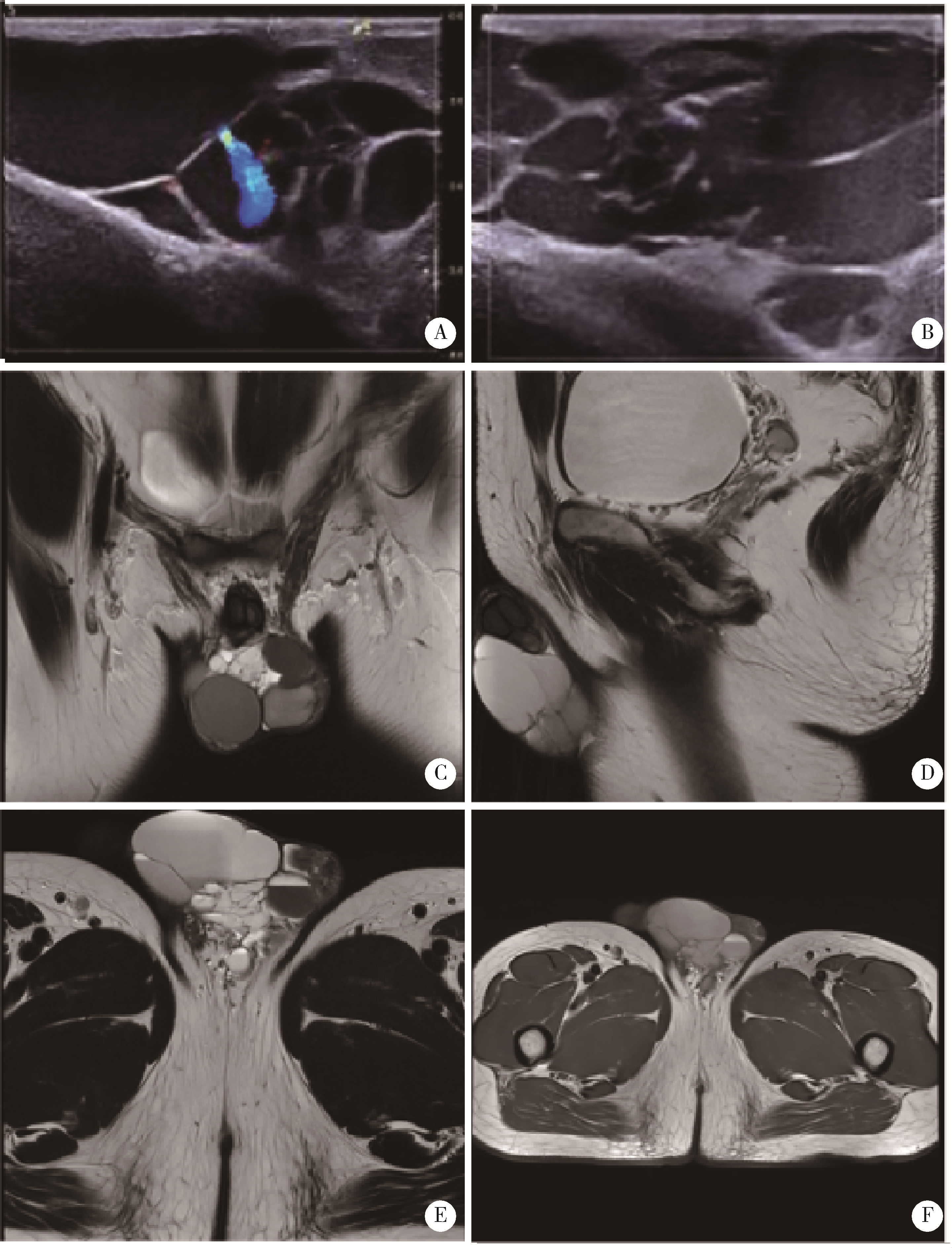
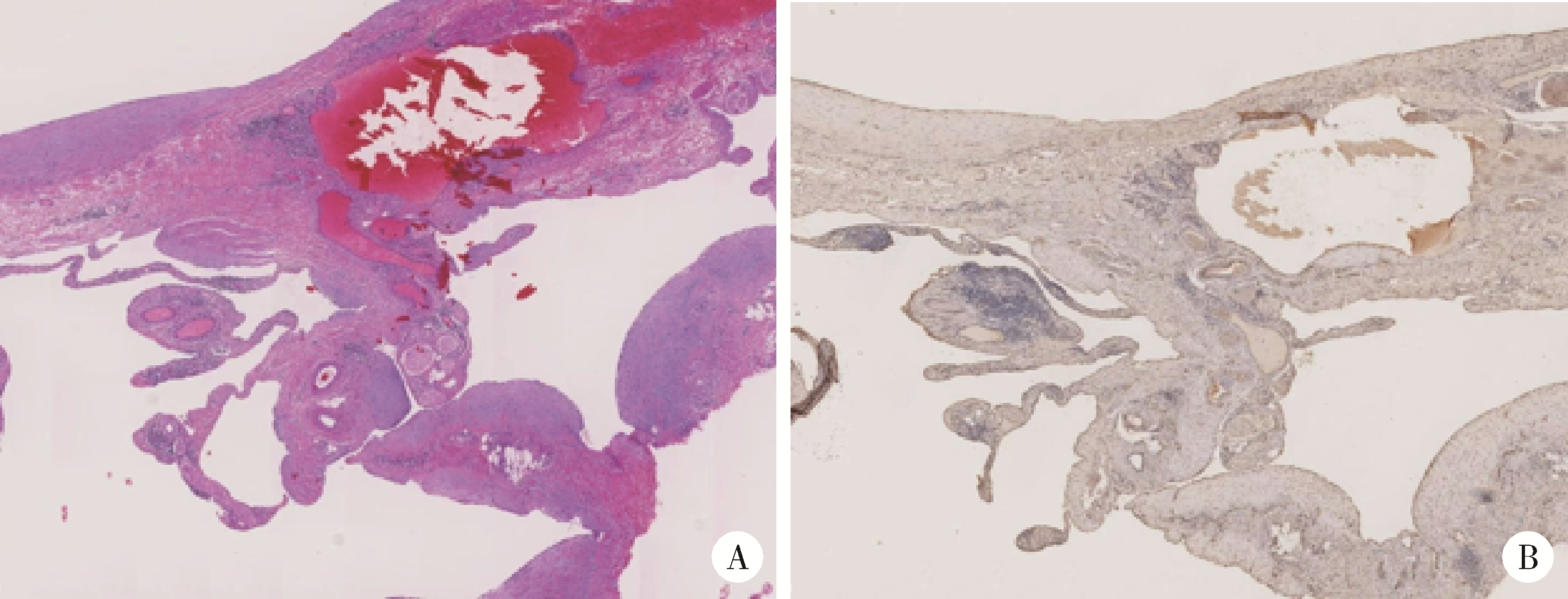
Lymphangioma is a congenital vascular malformation arising from aberrant lymphatic deve-lopment, with a reported incidence of approximately 1 in 4 000. The condition predominates in the pedia-tric population, with the head and neck representing the most frequently affected anatomical regions. By contrast, scrotal lymphangioma in adults is exceedingly rare and remains highly susceptible to misdiagnosis or diagnostic omission in clinical settings. Here, we report a case of cystic lymphangioma of the penis in a 42-year-old male who presented with a mass at the penile root persisting for over 30 years, with marked enlargement over the preceding six months. Physical examination revealed a cystic mass measuring approximately 7 cm×4 cm×5 cm on the left aspect of the penile root, with a positive transillumination sign. Bilateral testes, epididymides, and spermatic cords were unremarkable. Ultrasonography demonstrated an irregular cystic anechoic lesion at the penoscrotal junction with well-defined margins. Magnetic resonance imaging (MRI) revealed a multilocular cystic lesion at the scrotal root measuring approximately 5.6 cm×3.8 cm×6.3 cm. Both imaging modalities were consistent with cystic lymphangioma. The patient underwent complete surgical resection under epidural anesthesia. Intraoperatively, the mass was confirmed to be multilocular and contained taupe-colored fluid. Histopathological analysis revealed endothelial cell lining of the cyst wall, and immunohistochemical staining was positive for CD31, establishing the definitive diagnosis of cystic lymphangioma. Genomic profiling identified a somatic heterozygous missense mutation in the PIK3CA gene at codon E545 (GAG>GCG), consistent with the established pathogenic mechanism underlying lymphangioma. Mechanistically, somatic PIK3CA mutations activate downstream signaling cascades, driving aberrant proliferation of lymphatic endothelial cells and culminating in cystic lesion formation. Clinically, lymphangioma typically manifests as a painless, slowly enlarging cystic mass. Definitive diagnosis relies on integrated imaging and histopathological evaluation, with differential diagnoses encompassing indirect inguinal hernia, hydrocele, and varicocele. Complete surgical resection remains the first-line therapeutic strategy and is associated with a substantially reduced recurrence rate. For patients deemed ineligible for surgery, sclerotherapy or molecularly targeted agents, including sirolimus and alpelisib, represent viable alternatives. The patient achieved an uneventful postoperative recovery with no evidence of recurrence at one-month follow-up. Although adult scrotal cystic lymphangioma is a rare clinical entity, it exhibits characteristic clinical, radiological, and pathological features that, in conjunction with molecular genetic analysis, enable accurate diagnosis. Early complete resection confers a favorable prognosis. Heightened clinical awareness and standardized management of lymphangioma at atypical anatomical sites are warranted.

Key words: Penis, Cystic lymphangioma, Lymphatic malformation

CLC Number: 

  • R737.27

Figure 1

Ultrasonography and MRI of cystic lymphangioma A, B, ultrasound revealed an irregular cystic anechoic area at the penoscrotal junction with clear margins; C-F, MRI revealed a multilocular cystic lesion at the scrotal root with clear boundaries. MRI, magnetic resonance imaging."

Figure 2

Histological features of cystic lymphangioma. A, cystic spaces lined by endothelial cells in dermis (HE ×100); B, CD31 immunostaining highlights the endothelial lining of the cysts (IHC ×100). HE, immunostaining; IHC, immunohistochemistry."

Figure 3

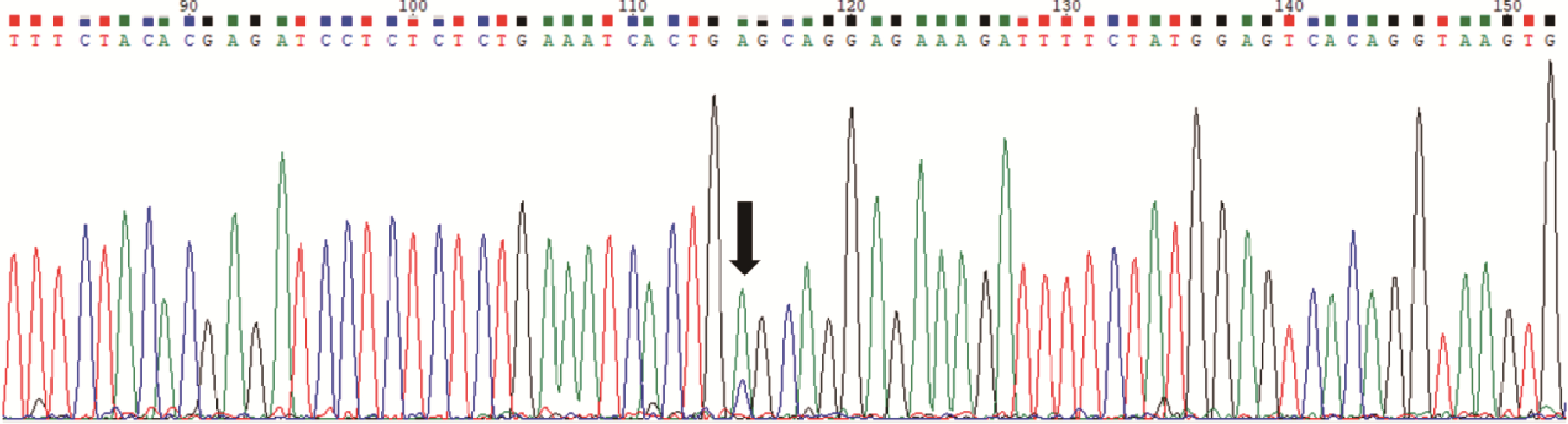
Genetic sequencing results of cystic lymphangioma Dual green and blue peaks at position 115 indicated a heterozygous PIK3CA exon 545 mutation(GAG>GCG) in somatic cells of the patient."

1
中华医学会整形外科分会血管瘤脉管畸形学组, 林晓曦. 血管瘤与脉管畸形诊疗指南(2024版)[J]. 组织工程与重建外科, 2024, 20 (1): 1- 50.
2
Karkkainen MJ , Haiko P , Sainio K , et al. Vascular endothelial growth factor C is required for sprouting of the first lymphatic vessels from embryonic veins[J]. Nat Immunol, 2004, 5 (1): 74- 80.

doi: 10.1038/ni1013
3
Hägerling R , Pollmann C , Andreas M , et al. A novel multistep mechanism for initial lymphangiogenesis in mouse embryos based on ultramicroscopy[J]. EMBO J, 2013, 32 (5): 629- 644.

doi: 10.1038/emboj.2012.340
4
Petrova TV , Karpanen T , Norrmén C , et al. Defective valves and abnormal mural cell recruitment underlie lymphatic vascular failure in lymphedema distichiasis[J]. Nat Med, 2004, 10 (9): 974- 981.

doi: 10.1038/nm1094
5
Martin-Almedina S , Martinez-Corral I , Holdhus R , et al. EPHB4 kinase-inactivating mutations cause autosomal dominant lymphatic-related hydrops fetalis[J]. J Clin Invest, 2016, 126 (8): 3080- 3088.

doi: 10.1172/JCI85794
6
Zhang G , Brady J , Liang WC , et al. EphB4 forward signalling regulates lymphatic valve development[J]. Nat Commun, 2015, 6, 6625.

doi: 10.1038/ncomms7625
7
Mäkinen T , Adams RH , Bailey J , et al. PDZ interaction site in ephrinB2 is required for the remodeling of lymphatic vasculature[J]. Genes Dev, 2005, 19 (3): 397- 410.

doi: 10.1101/gad.330105
8
Mäkinen T , Boon LM , Vikkula M , et al. Lymphatic malformations: Genetics, mechanisms and therapeutic strategies[J]. Circ Res, 2021, 129 (1): 136- 154.

doi: 10.1161/CIRCRESAHA.121.318142
9
Kunimoto K , Yamamoto Y , Jinnin M . ISSVA classification of vascular anomalies and molecular biology[J]. Int J Mol Sci, 2022, 23 (4): 2358.

doi: 10.3390/ijms23042358
10
Luks VL , Kamitaki N , Vivero MP , et al. Lymphatic and other vascular malformative/overgrowth disorders are caused by somatic mutations in PIK3CA[J]. J Pediatr, 2015, 166 (4): 1048- 1054.

doi: 10.1016/j.jpeds.2014.12.069
11
Osborn AJ , Dickie P , Neilson DE , et al. Activating PIK3CA alleles and lymphangiogenic phenotype of lymphatic endothelial cells isolated from lymphatic malformations[J]. Hum Mol Genet, 2015, 24 (4): 926- 938.

doi: 10.1093/hmg/ddu505
12
Janardhan HP , Saheera S , Jung R , et al. Vascular and lymphatic malformations: Perspectives from human and vertebrate studies[J]. Circ Res, 2021, 129 (1): 131- 135.

doi: 10.1161/CIRCRESAHA.121.319587
13
Elluru RG , Balakrishnan K , Padua HM . Lymphatic malformations: Diagnosis and management[J]. Semin Pediatr Surg, 2014, 23 (4): 178- 185.

doi: 10.1053/j.sempedsurg.2014.07.002
14
Rodriguez-Laguna L , Agra N , Ibañez K , et al. Somatic activating mutations in PIK3CA cause generalized lymphatic anomaly[J]. J Exp Med, 2019, 216 (2): 407- 418.

doi: 10.1084/jem.20181353
15
Martinez-Corral I , Zhang Y , Petkova M , et al. Blockade of VEGF-C signaling inhibits lymphatic malformations driven by oncogenic PIK3CA mutation[J]. Nat Commun, 2020, 11, 2869.

doi: 10.1038/s41467-020-16496-y
16
Churchill P , Otal D , Pemberton J , et al. Sclerotherapy for lymphatic malformations in children: A scoping review[J]. J Pediatr Surg, 2011, 46 (5): 912- 922.

doi: 10.1016/j.jpedsurg.2011.02.027
17
Ghaffarpour N , Petrini B , Svensson L , et al. Patients with lymphatic malformations who receive the immunostimulant OK-432 experience excellent long-term outcomes[J]. Acta Paediatr, 2015, 104 (11): 1169- 1173.

doi: 10.1111/apa.13086
[1] Hai-long HE,Qing LI,Tao XU,Xiao-wei ZHANG. Treatment of adult-acquired buried penis with suprapubic liposuction combined with modified Devine operation [J]. Journal of Peking University (Health Sciences), 2022, 54(4): 741-745.
[2] Zhong-cheng XIN,Bi-cheng YANG,Meng LI,Yi-ming YUAN,Wan-shou CUI,Yuan TANG,Dong FANG,Wei-dong SONG. Appllication of human acellular dermal matrix in surgical treatment of genitourinary disease [J]. Journal of Peking University(Health Sciences), 2019, 51(4): 778-782.
[3] Ting-min XU,Xiao-wei ZHANG,Guo-xi ZHANG,Wen-jun BAI,Qing LI,Tao XU. Complications and management for penile augmentation with acellular dermal matrix [J]. Journal of Peking University(Health Sciences), 2019, 51(4): 636-640.
[4] BAO Zheng-qing, FANG Dong,YUE Cai-bo, CAI Lin, WANG Tian-yu, LI Xue-song, ZHOU Li-qun. Primary penoscrotal extra-mammary Paget’s disease: analysis of 22 cases [J]. Journal of Peking University(Health Sciences), 2016, 48(4): 638-642.
[5] YANG Jun, WANG Tao, ZHANG Yan, XU Hua, WANG Shao-Gang, LIU Ji-Hong, YE Zhang-Qun. Concentration of basic fibroblast growth factor in plasma and cavernous corpus of aged rat [J]. Journal of Peking University(Health Sciences), 2015, 47(4): 582-585.
Viewed
Full text


Abstract

Cited

  Shared   
  Discussed   
No Suggested Reading articles found!