Journal of Peking University (Health Sciences) ›› 2020, Vol. 52 ›› Issue (4): 642-645. doi: 10.19723/j.issn.1671-167X.2020.04.008

Previous Articles     Next Articles

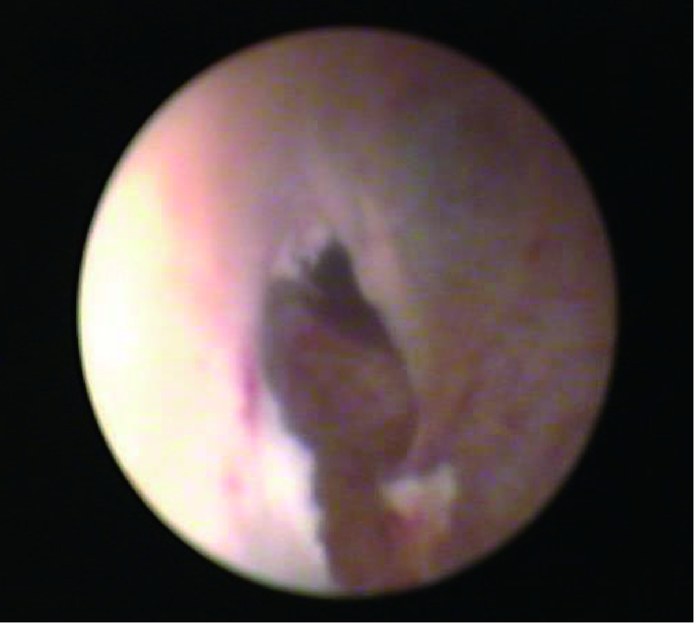
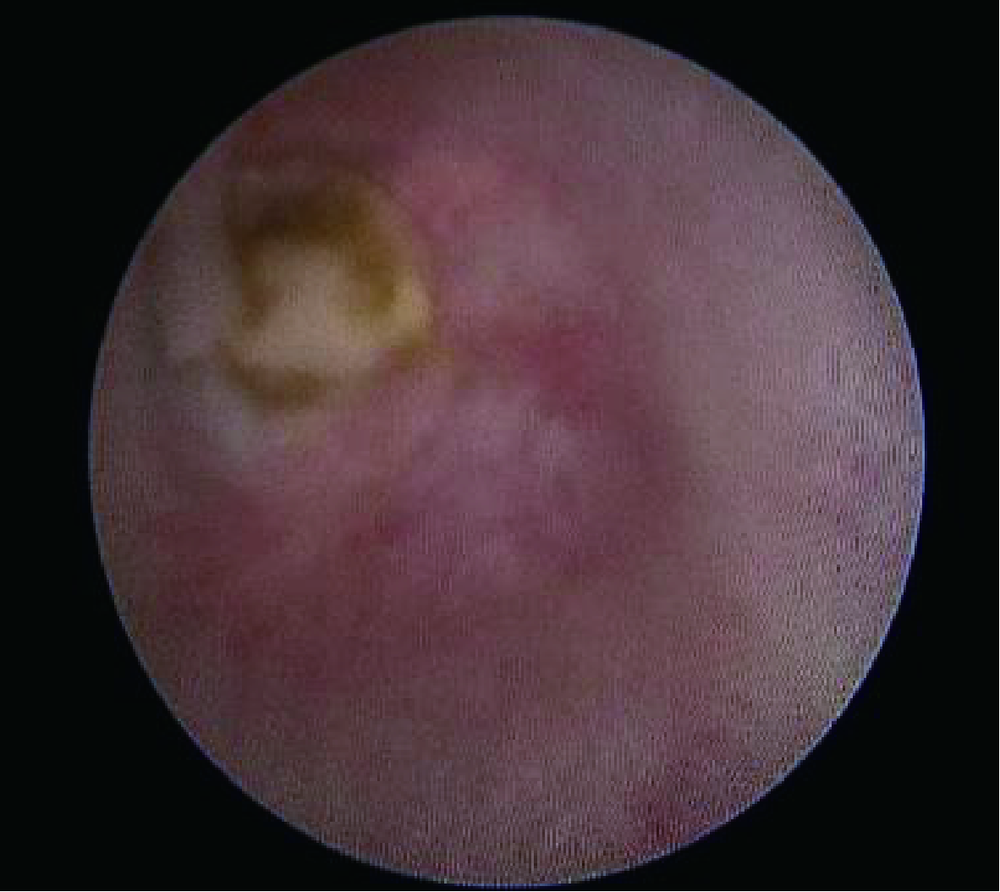
Transurethral seminal vesiculoscopy in treatment of oligoasthenozoospermia secondary incomplete ejaculatory duct obstruction: A report of 8 cases

Hong-bin WANG1,2,Lian-ming ZHAO3,(),Kai HONG3,Jia-ming MAO1,De-feng LIU1,Hao-cheng LIN3,Hui JIANG3   

  1. 1. Peking University Third Hospital Reproductive Center, Beijing 100191, China
    2. Department of Urology, Ningcheng County Central Hospital, Chifeng 024200, Inner Mongolia, China
    3. Department of Urology, Peking University Third Hospital, Beijing 100191, China
  • Received:2020-03-13 Online:2020-08-18 Published:2020-08-06
  • Contact: Lian-ming ZHAO E-mail:doctorzhaolm@163.com

Abstract:

Objective: To evaluate the utility of transurethral seminal vesiculoscopy with a slender ureteroscope in the treatment of severe oligoasthenozoospermia secondary incomplete ejaculatory duct obstruction (EDO). Methods: From March 2018 to September 2018, the clinical data of 8 patients with severe oligoasthenozoospermia secondary incomplete EDO treated by the technique of transurethral seminal vesiculoscopy in the Peking University Third Hospital Reproductive Center were analyzed. Preoperative routine included semen analysis, hormone determination, transrectal ultrasonography, pelvic magne-tic resonance examination and other examinations. All the patients were diagnosed with severe oligoasthenozoospermia secondary to incomplete EDO. All the patients were operated by the same surgeon with multiple cases of experience in transurethral surgery, and 1 year follow-up was conducted to evaluate the surgical effect. Results: The average age of the 8 patients was 29 years, and the average operation time was 32 min. Preoperative transrectal ultrasound indicated 6 cases of ejaculatory duct cyst or Mullerian cyst, 1 case of prostate calcification and bilateral seminal vesicle dilatation. The average maximum transverse diameter of the right seminal vesicle in pelvic MRI was 33.60 mm (24.63-42.28 mm), the average maximum transverse diameter of the left seminal vesicle was 32.85 mm (25.91-44.89 mm), the ave-rage maximum antero-posterior diameter was 27.99 mm (21.36-33.12 mm), the average maximum width of the seminal vesicle duct was 10.53 mm (5.93-19.39 mm). There were 5 cases of ejaculatory duct cyst, 2 cases of seminal vesicle hemorrhage, and 1 case of Mullerian cyst. The semen volume [(2.64±0.80) mL], the sperm concentration [(49.76±8.50)×106/mL], and the motility (grade a+b) [(25.76±6.48)%] in postoperation were significantly higher than those in preoperation [(1.46±0.50) mL, (28.78±5.17)×106/mL, and (2.88±0.93)%, P<0.05]. Two patients conceived naturally during the follow-up of 6 months after surgery. There were no severe complications, such as retrograde ejaculation, urinary incontinence or rectal injury. Conclusion: The technique of transurethral seminal vesiculoscopy is safe and effective for treating severe oligoasthenozoospermia secondary to incomplete EDO. However, due to the small sample size of this study, short follow-up time, and the uncertainty in seminal vesicle surgery, it still needs to be further confirmed by long-term follow-up studies with large samples.

Key words: Seminal vesiculoscope, Ejaculatory ducts obstruction, Oligospermia, Asthenozoospermia

CLC Number: 

  • R697

Table 1

Basic data of 8 EDO patients"

Items Data, x?±s
Age/years 29.0±4.5
Testicular size/mL 14.83±2.50
Sex hormones
FSH/(mIU/mL) 3.57±1.68
LH/(mIU/mL) 3.67±1.54
T/(nmol/L) 11.59±3.43
Pelvic MRI
Antero-posterior diameter of seminal vesicle /mm 32.85±7.64
Seminal vesicle diameter/mm 10.68±2.24

Figure 1

Intraoperative wall rupture followed by dilation"

Figure 2

One yellow stone was seen during the operation"

Table 2

Comparison of the semen quality between pre- and postoperative in 8 EDO patients"

Items Preoperative, x?±s (n=8) Postoperative, x?±s P value*
3 months (n=8) 6 months (n=6) 9 months (n=4)
Semen volume/mL 1.46±0.52 1.86±0.43 2.64±0.86 2.57±0.55 0.043
pH value 6.8±0.4 7.0±0.7 7.3±0.6 7.2±0.4 0.046
Sperm count/(×106/mL) 28.78±5.17 32.65±6.32 49.76±8.50 52.67±5.88 0.012
Sperm motility/% 2.88±0.93 8.69±3.23 25.76±6.48 32.56±8.34 0.001
[1] Wein AJ. Campbell-Walsh Urology[M]. 10th ed. Philadelphia PA: Elsevier Inc, 2012: 673-674.
[2] 吴宏飞. 射精管梗阻[J]. 中华男科学杂志, 2010,16(1):3-9.
[3] Hellerstein DK, Meacham RB, Lipshultz LI, et al. Transrectal ultrasound and partial ejaculatory duct obstruction in male infertility[J]. Urology, 1992,39(5):449-452.
[4] Engin G. Transrectal US-guided seminal vesicle aspiration in the diagnosis of partial ejaculatory duct obstruction[J]. Diagn Interv Radiol, 2012,18(5):488-495.
[5] Xu B, Niu X, Wang Z, et al. Novel methods for the diagnosis and treatment of ejaculatory duct obstruction[J]. BJU Int, 2010,108(2):263-266.
pmid: 20950310
[6] 谢军, 关登海, 陈光耀, 等. 经尿道射精管口电切术治疗射精管梗阻性无(少)精子症[J]. 黑龙江医学, 2012,36(6):445-446.
[7] 邓春华, 丘少鹏, 孙祥宙, 等. 经尿道射精管口电切术治疗射精管梗阻性无精子症[J]. 中华外科杂志, 2005,43(22):1464-1466.
[8] Yurdakul T, Gokce G, Kilic O, et al. Transurethral resection of ejaculatory ducts in the treatment of complete ejaculatory duct obstruction[J]. Int Urol Nephrol, 2008,40(2):369-372.
pmid: 17899434
[9] Cheng G, Liu B, Song Z, et al. A novel surgical management for male infertility secondary to midline prostatic cyst[J]. BMC Urology, 2015,15(1):18.
[10] 涂响安, 赵良运, 赵亮, 等. 经尿道射精管切开术治疗射精管梗阻的效果(附60例报告)[J]. 北京大学学报(医学版), 2011,43(4):559-561.
[1] DAI Xiao-wei, XU Ying, ZHENG Lian-wen, LI Ling-yun, LI Dan-dan1 TAN Xin, GAO Fei, WANG Yan, WU Gui-jie. Analysis of chromosome in 1 324 patients with oligozoospermia or azoosperm [J]. Journal of Peking University(Health Sciences), 2018, 50(5): 774-777.
Viewed
Full text


Abstract

Cited

  Shared   
  Discussed   
[1] . [J]. Journal of Peking University(Health Sciences), 2009, 41(4): 456 -458 .
[2] . [J]. Journal of Peking University(Health Sciences), 2009, 41(2): 125 -128 .
[3] . [J]. Journal of Peking University(Health Sciences), 2009, 41(2): 135 -140 .
[4] . [J]. Journal of Peking University(Health Sciences), 2009, 41(2): 158 -161 .
[5] . [J]. Journal of Peking University(Health Sciences), 2009, 41(2): 217 -220 .
[6] . [J]. Journal of Peking University(Health Sciences), 2009, 41(1): 52 -55 .
[7] . [J]. Journal of Peking University(Health Sciences), 2009, 41(1): 109 -111 .
[8] . [J]. Journal of Peking University(Health Sciences), 2009, 41(3): 297 -301 .
[9] . [J]. Journal of Peking University(Health Sciences), 2009, 41(5): 505 -515 .
[10] . [J]. Journal of Peking University(Health Sciences), 2009, 41(5): 516 -520 .