Journal of Peking University (Health Sciences) ›› 2020, Vol. 52 ›› Issue (5): 828-835. doi: 10.19723/j.issn.1671-167X.2020.05.006

Previous Articles     Next Articles

Study of etiology and esophageal motility characteristics of esophagogastric junction outlet obstruction patients

Kun WANG,Zhi-jie XU,Ying GE,Zhi-wei XIA,Li-ping DUAN()   

  1. Department of Gastroenterology, Peking University Third Hospital, Beijing 100191, China
  • Received:2020-02-11 Online:2020-10-18 Published:2020-10-15
  • Contact: Li-ping DUAN E-mail:duanlp@bjmu.edu.cn

RICH HTML


Abstract:

Objective: To analyze the causes of the esophagogastric junction outlet obstruction (EGJOO) patients, to discuss the differences of the clinical manifestation and esophageal motility characteristics between the anatomic EGJOO (A-EGJOO) and functional EGJOO (F-EGJOO) subgroups, and to search the diagnostic values of the specific metrics for differentiating the subgroups of EGJOO patients. Methods: For the current retrospective study, all the patients who underwent the esophageal high resonance manometry test were retrospectively analyzed from Jan 2012 to Oct 2018 in Peking University Third Hospital. The EGJOO patients were enrolled in the following research. The clinical characteristics, such as symptoms and causes of the patients were studied. Then the patients were divided into two subgroups as A-EGJOO subgroup and F-EGJOO subgroup. The clinical symptoms and the main manometry metrics were compared between these two subgroups. The significant different metrics between the two groups were selected to draw receiver operating characteristic (ROC) curves and the diagnostic values were analyzed in differentiating the A-EGJOO and F-EGJOO subgroups. Results: The most common symptom of EGJOO was chest pain or chest discomfort (30.63%), then the dysphagia (29.73%), and acid regurgitation/heartburn (27.03%). Non-erosive reflux disease (36.04%) was the most popular cause for EGJOO, then the reflux esophagitis (17.12%). Besides the intra-EGJOO and extra-EGJOO lesions, the connective tissue disease (6.31%) and central nervous diseases (2.70%) were found to be the etiology of EGJOO. The causes of the rest 19 EGJOO were unknown. A-EGJOO patients presented significantly higher intra bolus pressure (IBP) than that of F-EGJOO [6.80 (5.20, 9.20) mmHg vs. 5.10 (3.10, 7.60) mmHg, P=0.016]. The area under curve of IBP was 0.637. When IBP≥5.15 mmHg, the sensitivity was 78.60% and specificity 50.70% to differentiate A- or F-EGJOO. Conclusion: Chest pain or chest discomfort was the most common symptom in EGJOO patients. Besides the intraluminal structural disorders, the extra-luminal causes were found in EGJOO patients. A-EGJOO presented higher IBP than that of F-EGJOO patients. The cutoff value of IBP to differentiate A-EGJOO from EGJOO was 5.15 mmHg with sensitivity 78.06% and specificity 50.70%. However for the low area under curve, the diagnostic value of IBP was limited.

Key words: Esophageal motility disorders, Esophagogastric junction, Gastroesophageal reflux

CLC Number: 

  • R571

Figure 1

EGJOO patients data review strategy EGJOO, esophagogastric junction outlet obstruction; A-EGJOO, anatomic EGJOO; F-EGJOO, functional EGJOO; ECG, electrocardiogram; PPI, proton pump inhibitors; NERD, non-erosive reflux disease; CNS, central nervous system."

Table 1

Etiology of EGJOO patients"

Causes Cases (n) Percent (n=111) Age/years, x-±s Gender (Male ∶Female)
Reflux esophagitis 19 17.12% 57.42±13.35 8 ∶11
Other benign diseases in EGJ 14 12.61% 58.86±16.65 5 ∶9
Hiatus hernia 3 2.70%
Esophageal diverticulum near EGJ 3 2.70%
Esophageal venous aneurysm near EGJ 1 0.90%
Esophageal leiomyoma near EGJ 2 1.80%
Eosinophilic esophagus 1 0.90%
Severe fungous esophagitis 2 1.80%
Carditis 2 1.80%
Achalasia 4 3.60% 49.50±7.85 3 ∶1
Malignant tumor 5 4.50% 57.20±7.82 2 ∶3
Liver cancer 1 0.90%
Mediastinal mass 2 1.80%
Cardiac cancer 1 0.90%
Gastric fundus cancer 1 0.90%
NERD 40 36.04% 51.85±13.73 11 ∶29
Central nervous system disorders 3 2.70% 57.67±3.51 0 ∶3
Syringomyelia 1 0.90%
Cerebellar tonsillar hernia 2 1.80%
Connective tissue disease 7 6.31% 48.00±14.91 0 ∶7
Systemic sclerosis 5 4.50%
Dermatomyositis 1 0.90%
Undetermined type 1 0.90%
Unidentified etiology 19 17.12% 54.42±11.53 5 ∶14

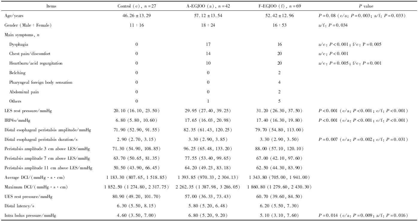
Table 2

Cmparison of esophageal motility metrics between A-EGJOO and F-EGJOO patients"

Figure 2

Esophageal pressure topographies of A-EGJOO and F-EGJOO patients A, A-EGJOO pressure topography during wet swallow; B, F-EGJOO pressure topography during wet swallow. Abbreviations as in Figure 1."

Table 3

Comparison of esophageal motility metrics among RE, NERD, A-EGJOONG, and F-EGJOONG patients"

Items RE (r), n=19 NERD (n), n=40
LES rest pressure/mmHg 29.20 (27.6, 34.95) 30.90 (28.00, 35.80)
IRP4s/mmHg 17.10 (16.00, 18.55) 16.95 (16.00, 18.93)
Distal esophageal peristalsis amplitude/mmHg 81.10 (58.90, 117.20) 78.95 (51.95, 95.73)
Distal esophageal peristalsis duration/s 3.20 (2.75, 3.55) 3.30 (2.70, 3.50)
Peristalsis amplitude 3 cm above LES/mmHg 85.50 (65.65, 124.55) 83.05 (57.08, 113.48)
Peristalsis amplitude 7 cm above LES/mmHg 78.30 (47.70, 106.70) 60.40 (40.38, 94.25)
Peristalsis amplitude 11 cm above LES/mmHg 65.10 (47.80, 83.40) 55.90 (41.33, 81.73)
Average DCI/(mmHg·s·cm) 1 263.70 (973.25, 1 892.55) 1 181.75 (656.85, 1 710.28)
Maximum DCI/(mmHg·s·cm) 2 003.70 (1 335.60, 2 578.45) 1 613.15 (1 266.58, 2 399.05)
UES rest pressure/mmHg 58.00 (37.90, 78.50) 57.90 (37.60, 88.63)
Distal latency/s 6.00 (5.40, 6.65) 6.25 (5.80, 7.30)
Intra bolus pressure/mmHg* 7.80 (3.70, 8.75) 4.65 (2.95, 6.73)
Items A-EGJOONG (a), n=23 F-EGJOONG (f), n=29
LES rest pressure/mmHg 35.60 (27.25, 45.45) 31.70 (26.00, 38.00)
IRP4s/mmHg 18.60 (16.20, 30.35) 17.90 (17.20, 21.00)
Distal esophageal peristalsis amplitude/mmHg 83.00 (69.85, 121.75) 86.30 (58.10, 122.80)
Distal esophageal peristalsis duration/s 3.50 (3.00, 4.30) 3.30 (2.90, 3.70)
Peristalsis amplitude 3 cm above LES/mmHg 105.30 (64.45, 150.40) 100.40 (58.60, 138.50)
Peristalsis amplitude 7 cm above LES/mmHg 74.30 (61.75, 98.35) 79.90 (45.10, 113.10)
Peristalsis amplitude 11 cm above LES/mmHg 63.30 (50.60, 76.70) 64.40 (47.60, 91.80)
Average DCI/(mmHg·s·cm) 1 641.90 (957.25, 2 854.20) 1 443.50 (823.70, 2 168.90)
Maximum DCI/(mmHg·s·cm) 2 352.70 (1 542.20, 3 562.00) 2 141.00 (1 311.50, 2 952.90)
UES rest pressure/mmHg 50.70 (30.75, 68.90) 60.70 (45.50, 74.20)
Distal latency/s 5.70 (5.10, 6.30) 5.60 (5.00, 7.10)
Intra bolus pressure/mmHg* 6.40 (5.50, 11.65) 5.90 (4.40, 8.10)
[1] Kahrilas PJ, Bredenoord AJ, Fox M, et al. The Chicago classification of esophageal motility disorder, v3.0[J]. Neurogastroenterol Motil, 2015,27(2):160-174.
doi: 10.1111/nmo.12477 pmid: 25469569
[2] Clayton SB, Patel R, Richter JE. Functional and anatomic eso-phagogastic junction outflow obstruction: Manometry, timed barium esophagram findings, and treatment outcomes[J]. Clin Gastroenterol Hepatol, 2016,14(6):907-911.
doi: 10.1016/j.cgh.2015.12.041 pmid: 26792374
[3] Shin IS, Min YW, Rhee PL. Esophagogastric junction outflow obstruction transformed to type Ⅱ achalasia[J]. J Neurogastorne-terol Motil, 2016,22(2):344-345.
[4] Ihara E, Muta K, Fukaura K, et al. Diagnosis and treatment strategy of Achalasia subtypes and esophagogastric junction outflow obstruction based on high-resolution manometry[J]. Digestion, 2017,95(1):29-35.
doi: 10.1159/000452354 pmid: 28052278
[5] Kim HP, Vance RB, Shaheen NJ, et al. The prevalence and diagnostic utility of endoscopic features of eosinophilic esophagitis: A meta-analysis[J]. Clin Gastroenterol Hepatol, 2012,10(9):988-996.
doi: 10.1016/j.cgh.2012.04.019 pmid: 22610003
[6] Armstrong D, Bennett JR, Blum AL, et al. The endoscopic assessment of esophagitis: a progress report on observer agreement[J]. Gastroenterology, 1996,111(1):85-92.
doi: 10.1053/gast.1996.v111.pm8698230 pmid: 8698230
[7] Zerbib F, Des Varannea SB, Roman S, et al. Normal values and day-to-day variability of 24-h-ambulatory oesophageal impedance pH monitoring in a Belgian-French cohort of healthy subjects[J]. Aliment Pha rmacol Ther, 2005,22(10):1011-1021.
[8] Samo S, Qayed E. Esophagogastric junction outflow obstruction: Where are we now in diagnosis and management?[J]. World J Gastroenterol, 2019,25(4):411-417.
doi: 10.3748/wjg.v25.i4.411 pmid: 30700938
[9] Pérez-Fernández MT, Santander C, Marinero A, et al. Characte-rization and follow-up of esophagogastric junction outflow obstruction detected by high resolution manometry[J]. Neurogastroenterol Motil, 2016,28(1):116-126.
pmid: 26517978
[10] Schupack D, Katzka DA, Geno DM, et al. The clinical significance of esophagogastric junction outflow obstruction and hypercontractile esophagus in high resolution esophageal manometry[J]. Neruogastroenterol Motil, 2017,29(10):1-9.
[11] Ong AML, Namasivayam V, Wang YT. Evaluation of symptomatic esophagogastric junction outflow obstruction[J]. J Gastroenterol Hepatol, 2018,33(10):1745-1750.
doi: 10.1111/jgh.14155 pmid: 29660156
[12] Gyawali CP, Kushnir VM. High-resolution manometric characte-ristics help differentiate types of distal esophageal obstruction in patients with peristalsis[J]. Neurogastroenterol Motil, 2011,23(6):502-508, e197.
pmid: 21303431
[13] 王琨, 段丽萍, 夏志伟, 等. 基于高分辨食管压力测定及阻抗-pH监测的难治性烧心患者食管动力特点[J]. 中华医学杂志, 2014,94(34):2650-2655.
No related articles found!
Viewed
Full text


Abstract

Cited

  Shared   
  Discussed   
No Suggested Reading articles found!