Journal of Peking University (Health Sciences) ›› 2025, Vol. 57 ›› Issue (5): 1002-1004. doi: 10.19723/j.issn.1671-167X.2025.05.028

Previous Articles     Next Articles

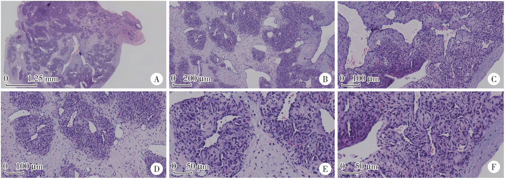
Left sided sternocleidomastoid interosseous intravascular papillary endothelial hyperplasia: A case report

Xiaodi XIAO1, Youchen XIA1, Jianying LIU2, Peng FU3,*()   

  1. 1. Department of Plastic Surgery, Peking University Third Hospital, Beijing 100191, China
    2. Department of Pathology, Peking University Third Hospital, Beijing 100191, China
    3. Department of Ultrasound, Peking University Third Hospital, Beijing 100191, China
  • Received:2022-12-09 Online:2025-10-18 Published:2025-09-05
  • Contact: Peng FU

RICH HTML


Abstract: Intravascular papillary endothelial hyperplasia (IPEH), also known as Masson tumor, is a rare vascular benign tumor of blood vessels. It may occur in any part of the body, especially the deep dermis and subcutaneous tissue of the head, neck, fingers and trunk. The imaging and histopathology of IPEH are similar to hemangiosarcoma, especially in the case of active vascular endothelial hyperplasia. IPEH is a reactive proliferative lesion of vascular intima. The etiology is still unclear. After some studies showed that IPEH was a benign lesion, few reports on the etiology of it were reported. IPEH is usually limited to the thrombotic vessels or lumens of vascular malformations, usually accompanied by a clear history of trauma. IPEH usually does not cause any symptoms. It looks like a slow-growing lump. Some cases have been reported with pain and swelling. Although IPEH is relatively rare, its accurate diagnosis is crucial because it may be similar to malignant angiogenic lesions in clinical practice. There were few reports of cases related to intravascular papillary endothelial hyperplasia located in the sternocleidomastoid muscle after reviewing the domestic and foreign literature in recent 10 years. This case reports that a young male, who was admitted to the hospital one month after finding a subcutaneous tumor in the left neck. After admittance, relevant preoperative examinations were completed. After multi-disciplinary discussion and elimination of surgical contraindications, a specific surgical plan was formulated. The tumor was removed under local anesthesia on the second day after admission. During the operation, it was found that the tumor was located between the sternocleidomastoid muscle bundles, and it was sent for pathologic examination. Paraffin section pathology was reported after operation. Histological examination showed that the morphology was consistent with vascular endothelial papillary hyperplasia. There were no related surgical complications and recurrence in the 3-month follow-up. The purpose of this paper is to provide clinicians with a certain understanding of this rare disease through the report of this case of IPEH, and to identify it in later clinical work, and at the same time, to avoid confusion with malignant diseases, such as hemangiosarcoma, leading to unnecessary treatment and increase the cost of treatment.

Key words: Hemangioendothelioma, Hyperplasia, Hemangiosarcoma, Differential diagnosis, Intravascular papillary endothelial hyperplasia

CLC Number: 

  • R730.262

Figure 1

Ultrasound imaging of the patient with IPEH A, a hypoechoic nodule with clear, incompressible borders was seen in a local superficial vein; B, color doppler imageing shows rich blood flow signal; C, adjacency of the nodule to the surrounding neck vessels.IPEH, intravascular papillary endothelial hyperplasia."

Figure 2

Intraoperative situation of the patient with IPEH A, tumor range and incision design line; B, herniation of a mass after blunt separation of the left sternocleidomastoid muscle; C, isolated mass specimen; D, wound condition after suture. IPEH, intravascular papillary endothelial hyperplasia."

Figure 3

Pathological microscopic features of IPEH A, full view of intravascular tumor (20×); B, vascular endothelial hyperplasia, fibrous tissue hyperplasia, fibrous interstitial hyperplasia and recanalized vessels (40×); C, papillary structure (200×); D, endothelial lining fissures and papillary hyperplasia (200×); E, no obvious polymorphism of endothelial cells (400×); F, papillary structure (400×).IPEH, intravascular papillary endothelial hyperplasia."

1
Han JW , Lee JH , Kim EK . Intravascular papillary endothelial hyperplasia (Masson' s hemangioma) of the face[J]. Arch Craniofac Surg, 2019, 20 (2): 109- 111.

doi: 10.7181/acfs.2018.02208
2
Guledgud MV , Patil K , Saikrishna D , et al. Intravascular papillary endothelial hyperplasia: Diagnostic sequence and literature review of an orofacial lesion[J]. Case Rep Dent, 2014, 2014, 934593.
3
Yadav M , Popli K , Bisoi AK , et al. Masson ' s hemangioma mimicking as leaking aortic pseudoaneurysm: An extremely rare presentation[J]. Aorta (Stamford), 2019, 7 (2): 59- 62.

doi: 10.1055/s-0039-1688925
4
Mignogna C , Barca I , Di Vito A , et al. Extravascular type of intravascular papillary endothelial hyperplasia mimicking parotid gland neoplasia and the possible role of ferritin in the pathogenesis: A case report[J]. Mol Clin Oncol, 2017, 6 (2): 193- 196.

doi: 10.3892/mco.2016.1117
5
Soares AB , Altemani A , Furuse C , et al. Intravascular papillary endothelial hyperplasia: Report of 2 cases and immunohistochemical study[J]. Oral Surg Oral Med Oral Pathol Oral Radiol Endod, 2008, 106 (5): 708- 711.

doi: 10.1016/j.tripleo.2008.08.011
6
Kim OH , Kim YM , Choo HJ , et al. Subcutaneous intravascular papillary endothelial hyperplasia: Ultrasound features and pathological correlation[J]. Skeletal Radiol, 2016, 45 (2): 227- 233.

doi: 10.1007/s00256-015-2281-7
7
Clearkin KP , Enzinger FM . Intravascular papillary endothelial hyperplasia[J]. Arch Pathol Lab Med, 1976, 100 (8): 441- 444.
8
Levere SM , Barsky SH , Meals RA . Intravascular papillary endothelial hyperplasia: A neoplastic "actor" representing an exag-gerated attempt at recanalization mediated by basic fibroblast growth factor[J]. J Hand Surg Am, 1994, 19 (4): 559- 564.

doi: 10.1016/0363-5023(94)90256-9
9
Hashimoto H , Daimaru Y , Enjoji M . Intravascular papillary endothelial hyperplasia: A clinicopathologic study of 91 cases[J]. Am J Dermatopathol, 1983, 5 (6): 539- 546.

doi: 10.1097/00000372-198312000-00004
10
Kim D , Israel H , Friedman M , et al. Intravascular papillary endothelial hyperplasia manifesting as a submandibular mass: An unusual presentation in an uncommon location[J]. J Oral Maxillofac Surg, 2007, 65 (4): 786- 790.

doi: 10.1016/j.joms.2005.11.095
[1] Chao ZUO, Guoli WANG, Kunlin YANG, Xinyan CHE, Yisen MENG, Kai ZHANG. Comparison of efficacy and safety of transurethral thulium fiber laser enucleation of prostate in patients with different prostate volumes [J]. Journal of Peking University (Health Sciences), 2025, 57(4): 711-716.
[2] Ning LIU, Libo MAN, Feng HE, Guanglin HUANG, Jianpo ZHAI. Correlation between urination intermittences and urodynamic parameters in benign prostatic hyperplasia patients [J]. Journal of Peking University (Health Sciences), 2025, 57(2): 328-333.
[3] Hua ZHONG, Yuan LI, Liling XU, Mingxin BAI, Yin SU. Application of 18F-FDG PET/CT in rheumatic diseases [J]. Journal of Peking University (Health Sciences), 2024, 56(5): 853-859.
[4] Xue-mei HA,Yong-zheng YAO,Li-hua SUN,Chun-yang XIN,Yan XIONG. Solid placental transmogrification of the lung: A case report and literature review [J]. Journal of Peking University (Health Sciences), 2023, 55(2): 357-361.
[5] Chieko MORIMOTO,Yi-qin WANG,Rong ZHOU,Jian-liu WANG. Clinical analysis of fertility-sparing therapy of patients with complex atypical hyperplasia and endometrial cancer [J]. Journal of Peking University (Health Sciences), 2022, 54(5): 936-942.
[6] HU Hao, XU Ke-Xin, ZHANG Xiao-Peng, FANG Zhi-Wei, CHEN Jing-Wen, HUO Fei, WANG Dong, WANG Xiao-Feng. Expression of brainderived neurotrophic factor in urine of patients with benign prostatic hyperplasia  complicated overactive bladder symptoms [J]. Journal of Peking University(Health Sciences), 2014, 46(4): 519-523.
Viewed
Full text


Abstract

Cited

  Shared   
  Discussed   
No Suggested Reading articles found!