Journal of Peking University (Health Sciences) ›› 2025, Vol. 57 ›› Issue (5): 1005-1009. doi: 10.19723/j.issn.1671-167X.2025.05.029

Previous Articles     Next Articles

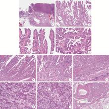
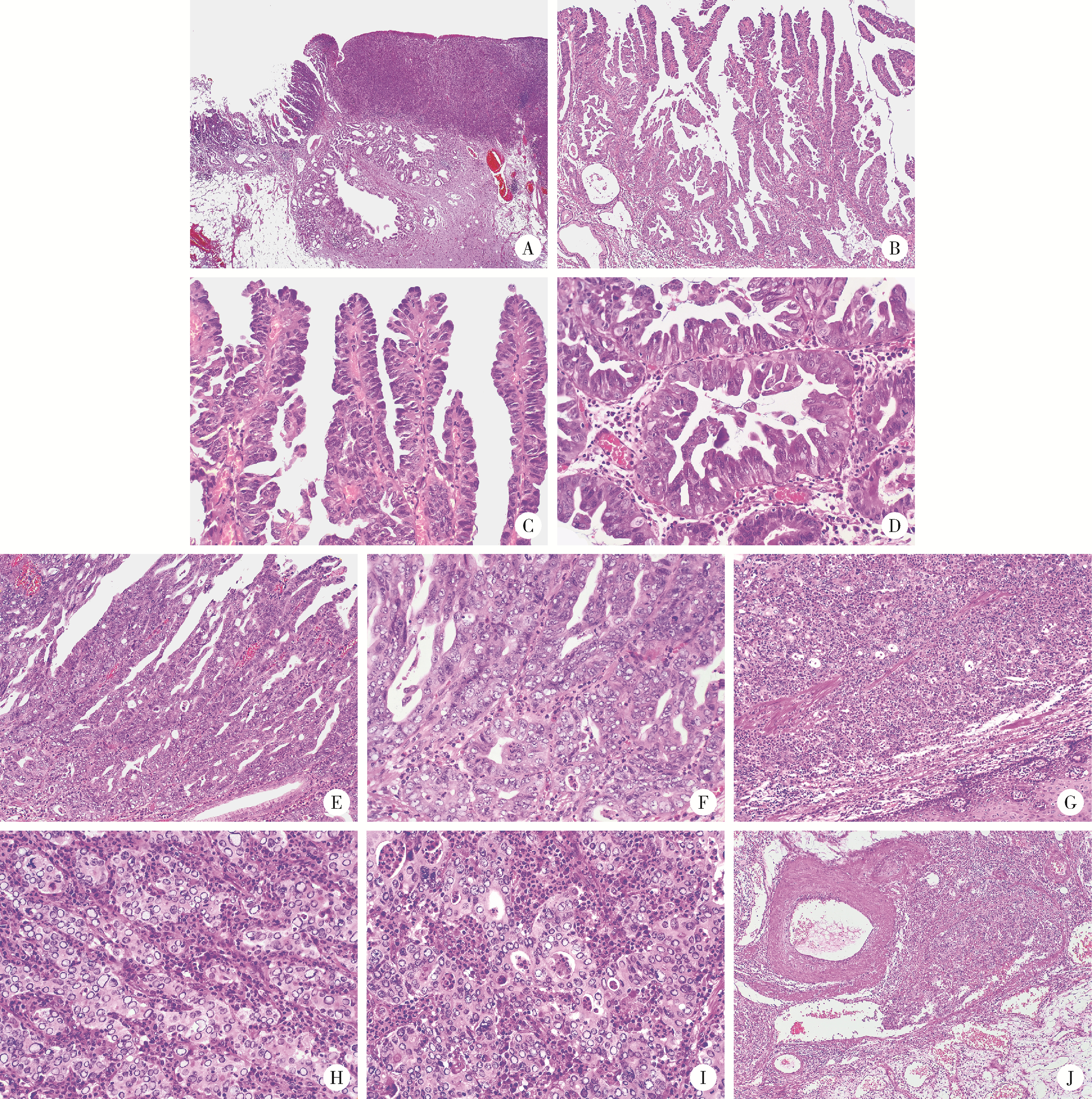
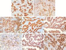
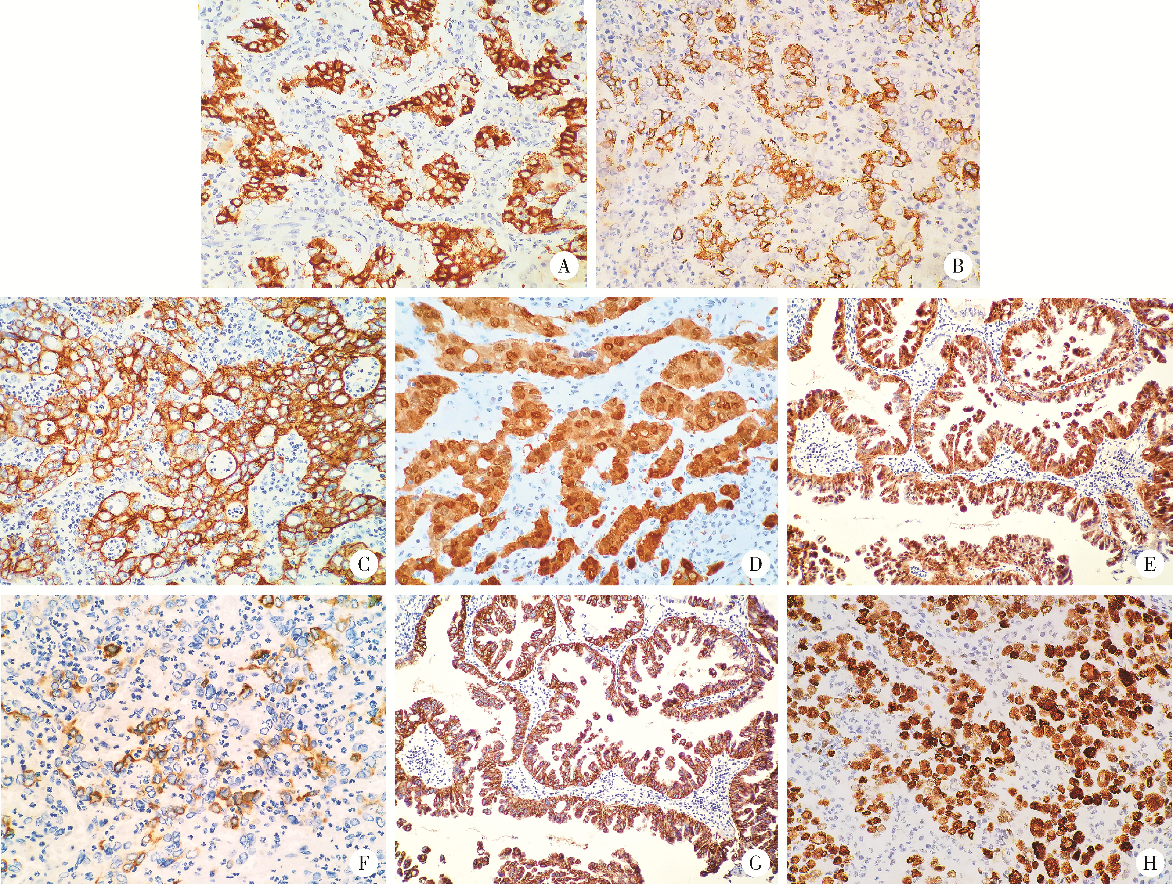
Neuroendocrine carcinoma with significantly vacuolar nucleus at the esophagogastric junction: A case report

Weihua HOU1, Shujie SONG2, Zhongyue SHI3, Lu LIU2, Mulan JIN3,*()   

  1. 1. Department of Pathology, Xiangcheng County People's Hospital, Xuchang 461700, Henan, China
    2. Department of Gastroenterology, 989 Hospital of Joint Logistic Support Force, Pingdingshan 467099, Henan, China
    3. Department of Pathology, Beijing Chaoyang Hospital, Capital Medical University, Beijing 100020, China
  • Received:2023-03-15 Online:2025-10-18 Published:2025-09-16
  • Contact: Mulan JIN
  • Supported by:
    the National Natural Science Foundation of China(62176168); Scientific Research Special Fund for the Joint Growth Plan of Chinese Early Gastrointestinal Cancer Physicians(GTCZ-2023-HN-01)

RICH HTML


Abstract: Neoplasms characterized by the expression of markers of neuroendocrine differentiation in neoplastic cells are defined as neuroendocrine neoplasms. A case of neuroendocrine carcinomas (NECs) with a small amount of papillary adenocarcinoma and significantly vacuolar nucleus at the esophagogastric junction was reported in this article. A 77-year-old male had dysphagia for one week. Endoscopy revealed early-stage esophagogastric junction carcinoma, and biopsy was diagnosed as poorly differentiated carcinoma. Endoscopic submucosal dissection was performed. Histologically, the papillary adenocarcinoma progresses from typically branching papillary structures (well-differentiated) to hyperplasia of the lining epithelium of the papilla to form a cribriform structure (moderately differentiated), to solid area lacking papillary structures (poorly differentiated). There was a continuous process, and during this process, the vacuoles in the nuclei of tumor cells showed progressive changes from mild to obvious and finally to significant vacuoles. The tumor was mainly composed of solid areas (about 95%), with single cell, large cell, round or oval to irregular nuclei, and significantly vacuolar nuclei, nuclei with larger vacuoles appeared in a loop, a few thin weakly basophilic or weakly eosinophilic fine particles could be seen in the vacuoles, and the vacuoles had rough edges. The nucleus chromatin at the outer edge of the vacuoles was fine particles, and mitosis was common (20-30/mm2), atypical mitosis could be seen, and nucleoli could be seen easily, the cytoplasm was weakly eosinophilic, and the boundaries of cells were unclear. The cells were arranged in a nested, trabecular, or diffuse sheet shape, with some arranged in a glandular tube shape. Tumor thrombus was found in the vein of submucosa; the interstitial tissue rich in capillaries within the tumor was accompanied by a large number of neutrophil infiltration. Immunohistochemical staining showed that the solid area of the tumor was positive for synaptophysin (Syn) and chromogranin A (CgA), while papillary adenocarcinoma was negative. Mucin 5AC (MUC5AC) was diffusely positive in papillary adenocarcinoma, while the proportion of positive cells in the solid area of the tumor was about 10% to 15%. In a word, this case showed the extreme situation of the vacuolar nuclear characteristics of NECs, extremely rare, in a sense, this case expanded the boundary of the morphological spectrum of NECs. Understanding the extreme vacuolar features of this nucleus is helpful to make a correct pathological diagnosis.

Key words: Esophagogastric junction, Papillary adenocarcinoma, Neuroendocrine carcinomas, Vacuolar nucleus

CLC Number: 

  • R735.2

Figure 1

Morphological characteristics of neuroendocrine carcinoma with significantly vacuolar nucleus at the esophagogastric junction (HE staining) A, the hierarchical structure of mucosa and submucosa existed; abnormal branched glands were seen in the tumor area, some were solid, and nodular glands and cystic expansion were seen in the submucosa; B, intramucosal tumor with branching papillary structure; C, D, the surface of the papillary and the glandular was lined with boot-like or cuboidal cells, and a few nuclei were slightly vacuolated; E, F, the lining epithelium of some papillae and glandular cord hyperplasia fused to form a cribriform structure; the nucleus was obviously vacuolated; G, the main body of the tumor is solid, with diffuse arrangement of cells and no involvement of the surface squamous epithelium; H, I, the cells of the solid area were structured with nests, cord, and glandular tubes, with significantly vacuolar changes in nuclei, atypical mitoses, and a large capillary network with a large number of neutrophils in the tumor stroma; J, cancer cell mass seen in submucosa veins. A, 20×; B, G, 100×; C-F, H, I, 400×; J, 40×. HE, hematoxylin and eosin."

Figure 2

Immunophenotypic characteristics of neuroendocrine carcinoma with significantly vacuolar nucleus at the esophagogastric junction (IHC staining) A, the cells of the solid area showed positive staining for chromogranin A (CgA); B, the cells of the solid area showed positive staining for synaptophy-sin (Syn); C, the tumor cells showed cytokeratin-pan (CKpan) positive for cytoplasmic membrane and negative for nucleus and intranuclear vacuoles; D, the cells of the solid area showed diffusely positive staining for P16; E, the papillary adenocarcinoma component showed diffusely positive staining for P16; F, the cells of the solid area showed partially positive for MUC5AC; G, the papillary adenocarcinoma component showed diffusely positive staining for MUC5AC; H, the cells of the solid area showed diffusely positive staining for p53. A, B, E, G, 200×; C, D, F, H, 400×. IHC, immunohistochemical."

1
Inzani F , Rindi G . Introduction to neuroendocrine neoplasms of the digestive system: Definition and classification[J]. Pathologica, 2021, 113 (1): 1- 4.

doi: 10.32074/1591-951X-227
2
Klimstra DS, Kloppel G, La Rosa S, et al. Classification of neuroendocrine neoplasms of the digestive system[M]//WHO classification of tumours editorial board. WHO classification of tumours, digestive system tumours. 5th ed. Lyon, France: IARC Press, 2019: 16-19.
3
Jacob A , Raj R , Allison DB , et al. An update on the management of mixed neuroendocrine-non-neuroendocrine neoplasms (MiNEN)[J]. Curr Treat Options Oncol, 2022, 23 (5): 721- 735.

doi: 10.1007/s11864-022-00968-y
4
La Rosa S , Sessa F , Uccella S . Mixed neuroendocrine-nonneuroendocrine neoplasms (MiNENs): Unifying the concept of a heterogeneous group of neoplasms[J]. Endocr Pathol, 2016, 27 (4): 284- 311.

doi: 10.1007/s12022-016-9432-9
5
Frizziero M , Chakrabarty B , Nagy B , et al. Mixed neuroendocrine non-neuroendocrine neoplasms: A systematic review of a controversial and underestimated diagnosis[J]. J Clin Med, 2020, 9 (1): 273.

doi: 10.3390/jcm9010273
6
Toor D , Loree JM , Gao ZH , et al. Mixed neuroendocrine-non-neuroendocrine neoplasms of the digestive system: A mini-review[J]. World J Gastroenterol, 2022, 28 (19): 2076- 2087.

doi: 10.3748/wjg.v28.i19.2076
7
Elpek GO . Mixed neuroendocrine-nonneuroendocrine neoplasms of the gastrointestinal system: An update[J]. World J Gastroenterol, 2022, 28 (8): 794- 810.

doi: 10.3748/wjg.v28.i8.794
8
Lam AK, Kumarasinghe MP. Adenocarcinoma of the oesophagus and oesophagus and oesophagogastric junction NOS[M]//WHO classification of tumours editorial board. WHO classification of tumours, digestive system tumours. 5th ed. Lyon, France: IARC Press, 2019: 38-43.
9
Tomita Y , Seki H , Matsuzono E , et al. Early gastric mixed neuroendocrine-non-neuroendocrine neoplasm with early poor prognosis after endoscopic submucosal dissection: A case report[J]. DEN Open, 2021, 2 (1): e10.
10
Ishida S , Akita M , Fujikura K , et al. Neuroendocrine carcinoma and mixed neuroendocrine-non-neuroendocrine neoplasm of the stomach: A clinicopathological and exome sequencing study[J]. Hum Pathol, 2021, 110, 1- 10.

doi: 10.1016/j.humpath.2020.12.008
11
La RS . Challenges in high-grade neuroendocrine neoplasms and mixed neuroendocrine/non-neuroendocrine neoplasms[J]. Endocr Pathol, 2021, 32 (2): 245- 257.

doi: 10.1007/s12022-021-09676-z
12
Guerrera LP , Suarato G , Napolitano R , et al. Mixed neuroendocrine non-neuroendocrine neoplasms of the gastrointestinal tract: A case series[J]. Healthcare (Basel), 2022, 10 (4): 708.
13
Jepsen DNM , Fiehn AK , Garbyal RS , et al. Immunohistochemical staining with neuroendocrine markers is essential in the diagnosis of neuroendocrine neoplasms of the esophagogastric junction[J]. Appl Immunohistochem Mol Morphol, 2021, 29 (6): 454- 461.

doi: 10.1097/PAI.0000000000000906
[1] Kun WANG,Zhi-jie XU,Ying GE,Zhi-wei XIA,Li-ping DUAN. Study of etiology and esophageal motility characteristics of esophagogastric junction outlet obstruction patients [J]. Journal of Peking University (Health Sciences), 2020, 52(5): 828-835.
Viewed
Full text


Abstract

Cited

  Shared   
  Discussed   
No Suggested Reading articles found!