Journal of Peking University (Health Sciences) ›› 2022, Vol. 54 ›› Issue (6): 1208-1213. doi: 10.19723/j.issn.1671-167X.2022.06.025
Previous Articles Next Articles
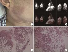
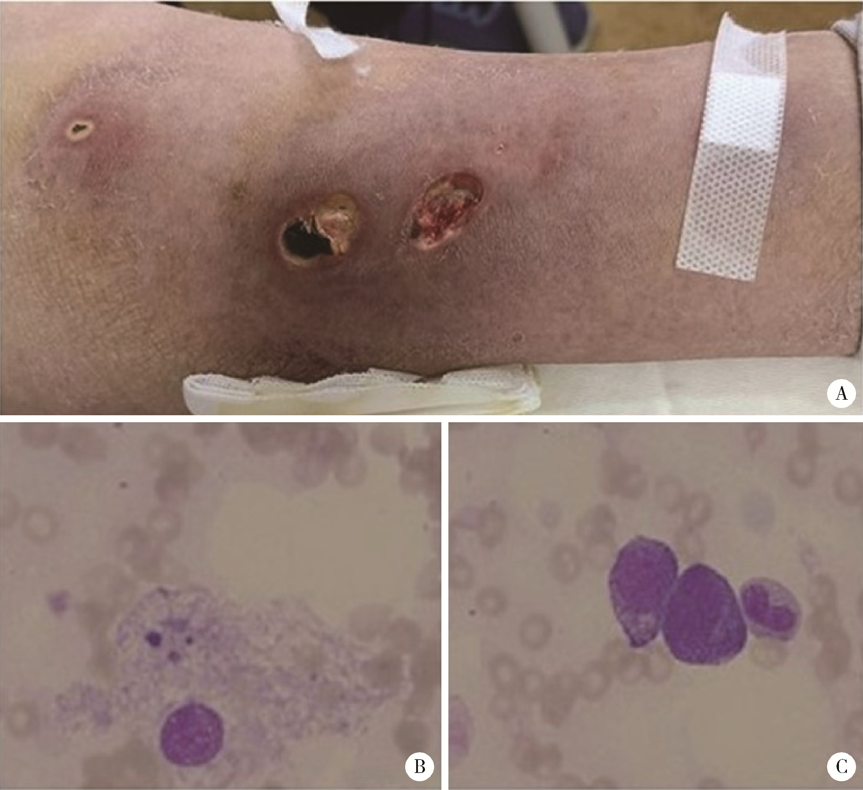
Ruxolitinib as an effective treatment for panniculitis associated hemophagocytic syndrome: A report of 2 cases and literature review
Gong-ming LI1,2,Yue-bo JIN2,Yu-zhou GAN2,Chen CHEN2,Yuan JIA2,Chun LI2,*( )
)
- 1. Department of Rheumatology, Linyi Traditional Chinese Medicine Hospital, Linyi 276003, Shandong, China
2. Department of Rheumatology and Immunology, Peking University People's Hospital, Beijing 100044, China
CLC Number:
- R55


| 1 |
中国医师协会血液科医师分会, 中华医学会儿科学分会血液学组, 噬血细胞综合征中国专家联盟. 中国噬血细胞综合征诊断与治疗指南(2022年版)[J]. 中华医学杂志, 2022, 102 (20): 1492- 1499.
doi: 10.3760/cma.j.cn112137-20220310-00488 |
| 2 |
Gupta P , Saikia U , Arora S , et al. Panniculitis: A dermatopathologist's perspective and approach to diagnosis[J]. Indian J Dermatopathol and Diagn Dermatol, 2016, 3, 29- 41.
doi: 10.4103/2349-6029.195224 |
| 3 |
Diaz Cascajo C , Borghi S , Weyers W . Panniculitis: Definition of terms and diagnostic strategy[J]. Am J Dermatopathol, 2000, 22 (6): 530- 549.
doi: 10.1097/00000372-200012000-00009 |
| 4 |
Aronson IK , Worobec SM . Cytophagic histiocytic panniculitis and hemophagocytic lymphohistiocytosis: An overview[J]. Dermatol Ther, 2010, 23 (4): 389- 402.
doi: 10.1111/j.1529-8019.2010.01339.x |
| 5 |
郭霞, 周晨燕, 李强. 结节性脂膜炎并发噬血细胞综合征1例[J]. 四川医学, 2005, 26 (12): 1372.
doi: 10.3969/j.issn.1004-0501.2005.12.098 |
| 6 |
Yi G , Huang Z , Huang Z , et al. Case report: Baricitinib as an alternative in the maintenance therapy for macrophage activation syndrome secondary to nodular panniculitis[J]. Front Immunol, 2022, 13, 914265.
doi: 10.3389/fimmu.2022.914265 |
| 7 |
Winkelmann RK , Bowie EJ . Hemorrhagic diathesis associated with benign histiocytic, cytophagic panniculitis and systemic histiocytosis[J]. Arch Intern Med, 1980, 140 (11): 1460- 1463.
doi: 10.1001/archinte.1980.00330220038015 |
| 8 |
Aronson IK , West DP , Variakojis D , et al. Fatal panniculitis[J]. J Am Acad Dermatol, 1985, 12 (3): 535- 551.
doi: 10.1016/S0190-9622(85)70076-X |
| 9 | Brisse E , Wouters CH , Matthys P . Hemophagocytic lymphohistiocytosis (HLH): A heterogeneous spectrum of cytokine-driven immune disorders[J]. Cytokine Growth Factor Rev, 2015, 26 (3): 263- 280. |
| 10 |
Das R , Guan P , Sprague L , et al. Janus kinase inhibition lessens inflammation and ameliorates disease in murine models of hemophagocytic lymphohistiocytosis[J]. Blood, 2016, 127 (13): 1666- 1675.
doi: 10.1182/blood-2015-12-684399 |
| 11 | 吴梦慧, 徐晓军. JAK1/2抑制剂芦可替尼治疗噬血细胞综合征的研究进展[J]. 医学研究杂志, 2021, 50 (11): 142- 146. |
| 12 |
中华医学会风湿病学分会. 结节性脂膜炎诊治指南(草案)[J]. 中华风湿病学杂志, 2004, 8 (4): 253- 255.
doi: 10.3760/j:issn:1007-7480.2004.04.017 |
| 13 |
Henter JI , Horne A , Aricó M , et al. HLH-2004: Diagnostic and therapeutic guidelines for hemophagocytic lymphohistiocytosis[J]. Pediatr Blood Cancer, 2007, 48 (2): 124- 131.
doi: 10.1002/pbc.21039 |
| 14 |
Kumakura S , Murakawa Y . Clinical characteristics and treatment outcomes of autoimmune-associated hemophagocytic syndrome in adults[J]. Arthritis Rheumatol, 2014, 66 (8): 2297- 2307.
doi: 10.1002/art.38672 |
| 15 |
Janka GE . Familial and acquired hemophagocytic lymphohistiocytosis[J]. Annu Rev Med, 2012, 63, 233- 246.
doi: 10.1146/annurev-med-041610-134208 |
| 16 |
Chandrakasan S , Filipovich AH . Hemophagocytic lymphohistiocytosis: Advances in pathophysiology, diagnosis, and treatment[J]. J Pediatr, 2013, 163 (5): 1253- 1259.
doi: 10.1016/j.jpeds.2013.06.053 |
| 17 | 雷玲, 田新平, 李春雨. 脂膜炎患者的临床特征及治疗随访分析[J]. 中华风湿病学杂志, 2009, 13 (1): 36- 38. |
| 18 |
Requena L , Sánchez Yus E . Panniculitis. Part Ⅱ. Mostly lobular panniculitis[J]. J Am Acad Dermatol, 2001, 45 (3): 325- 364.
doi: 10.1067/mjd.2001.114735 |
| 19 |
Gallardo F , Pujol RM . Subcutaneous panniculitic-like T-cell lymphoma and other primary cutaneous lymphomas with prominent subcutaneous tissue involvement[J]. Dermatol Clin, 2008, 26 (4): 529- 540.
doi: 10.1016/j.det.2008.05.008 |
| 20 |
李梦涛, 曾小峰, 张奉春, 等. 组织细胞吞噬性脂膜炎六例临床分析及文献复习[J]. 中华内科杂志, 2004, 43 (8): 576- 579.
doi: 10.3760/j.issn:0578-1426.2004.08.007 |
| 21 |
Berard CW , Cooper RA , Freireich EJ , et al. Disseminated histiocytosis associated with atypical lymphoid cells (lymphohistiocytosis)[J]. Cancer, 1966, 19 (10): 1429- 1437.
doi: 10.1002/1097-0142(196610)19:10<1429::AID-CNCR2820191016>3.0.CO;2-M |
| 22 |
Ambruso DR , Hays T , Zwartjes WJ , et al. Successful treatment of lymphohistiocytic reticulosis with phagocytosis with epipodophyllotoxin VP 16-213[J]. Cancer, 1980, 45 (10): 2516- 2520.
doi: 10.1002/1097-0142(19800515)45:10<2516::AID-CNCR2820451008>3.0.CO;2-V |
| 23 |
Chen RL , Hsu YH , Ueda I , et al. Cytophagic histiocytic panniculitis with fatal haemophagocytic lymphohistiocytosis in a paediatric patient with perforin gene mutation[J]. J Clin Pathol, 2007, 60 (10): 1168- 1169.
doi: 10.1136/jcp.2007.049551 |
| 24 |
Maschalidi S , Sepulveda FE , Garrigue A , et al. Therapeutic effect of JAK1/2 blockade on the manifestations of hemophagocytic lymphohistiocytosis in mice[J]. Blood, 2016, 128 (1): 60- 71.
doi: 10.1182/blood-2016-02-700013 |
| 25 |
Albeituni S , Verbist KC , Tedrick PE , et al. Mechanisms of action of ruxolitinib in murine models of hemophagocytic lymphohistiocytosis[J]. Blood, 2019, 134 (2): 147- 159.
doi: 10.1182/blood.2019000761 |
| 26 |
Wang H , Gu J , Liang X , et al. Low dose ruxolitinib plus HLH-94 protocol: A potential choice for secondary HLH[J]. Semin Hematol, 2020, 57 (1): 26- 30.
doi: 10.1053/j.seminhematol.2018.07.006 |
| 27 |
Wang J , Zhang R , Wu X , et al. Ruxolitinib-combined doxorubicin-etoposide-methylprednisolone regimen as a salvage therapy for refractory/relapsed haemophagocytic lymphohistiocytosis: A singlearm, multicentre, phase 2 trial[J]. Br J Haematol, 2021, 193 (4): 761- 768.
doi: 10.1111/bjh.17331 |
| 28 |
Zandvakili I , Conboy CB , Ayed AO , et al. Ruxolitinib as first-line treatment in secondary hemophagocytic lymphohistiocytosis: A second experience[J]. Am J Hematol, 2018, 93 (5): E123- E125.
doi: 10.1002/ajh.25063 |
| 29 | Gálvez Acosta S , Javalera Rincón M . Ruxolitinib as first-line therapy in secondary hemophagocytic lymphohistiocytosis and HIV infection[J]. Int J Hematol, 2020, 112 (3): 418- 421. |
| 30 | Bursi R , Cafaro G , Perricone C , et al. Contribution of Janus-kinase/signal transduction activator of transcription pathway in the pathogenesis of vasculitis: A possible treatment target in the upcoming future[J]. Front Pharmacol, 2021, 12, 635663. |
| 31 | Morgan AJ , Schwartz RA . Cutaneous polyarteritis nodosa: A comprehensive review[J]. Int J Dermatol, 2010, 49 (7): 750- 756. |
| [1] | Zhi-jun LUO,Jia-jia WU,You SONG,Chun-li MEI,Rong DU. Systemic lupus erythematosus associated macrophage activation syndrome with neuropsychiatric symptoms: A report of 2 cases [J]. Journal of Peking University (Health Sciences), 2023, 55(6): 1111-1117. |
| [2] | WANG Yu, HAO Yan-Jie, ZHAO Juan, ZHANG Zhuo-Li. Systemic lupus erythmatosus and panniculitis presenting as multiple ulcers: one case report [J]. Journal of Peking University(Health Sciences), 2015, 47(2): 352-354. |
|
||