Journal of Peking University (Health Sciences) ›› 2025, Vol. 57 ›› Issue (1): 73-77. doi: 10.19723/j.issn.1671-167X.2025.01.011
Previous Articles Next Articles
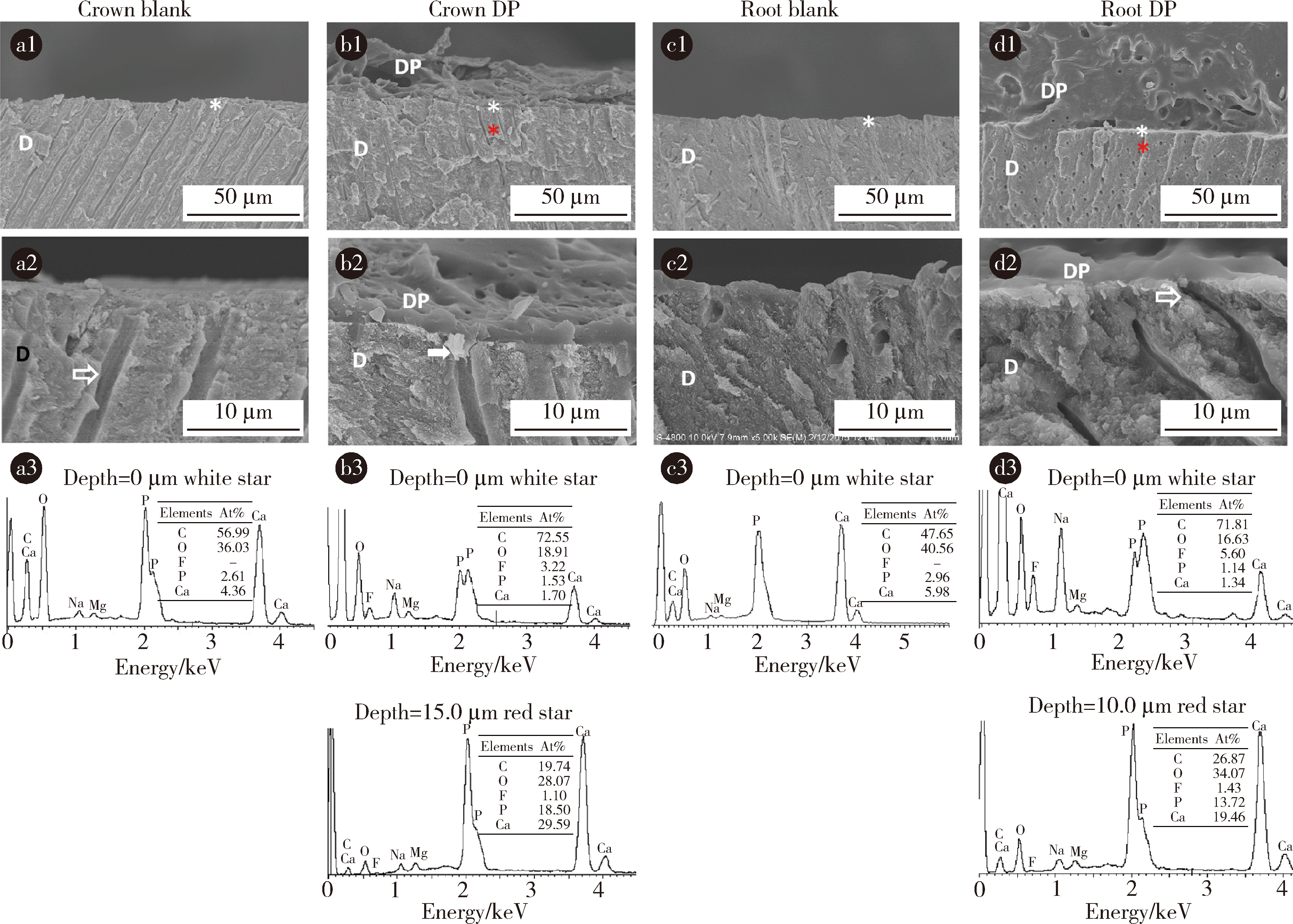
Durability of the anti-demineralization effects of fluoride varnish on dental root surfaces: An in vitro study
Hongyan TIAN1, Xue CAI2,*( ), Xiaoyan WANG2
- 1. First Clinical Division, Peking University School and Hospital of Stomatology & National Center for Stomatology & National Clinical Research Center for Oral Diseases & National Engineering Research Center of Oral Biomaterials and Digital Medical Devices, Beijing 100081, China
2. Department of Cariology and Endodontology, Peking University School and Hospital of Stomatology & National Center for Stomatology & National Clinical Research Center for Oral Diseases & National Engineering Research Center of Oral Biomaterials and Digital Medical Devices, Beijing 100081, China
CLC Number:
- R783.1


| 1 | 冯希平. 中国居民口腔健康状况——第四次中国口腔健康流行病学调查报告[C]//中华口腔医学会口腔预防医学专业委员会. 2018年中华口腔医学会第十八次口腔预防医学学术年会论文汇编. 西安: [出版者不详], 2018: 13-14. |
| 2 | 台保军. 中国居民口腔健康状况及防控策略第四次全国口腔健康流行病学调查结果解读[C]//中华口腔医学会老年口腔医学专业委员会. 第十三次全国老年口腔医学学术年会论文汇编. 武汉: [出版者不详], 2018: 16-17. |
| 3 |
Cai J , Palamara J , Manton DJ , et al. Status and progress of treatment methods for root caries in the last decade: A literature review[J]. Aust Dent J, 2018, 63 (1): 34- 54.
doi: 10.1111/adj.12550 |
| 4 | 管兆兰, 钱雅婧, 王威. 高频率使用多乐氟预防正畸患者牙脱矿的效果研究[J]. 口腔医学, 2023, 43 (2): 141- 144. |
| 5 | 闻静, 周春华, 许妍. 光学相干断层成像检测牙釉质表层脱矿与再矿化的实验研究[J]. 北京口腔医学, 2019, 27 (3): 143- 146. |
| 6 | 杨茜, 付梦辰, 王慧慧, 等. Duraphat与Fluor Protector抑制固定正畸矫治中牙釉质脱矿的比较研究[J]. 口腔医学, 2019, 39 (11): 1022- 1026. |
| 7 | Jiang W , Wang G , Wu W , et al. The effect of calcium phosphate ion clusters in enhancing enamel conditions versus Duraphat and Icon[J]. Aust Endod J, 2023, 49 (Suppl 1): 46- 57. |
| 8 | Casimiro-Iriarte SA , Chiok-Ocana LR . Fluoride release from fluo-ride varnishes exposed to commonly consumed beverages: An in vitro study[J]. J Clin Exp Dent, 2023, 15 (3): e187- e194. |
| 9 | 舒泉湧, 麻纪斌, 邢建峰, 等. 多乐氟中氟含量及体外释放度研究[J]. 合成材料老化与应用, 2017, 46 (2): 79- 81. |
| 10 |
Hosoya Y , Ando S , Yamaguchi K , et al. Quality of the interface of primary tooth dentin bonded with antibacterial fluoride-releasing adhesive[J]. J Dent, 2010, 38 (5): 423- 430.
doi: 10.1016/j.jdent.2010.02.001 |
| 11 |
Ma S , Imazato S , Chen J , et al. Effects of a coating resin containing S-PRG filler to prevent demineralization of root surfaces[J]. Dent Mater J, 2012, 31 (6): 909- 915.
doi: 10.4012/dmj.2012-061 |
| 12 | Carey CM . Focus on fluorides: Update on the use of fluoride for the prevention of dental caries[J]. J Evid Based Dent Pract, 2014, 14 (Suppl): 95- 102. |
| 13 |
Joves GJ , Inoue G , Nakashima S , et al. Mineral density, morphology and bond strength of natural versus artificial caries-affected dentin[J]. Dent Mater J, 2013, 32 (1): 138- 143.
doi: 10.4012/dmj.2012-243 |
| 14 |
Arends J , Duschner H , Ruben JL . Penetration of varnishes into demineralized root dentine in vitro[J]. Caries Res, 1997, 31 (3): 201- 205.
doi: 10.1159/000262399 |
| 15 | Asian J , Quenta E , Castillo J . Do viscosity and wettability of fluoride varnishes affect their fluoride release?[J]. J Clin Exp Dent, 2021, 13 (3): e221- e226. |
| 16 |
Cochrane NJ , Shen P , Yuan Y , et al. Ion release from calcium and fluoride containing dental varnishes[J]. Aust Dent J, 2014, 59 (1): 100- 105.
doi: 10.1111/adj.12144 |
| 17 |
Ekstrand K , Martignon S , Holm-Pedersen P . Development and evaluation of two root caries controlling programmes for home-based frail people older than 75 years[J]. Gerodontology, 2008, 25 (2): 67- 75.
doi: 10.1111/j.1741-2358.2007.00200.x |
| [1] | TIAN Hong-yan, YU Peng, YUAN Chong-yang, ZHANG Wei, QIU Yue-xiu, LI De-hui, LIANG Xin-jie, WANG Xiao-yan. Durability of protective effect of resin-based coating material on root surface [J]. Journal of Peking University(Health Sciences), 2016, 48(5): 889-893. |
|
||