
1 资料与方法
1.1 研究对象
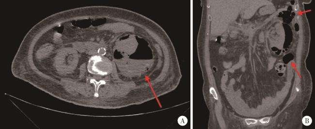
图1 1例左肾EPN患者的CT平扫结果,分级ⅢB级Figure 1 Non-contrast CT images of a patient with left renal EPN, classified as grade ⅢB A, axial CT image demonstrated extensive perirenal gas and fluid accumulation with an air-fluid level (red arrow); B, coronal CT image revealed signi-ficant retroperitoneal gas (red arrow). EPN, emphysematous pyelonephritis. |
1.2 治疗方法
1.3 统计学分析
2 结果
表1 患者就诊时主要实验室检查结果(n=13)Table 1 Key laboratory results of patients at admission(n=13) |
| Parameters | Value |
| White blood cell/(×109/L),${\bar x}$±s | 13.82 ± 5.92 |
| Neutrophil/(×109/L),${\bar x}$±s | 11.68 ± 5.09 |
| Platelet/(×109/L),${\bar x}$±s | 326.25 ± 177.69 |
| Hemoglobin/(g/L),${\bar x}$±s | 92.67 ± 23.42 |
| C-reactive protein/(mg/L),${\bar x}$±s | 186.74 ± 101.92 |
| Procalcitonin/(μg/L), M(P25,P75) | 3.97 (0.19, 11.46) |
| Serum creatinine/(mmol/L),${\bar x}$±s | 146.75 ± 95.55 |
| Blood glucose/(mmol/L),M(P25,P75) | 8.20 (6.08, 15.93) |
表2 患者致病菌分布(n=13)Table 2 Pathogens distribution in patients(n=13) |
| Pathogens | Cases, n (%) |
| Escherichia coli | 5 (38.5) |
| Klebsiella pneumoniae | 5 (38.5) |
| Culture-negative | 2 (15.3) |
| No culture performed | 1 (7.7) |
表3 患者各种手术方式统计(n=12)Table 3 Surgical treatment statistics for surgical patients(n=12) |
| Surgical procedure | Cases, n (%) |
| Minimally invasive surgery | 7 (58.3) |
| PCN | 2 (28.7) |
| DJ stent placement | 2 (28.7) |
| PCD | 1 (14.2) |
| Initial DJ stent placement → PCN | 1 (14.2) |
| PCN+PCD | 1 (14.2) |
| Open drainage | 5 (41.7) |
| Perirenal VSD | 3 (60.0) |
| Perirenal VSD + PCN | 1 (20.0) |
| Perirenal VSD + DJ stent placement | 1 (20.0) |
PCN, percutaneous nephrostomy; DJ, double J; PCD, percu-taneous drainage; VSD, vacuum sealing drainage. |
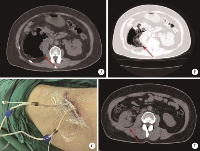
图2 1例右肾EPN患者的术前、术后及出院前的临床资料,分级ⅢA级Figure 2 Clinical data of a patient with right renal EPN at preoperative, postoperative, and pre-discharge stages, classified as grade ⅢA A, preoperative abdominal window CT image showed extensive perirenal gas accumulation and complete disappearance of the kidney (red arrow); B, preoperative lung window CT image demonstrated the "vanished kidney" (red arrow) and massive intraparenchymal gas within the renal tissue; C, postoperative incision photo following VSD surgery; D, abdominal CT image prior to discharge revealed near-normal restoration of renal morphology, with no abscesses or gas in the renal parenchyma or perirenal region (red arrow). VSD, vacuum sealing drainage; EPN, emphysematous pyelonephritis. |