HDAC2-mediated H3K27 acetylation promotes the proliferation and migration of hepatocellular carcinoma cells

  • Shaohai TANG 1 ,
  • Baoming YANG 1 ,
  • Jiankun LI 1 ,
  • Lili ZHAO 2 ,
  • Yifan WANG 3 ,
  • Shunxiang WANG , 1, *
Expand
  • 1. Department of Hepatobiliary Surgery, the Fourth Hospital of Hebei Medical University, Shijiazhuang 050000, China
  • 2. College of Pharmacy, Hebei Medical University, Shijiazhuang 050000, China;
  • 3. Department of Colorectal Surgery, the Fourth Hospital of Hebei Medical University, Shijiazhuang 050000, China
WANG Shunxiang, e-mail,

Received date: 2024-06-28

  Online published: 2025-06-18

Supported by

the Natural Science Foundation Project of Hebei Province(H2020206455)

the 2023 Annual Medical Science Research Project Plan of Hebei Province(20230875)

the Key Medical Science Research Project Plan of Hebei Province in 2018(20180537)

the 2015 and 2016 Hebei Provincial Government' s Plan for Funding the Cultivation of Outstanding Clinical Medicine Talents and Basic Research Projects(361006)

Copyright

All rights reserved. Unauthorized reproduction is prohibited.

Abstract

Objective: To explore the specific mechanism of histone deacetylase 2 (HDAC2) mediated histone H3 lysine 27 acetylation (H3K27ac) modification in promoting the proliferation and migration of hepatocellular carcinoma cells. Methods: Samples of 40 cases of hepatocellular carcinoma and paracancerous tissues resected from January 2021 to January 2023 were collected. The expressions of HDAC2 and H3K27ac in hepatocellular carcinoma, paracancerous tissues and cell lines were detected by immunohistochemistry and Western blotting. The correlation between the expression levels of HDAC2 and H3K27ac and the relationship between HDAC2 expression and clinicopathological characteristics of patients with hepatocellular carcinoma were analyzed. The proliferation, migration and invasion of Hep3B and HepG2 cells were determined by MTS, clone formation, scratch and Transwell experiments. The acetylation of H3K27 mediated by HDAC2 was verified by Western blotting, real-time fluorescence quantitative PCR (qRT-PCR) and chromatin immunoprecipitation high-throughput sequencing (ChIP-seq). In vivo xenotransplantation experiment, the tumorigenicity of cells in each group was measured, and the expression of proteins related to phosphoinositide 3-kinases/phosphatase and tensin homolog deleted on chromosome ten/protein kinase B/mammalian target of rapamycin (PI3K/PTEN/AKT/mTOR) signal pathway was detected. Results: High expression of HDAC2 and low expression of H3K27ac were found in hepatocellular carcinoma tissues and cell lines (P < 0.05), and there was a negative correlation between them (r=-0.477, P=0.002). The expression of HDAC2 was related to tumor size, hepatitis B virus infection, TNM stage and portal vein tumor thrombus (P < 0.05). Compared with the sh-NC group of Hep3B and HepG2 cells, the proliferation, clone formation, migration and invasion ability of sh-HDAC2 group were decreased (P < 0.05). Compared with the Empty group, the HDAC2 group exhibited increased expression levels and activity of HDAC2, as well as enhanced cell proliferation, clone formation, migration, invasion ability, tumor volume and mass in vivo, and elevated expression levels of p-PI3K, p-AKT, and p-mTOR (P < 0.05). Conversely, the enrichment and expression levels of H3K27ac, along with the expression level of PTEN, were decreased (P < 0.05). In the iHDAC2 group, the expression levels and activity of HDAC2, as well as the proliferation, clone formation, migration, invasion ability, tumor volume and mass in vivo, and expression levels of p-PI3K, p-AKT, and p-mTOR were reduced (P < 0.05). Additionally, the expression levels of H3K27ac and PTEN were increased (P < 0.05). To validate the involvement of the PI3K/PTEN/AKT/mTOR signaling pathway in HDAC2-mediated regulation of malignant behaviors in liver cancer cells through H3K27ac, the PI3K activator 740Y-P was introduced. Compared with the iHDAC2 group, the iHDAC2+740Y-P group exhibited increased proliferation, clone formation, migration, invasion ability, tumor volume and mass in vivo, and elevated expression levels of p-PI3K, p-AKT, and p-mTOR (P < 0.05). Conversely, the expression level of PTEN was decreased (P < 0.05). Conclusion: HDAC2 initiates PI3K/PTEN/AKT/mTOR signal pathway by mediating H3K27 acetylation, which promotes the occurrence and development of hepatocellular carcinoma.

Cite this article

Shaohai TANG , Baoming YANG , Jiankun LI , Lili ZHAO , Yifan WANG , Shunxiang WANG . HDAC2-mediated H3K27 acetylation promotes the proliferation and migration of hepatocellular carcinoma cells[J]. Journal of Peking University(Health Sciences), 2025 , 57(5) : 884 -894 . DOI: 10.19723/j.issn.1671-167X.2025.05.012

肝癌发病率在全球范围内上升,其相关死亡率约占全球癌症相关死亡的8%[1-3]。肝细胞癌约占肝癌病例的90%,病毒性肝炎、酒精性肝炎、非酒精性脂肪性肝炎、吸烟、糖尿病等为其常见病因[4-5]。尽管如今已研发出索拉非尼、仑伐替尼等多种肝癌的靶向治疗药物,但肝癌患者的总生存期并未出现明显变化[6-7],因此,深入了解促进肝癌发生的分子机制有利于研发更加高效的肝癌治疗药物。表观遗传机制是肝脏协调实体器官、控制其再生以及适应环境变化的能力的基础[8]。在真核细胞中,每个核小体由4对组蛋白(histone,H)H2A、H2B、H3、H4与包裹着组蛋白的一段DNA组成,其中组蛋白H3第27位赖氨酸乙酰化(histone H3 lysine 27 acetylation,H3K27ac)是影响染色质结构和基因表达的最常见的表观遗传修饰之一,通常位于转录起始点,与活性增强子相互作用促进基因表达[9-10]。组蛋白去乙酰化酶(histone deacetylases,HDACs)介导组蛋白去乙酰化,且具有修饰部分转录因子、信号转导介质等不同类型的非组蛋白活性的功能,在多种癌症中表达异常[11-12]。磷脂酰肌醇-3激酶/第10号染色体磷酸酶和张力蛋白同源缺失基因/蛋白激酶B/哺乳动物雷帕霉素靶蛋白(phosphoinositide 3-kinases/phosphatase and tensin homolog deleted on chromosome ten/protein kinase B/mammalian target of rapamycin,PI3K/PTEN/AKT/mTOR)信号通路是许多癌症组织中异常激活最频繁的通路之一,近期研究显示,同时抑制HDACs与PI3K/PTEN/AKT/mTOR信号通路的多靶点抑制剂抗肿瘤效果较单一靶点抑制剂明显提升,但其分子层面的具体作用机制尚未明确[13-15]。本研究通过体内外实验观察HDAC2对肝癌细胞增殖、迁移的影响,验证HDAC2介导H3K27ac与HDAC2影响肝癌细胞增殖、迁移的相关分子机制。

1 材料与方法

1.1 组织样本

收集2021年1月至2023年1月河北医科大学第四医院经手术切除的40例肝癌样本与对应癌旁正常肝组织,术后接受组织病理学诊断鉴定[16]。患者年龄(59.02±10.61)岁(38~73岁),其中男性21例,女性19例。本研究开始前已经河北医科大学第四医院伦理委员会审查批准(HBDS-2020-1126),所有患者均书面知情同意自愿提供组织样本。

1.2 实验动物

48只SPF级4周龄健康雄性BALB/c裸鼠,体重(30±5) g,购自河北医科大学实验动物公共服务平台[动物合格证号SCXK(冀)2020-001],适应性饲养1周后开展实验,实验所有操作符合3R原则。

1.3 实验细胞

人肝癌细胞Hep3B(货号BH-X5108)、SMMC7721(货号BH-X0774)、Huh7(货号BH-X5107)、HepG2(货号P-X753)、MHCC-97H(货号P-X853)和人正常肝细胞(L02细胞,货号BH-X5090) 均购自上海博湖生物科技有限公司。

1.4 主要试剂

DMEM培养基(货号11965092)、胎牛血清(货号16140063)、链霉素/青霉素(货号15070063)、NE-PER细胞核和细胞质提取试剂(货号78835)均购自美国Thermo Fisher公司,PI3K激活剂740Y-P(货号HAS-S7865)购自深圳海思安生物技术有限公司,慢病毒表达载体的构建由昆山远慕生物科技有限公司完成,ChIP试剂盒(货号BYEK-P-2002-1)购自南京北鱼生物科技有限公司,高通量测序由上海普阳生物科技有限公司完成,MTS试剂盒(货号15711)购自西安百萤生物科技有限公司,Matrigel基质胶(货号354234)购自上海研卉生物科技有限公司;HDAC2活性检测试剂盒(货号GMS50082.3.2)购自深圳子科生物科技有限公司,兔H3K27ac(货号ab4729)、兔HDAC1(货号ab280198)、兔HDAC2(货号ab32117)、兔HDAC3(货号ab32369)、兔HDAC8(货号ab187139)、兔p-Rac1(货号ab247485) 均购自英国Abcam公司,兔p-AKT(货号4060)、兔p-NF-κB(货号8242)均购自美国CST公司;其他试剂均为市售分析纯。

1.5 实验方法

1.5.1 细胞培养、分组与转染

复苏Hep3B、SMMC7721、Huh7、HepG2、MHCC-97H和L02细胞,使用含10%(体积分数)胎牛血清与100 g/L链霉素/青霉素的DMEM培养基,于37 ℃、5% CO2、饱和湿度的电热恒温培养箱中进行培养。Hep3B、HepG2细胞分为sh-NC组(转染sh-NC)、sh-HDAC2组(转染sh-HDAC2)、空质粒组(转染dCas9-空质粒)、HDAC2组(转染dCas9-HDAC2)、iHDAC2组(转染dCas9-失活HDAC2)、iHDAC2+740Y-P组(转染dCas9-失活HDAC2,培养基加入30 mmol/L 740Y-P),按照分组名称进行慢病毒转染、加药培养,构建稳定表达的细胞株。

1.5.2 免疫组化检测

取部分患者肝癌、癌旁组织制成石蜡切片,经脱蜡、梯度酒精水化、抗原修复、封闭,加入稀释度(体积比)为1 ∶ 100的HDAC2、H3K27ac抗体,4 ℃环境下孵育过夜,洗涤后加入生物素标记的二抗,孵育30 min,加入链霉亲和素酶,孵育30 min,经显色、复染、脱水、透明后用中性树胶封片,于光学显微镜下拍摄实验结果,每张切片随机选择5个不同视野,用ImageJ软件进行图像分析,取平均值作为各抗体相对表达量。

1.5.3 Western blotting检测

收集各组细胞,加入适量RIPA裂解液充分裂解后4 ℃、10 000 r/min离心10 min,收集上清液定量并制样。经凝胶电泳、转膜,剪取目的条带,加入封闭液室温封闭1 h。加入1 ∶ 500稀释度(体积比)的一抗,4 ℃过夜。洗膜,加入1 ∶ 5 000稀释(体积比)的对应二抗,室温孵育2 h,洗膜,加入增强化学发光液,避光反应5 min,收集荧光结果。

1.5.4 MTS检测

各组细胞以2 000个/孔的密度接种于96孔板上,于培养24、48、72 h进行MTS检测,每孔加入40 μL MTS试剂,避光培养4 h,于490 nm波长下检测光密度。

1.5.5 克隆形成实验

各组细胞接种于6孔板中,接种密度2 000个/孔,正常培养10 d,弃去培养基,甲醇固定30 min,1%(质量分数)结晶紫染色20 min,统计克隆数量。

1.5.6 划痕实验

各组细胞接种于6孔板中,接种密度1×10⁶个/孔,观察细胞覆盖率达到90%,用200 μL移液器枪头作划痕,清去脱落的细胞,加入无血清培养基培养24 h。于划痕后即刻(0 h)和培养24 h后拍照,测量划痕宽度,计算细胞迁移率。

1.5.7 Transwell实验

无血清DMEM/F12培养基与Matrigel基质胶按1 ∶ 3(体积比)的比例稀释,混匀后均匀涂抹于小室上室膜底部,放入培养箱中过夜,次日置于紫外线灯下照射30 min。各组细胞制成细胞悬液,接种于Transwell小室上室,下室中加入含10%(体积分数)胎牛血清的DMEM培养基,正常培养24 h。4%(体积分数)多聚甲醛固定下室细胞30 min,结晶紫染色,于光学显微镜下观察,计算细胞侵袭率=各组侵袭细胞数/对照组侵袭细胞数×100%。

1.5.8 实时荧光定量PCR(real-time fluorescence quantitative PCR,qRT-PCR)检测

使用TRIzol试剂提取空质粒组、HDAC2组、iHDAC2组Hep3B和HepG2细胞总RNA,经反转录获得cDNA。使用1 μg cDNA模板、上下游引物各0.5 μg和SYBR Premix Ex Taq Ⅱ试剂盒配制20 μL PCR反应体系,引物序列见表 1。应用2-ΔΔCt法计算HDAC2相对表达水平。
表1 引物序列

Table 1 Primer sequence

Gene Primer sequences (5′-3′)
HDAC2 Forward:ATGGCGTACAGTCAAGGAGGC
Reverse:AAATCAGAACAGCTCAGCAAC
GAPDH Forward:ATGGGGAAGGTGAAGGTCGG
Reverse:TTACTCCTTGGAGGCCATGT

HDAC, histone deacetylase; GAPDH, glyceraldehyde-3-phosphatedehydrogenase.

1.5.9 HDAC2活性检测

使用NE-PER细胞核和细胞质提取试剂提取空质粒组、HDAC2组、iHDAC2组Hep3B、HepG2细胞核提取物,测定蛋白浓度后使用HDAC2活性检测试剂盒进行检测,于405 nm波长下检测光密度。

1.5.10 染色质免疫共沉淀-高通量测序(chromatin immunoprecipitation high-throughput sequencing,ChIP-seq)

空质粒组、HDAC2组、iHDAC2组Hep3B、HepG2细胞加入1%(体积分数)甲醛固定10 min,加入0.125 mol/min甘氨酸终止反应。加入含蛋白酶抑制剂的裂解液,冰上孵育10 min,刮取细胞,4 ℃、2 500 r/min离心5 min。加入细胞核裂解液,冰上孵育10 min,使用Covaris S220超声波DNA破碎仪进行染色质片段化处理。加入H3K27ac抗体,置于4 ℃环境下过夜。加入蛋白A免疫沉淀磁珠,4 ℃孵育4 h,收集磁珠沉淀,经低盐缓冲液、高盐缓冲液、氯化锂缓冲液、TE缓冲液清洗后,加入ChIP洗脱缓冲液洗脱磁珠染色质,经纯化、定量后进行高通量测序。

1.5.11 体内异种移植实验

48只裸鼠根据接种不同肿瘤细胞平均分为两部分,每部分24只,根据接种细胞不同分为空质粒组、HDAC2组、iHDAC2组、iHDAC2+740Y-P组,每组6只。于裸鼠左侧腹部皮下接种各组细胞,细胞悬液密度为2×10⁶个/mL。接种当日记为0 d,每3天测量一次肿瘤体积,计算公式:体积=1/2(长×宽2)。21 d后完整取出肿瘤,称重记录。

1.6 统计学分析

使用SPSS 24.0软件,所有结果独立重复实验3次。计量资料经Shapiro-Wilk检验符合正态分布,以$\bar x \pm s$表示,组间比较采用t检验,多组间比较采用单因素方差分析,结合Tukey事后检验确定具体组别间的差异,计数资料以n(%)表示,组间比较采用卡方检验。采用Pearson相关系数分析HDAC2与H3K27ac表达水平的相关性,P<0.05认为差异有统计学意义。

2 结果

2.1 HDAC2、H3K27ac在肝癌中的表达情况与HDAC2表达和肝癌临床病理特征的关系
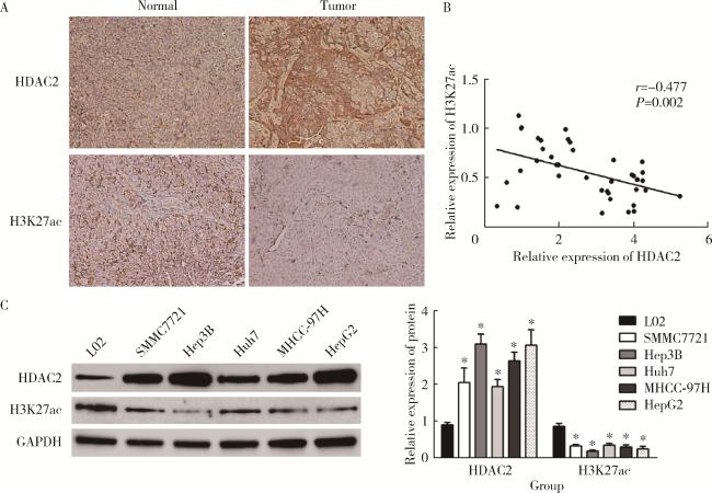
免疫组化检测结果显示,HDAC2在肝癌组织中高表达,H3K27ac呈低表达;相关性分析结果显示,HDAC2与H3K27ac表达水平呈负相关(r=-0.477,P=0.002)。以HDAC2免疫组化检测相对表达水平的中位数2.94将肝癌患者分为低表达组与高表达组,年龄、性别、甲胎蛋白、组织学分级、肝硬化与肝癌组织HDAC2表达水平无相关性(P>0.05),肿瘤大小、感染乙肝病毒、TNM分期、门静脉癌栓与肝癌组织HDAC2表达水平存在相关性(P<0.05)。Western blotting检测结果显示,与正常肝细胞L02相比,Hep3B、SMMC7721、Huh7、MHCC-97H、HepG2细胞HDAC2表达水平均升高(P<0.05),H3K27ac表达水平均下降(P<0.05),其中Hep3B与HepG2细胞HDAC2相对表达水平最高,H3K27ac相对表达水平最低(图 1表 2),因此选择Hep3B、HepG2细胞进行后续实验。
图1 HDAC2、H3K27ac在肝癌中的表达情况

Figure 1 Expression of HDAC2 and H3K27ac in hepatocellular carcinoma

A, immunohistochemical detection of HDAC2 and H3K27ac expression in hepatocellular carcinoma; B, correlation analysis of HDAC2 and H3K27ac expression in hepatocellular carcinoma; C, detection of HDAC2 and H3K27ac expression in hepatocellular carcinoma cell line by Western blotting. HDAC, histone deacetylase; H3K27ac, histone H3 lysine 27 acetylation; GAPDH, glyceraldehyde-3-phosphatedehydrogenase. *P < 0.05, vs. L02 cell.

表2 HDAC2表达水平与肝癌患者临床病理特征间的关系

Table 2 Relationship between HDAC2 expression level and clinicopathological features of patients with hepatocellular carcinoma

Items Total HDAC2 expression χ2 P
High (n=20) Low (n=20)
Age/years
  ≥59 25 (62.50) 14 (35.00) 11 (27.50) 0.960 0.327
  <59 15 (37.50) 6 (15.00) 9 (22.50)
Gender
  Male 21 (52.50) 13 (32.50) 8 (20.00) 2.506 0.113
  Female 19 (47.50) 7 (17.50) 12 (30.00)
Tumor size/cm
  ≥3 28 (70.00) 17 (42.50) 11 (27.50) 4.286 0.038
  <3 12 (30.00) 3 (7.50) 9 (22.50)
HBV infection
  Positive 30 (75.00) 19 (47.50) 11 (27.50) 8.533 0.003
  Negative 10 (25.00) 1 (2.50) 9 (22.50)
Alpha-fetoprotein/(μg/L)
  ≥400 27 (67.50) 12 (30.00) 15 (37.50) 1.026 0.311
  <400 13 (32.50) 8 (20.00) 5 (12.50)
Histologic grade
  Well and moderate 25 (62.50) 10 (25.00) 15 (37.50) 2.667 0.102
  Low 15 (37.50) 10 (25.00) 5 (12.50)
TNM stage
  Ⅰ to Ⅱ 19 (47.50) 6 (15.00) 13 (32.50) 4.912 0.027
  Ⅲ to Ⅳ 21 (52.50) 14 (35.00) 7 (17.50)
Portal vein tumor thrombus
  Yes 11 (27.50) 9 (22.50) 2 (5.00) 6.144 0.013
  No 29 (72.50) 11 (27.50) 18 (45.00)
Cirrhosis
  Yes 15 (37.50) 6 (15.00) 9 (22.50) 0.960 0.327
  No 25 (62.50) 14 (35.00) 11 (27.50)

Data are n(%). HDAC, histone deacetylase; HBV, hepatitis B virus.

2.2 抑制HDAC2对肝癌细胞增殖、迁移和侵袭的影响
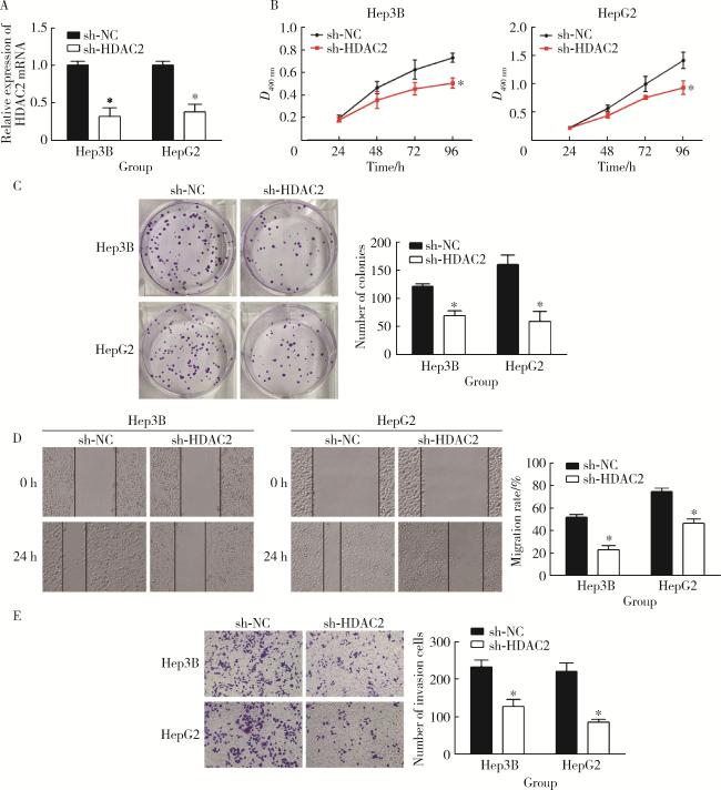
qRT-PCR检测结果显示,与Hep3B、HepG2细胞sh-NC组相比,sh-HDAC2组HDAC2 mRNA表达水平降低(P<0.05),提示转染成功。MTS、克隆形成、划痕、Transwell实验发现,与Hep3B、HepG2细胞sh-NC组相比,sh-HDAC2组增殖、克隆形成、迁移、侵袭能力降低(P<0.05),表明抑制HDAC2表达减弱了肝癌细胞Hep3B、HepG2的恶性生物学行为(图 2)。
图2 抑制HDAC2对肝癌细胞增殖、迁移和侵袭的影响

Figure 2 Effects of inhibition of HDAC2 on proliferation, migration and invasion of hepatocellular carcinoma cells

A, qRT-PCR assay results; B, MTS assay result; C, clone formation experiment results; D, wound scratch assay results; E, transwell assay results. HDAC, histone deacetylase; GAPDH, glyceraldehyde-3-phosphatedehydrogenase. *P < 0.05, vs. sh-NC group.

2.3 HDAC2介导H3K27ac修饰
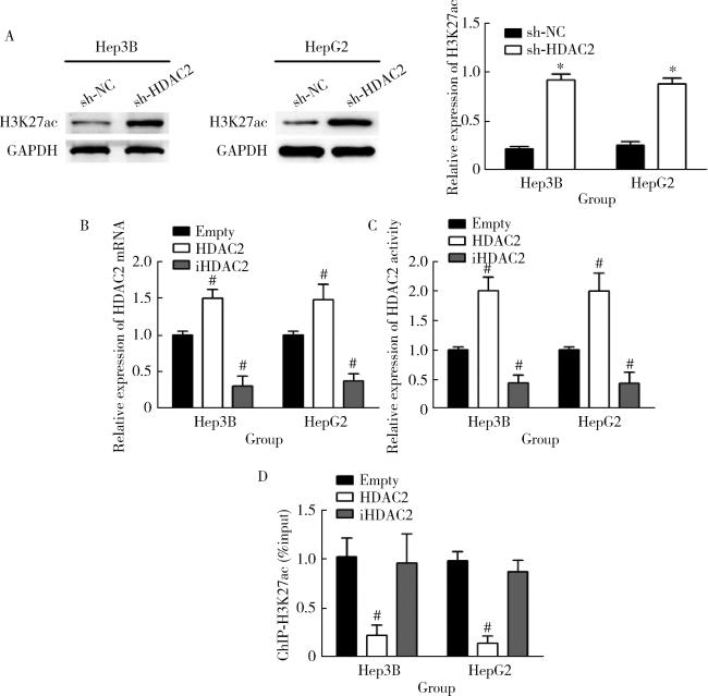
为探究HDAC2是否参与调控H3K27的乙酰化修饰过程,进一步比较Hep3B、HepG2细胞sh-HDAC2组(HDAC2特异性敲低)与sh-NC组(阴性对照组)的H3K27ac表达水平,Western blotting检测发现,与Hep3B、HepG2细胞sh-NC组相比,sh-HDAC2组H3K27ac表达水平提升(P<0.05),提示HDAC2可能具有抑制H3K27ac的作用。通过qRT-PCR、HDAC2活性检测验证HDAC2在细胞中的表达水平和活性变化发现,与Hep3B、HepG2细胞空质粒组(转染dCas9空质粒)相比,HDAC2组(转染dCas9-HDAC2)的HDAC2表达水平、活性升高(P<0.05),iHDAC2组(转染dCas9-失活HDAC2)的HDAC2表达水平、活性降低(P<0.05)。ChIP-seq检测结果显示,与Hep3B、HepG2细胞空质粒组相比,HDAC2组H3K27ac富集程度明显降低(P<0.05),提示HDAC2可能对H3K27ac具有抑制作用(图 3)。
图3 HDAC2介导H3K27ac修饰

Figure 3 Acetylation of H3K27 mediated by HDAC2

A, Western blotting assay result; B, qRT-PCR assay results; C, result of HDAC2 activity detection; D, ChIP-seq assay results. HDAC, histone deacetylase; H3K27ac, histone H3 lysine 27 acetylation; GAPDH, glyceraldehyde-3-phosphatedehydrogenase. *P < 0.05, vs. sh-NC group. # P < 0.05, vs. empty group.

2.4 HDAC2介导H3K27ac修饰启动PI3K/PTEN/AKT/mTOR信号通路促进肝癌细胞增殖、迁移、侵袭

MTS、克隆形成、划痕、Transwell实验发现,与Hep3B、HepG2细胞空质粒组相比,HDAC2组增殖、克隆形成、迁移、侵袭能力升高(P<0.05),iHDAC2组增殖、克隆形成、迁移、侵袭能力降低(P<0.05)。引入PI3K激活剂740Y-P验证PI3K/PTEN/AKT/mTOR信号通路是否参与HDAC2介导H3K27ac修饰的肝癌细胞恶性行为调控,与iHDAC2组相比,iHDAC2+740Y-P组增殖、克隆形成、迁移、侵袭能力升高(P<0.05)。进一步验证HDAC2对肝癌细胞体内生长的影响,体内异种移植实验结果显示,与Hep3B、HepG2细胞空质粒组相比,HDAC2组肿瘤生长速度提升,体积、质量增大(P<0.05),iHDAC2组肿瘤生长速度下降,体积、质量减小(P<0.05);与iHDAC2组相比,iHDAC2+740Y-P组肿瘤生长速度提升,体积、质量增大(P<0.05)。Western blotting检测发现,与Hep3B、HepG2细胞空质粒组相比,HDAC2组H3K27ac、PTEN表达水平降低(P<0.05),p-PI3K、p-AKT、p-mTOR表达水平升高(P<0.05),iHDAC2组H3K27ac、PTEN表达水平升高(P<0.05),p-PI3K、p-AKT、p-mTOR表达水平降低(P<0.05);与iHDAC2组相比,iHDAC2+740Y-P组PTEN表达水平降低(P<0.05),p-PI3K、p-AKT、p-mTOR表达水平升高(P<0.05,图 45)。
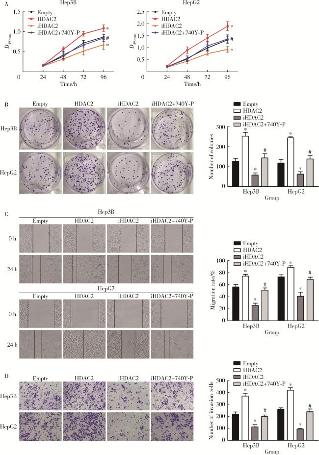
图4 体外细胞实验验证HDAC2介导H3K27ac修饰促进肝癌细胞增殖、迁移和侵袭

Figure 4 In vitro cell experiments show that HDAC2-mediated H3K27 acetylation promotes the proliferation, migration and invasion of hepatocellular carcinoma cells

A, MTS assay result; B, clone formation experiment results; C, wound scratch assay results; D, transwell assay results. HDAC, histone deacetylase; H3K27ac, histone H3 lysine 27 acetylation; GAPDH, glyceraldehyde-3-phosphatedehydrogenase. *P < 0.05, vs. empty group. #P < 0.05, vs. iHDAC2 group.

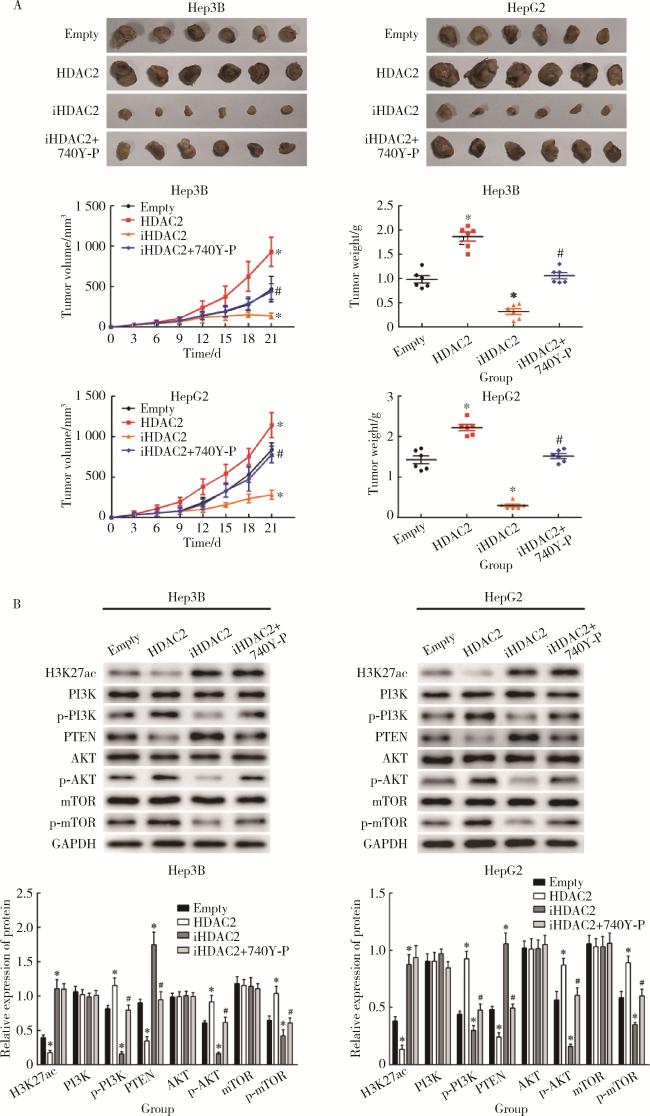
图5 体内细胞实验验证HDAC2介导H3K27ac修饰启动PI3K/PTEN/AKT/mTOR信号通路增强肝癌细胞成瘤能力

Figure 5 In vivo cell experiments show that HDAC2-mediated H3K27 acetylation initiates PI3K/PTEN/AKT/mTOR signal pathway to enhance the tumorigenicity of hepatocellular carcinoma cells

A, experimental results of xenotransplantation in vivo; B, Western blotting assay results of transplanted tumor. HDAC, histone deacetylase; GAPDH, glyceraldehyde-3-phosphatedehydrogenase. H3K27ac, histone H3 lysine 27 acetylation; PI3K/PTEN/AKT/mTOR, phosphoinositide 3-kinases/phosphatase and tensin homolog deleted on chromosome ten/protein kinase B/mammalian target of rapamycin. *P < 0.05, vs. empty group. #P < 0.05, vs. iHDAC2 group.

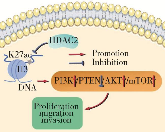
2.5 HDAC2介导H3K27ac修饰促进肝癌进展的作用机制

HDAC2在肝癌组织与细胞系中表达升高,其表达水平与肿瘤大小、感染乙肝病毒、TNM分期、门静脉癌栓相关。抑制HDAC2表达能够减弱Hep3B、HepG2细胞增殖、迁移、侵袭能力;HDAC2介导H3K27ac修饰,通过启动PI3K/PTEN/AKT/mTOR信号通路促进Hep3B、HepG2细胞增殖、迁移、侵袭(图 6)。
图6 HDAC2介导H3K27ac修饰促进肝癌进展的作用机制

Figure 6 Specific mechanism of HDAC2-mediated H3K27 acetylation modification in promoting the progression of hepatocellular carcinoma

HDAC, histone deacetylase; H3K27ac, histone H3 lysine 27 acetylation; PI3K/PTEN/AKT/mTOR, phosphoinositide 3-kinases/phosphatase and tensin homolog deleted on chromosome ten/protein kinase B/mammalian target of rapamycin.

3 讨论

表观遗传修饰在基因转录激活、沉默等过程中起着主要的调节作用,组蛋白的乙酰化水平可对细胞核稳定性、染色质结构、基因表达、生理功能等多个方面产生重要影响[17-18]。HDACs促进组蛋白去乙酰化,在赖氨酸表面重建正电荷,增加与带负电荷的DNA表面的结合亲和力,组蛋白与DNA相互作用形成紧密的、功能不活跃的染色质,进而抑制基因转录[19]。HDACs与组蛋白乙酰转移酶互为拮抗蛋白酶,负责平衡核小体组蛋白乙酰化和去乙酰化的相对水平,对细胞增殖、分化、细胞周期、免疫逃避、凋亡等病理生理过程的调控具有重要意义[20-21]。已有研究表明,HDAC2在肝癌中呈高表达,HDAC2高表达患者生存期较低表达患者明显缩短[22]。Ma等[23]研究发现,HDAC2可通过调节细胞周期影响肝癌进展。本研究同样检测到HDAC2在肝癌组织、细胞系中高表达,高表达患者肿瘤尺寸较低表达患者更大,乙肝病毒感染率、TNM Ⅲ~Ⅳ期比例、门静脉癌栓发生率更高。尽管目前尚未有直接证据表明HDAC2与乙肝病毒感染之间存在直接的因果关系,但相关研究发现了HDAC抑制剂在治疗乙肝病毒感染中的潜在作用[24],还需更多的科学实验和临床数据来揭示其潜在的机制和应用价值。本研究细胞实验结果显示,抑制HDAC2表达降低了Hep3B、HepG2细胞的增殖、迁移、侵袭能力与H3K27ac水平,推测HDAC2表达水平可用作预测肝癌患者预后水平的预测因子。免疫组化、Western blotting检测结果显示,H3K27ac在肝癌组织、细胞系中低表达;相关性分析结果显示,HDAC2与H3K27ac表达负相关。建立稳定表达dCas9-HDAC2的Hep3B、HepG2细胞株,通过ChIP-seq检测量化H3K27ac水平,发现HDAC2组较空质粒组H3K27ac富集程度显著下降,而转染失活HDAC2的iHDAC2组H3K27ac富集程度无明显差异,推测H3K27的去乙酰化过程需要HDAC2的参与。过表达HDAC2能够促进Hep3B、HepG2细胞增殖、迁移等多种恶性生物学行为,提高体内成瘤能力与PI3K/AKT信号通路磷酸化水平,降低H3K27ac、PTEN表达,iHDAC2组这些方面的表现则相反,提示HDAC2在促进肝癌细胞Hep3B、HepG2恶性行为方面扮演着重要角色。
PI3K/PTEN/AKT/mTOR信号通路是调节代谢、细胞增殖、存活等过程的关键信号通路,其中PTEN是PI3K/AKT信号通路的关键负调控因子,多数情况下发挥抑癌作用,在多种癌症中表达水平降低[25-27]。Wang等[28]研究发现,肝细胞癌Huh7细胞中异常激活的PI3K/AKT信号通路可赋予Huh7细胞对化疗药物高度耐药性,并通过抑制PTEN表达获得凋亡抵抗,而过表达PTEN可激活线粒体凋亡途径促进细胞内源性凋亡。已有研究表明,预后不良的肝癌患者HDAC2表达水平与PTEN负相关[29]。Mao等[30]研究显示,沉默HDAC2后其下游差异表达基因包含PI3K/AKT信号通路。Gui等[31]通过基因富集分析、泛素化检测等实验观察到,肝癌PTEN泛素化水平较高,进而促进AKT磷酸化累积,增强肝癌细胞的化疗耐药性。为了验证PI3K/PTEN/AKT/mTOR信号通路是否参与HDAC2介导的H3K27乙酰化修饰对肝癌细胞恶性行为的调控,本研究设置了转染失活HDAC2同时使用PI3K激活剂740Y-P处理的iHDAC2+740Y-P组。与iHDAC2组相比,iHDAC2+740Y-P组细胞增殖、克隆形成、迁移、侵袭能力均有所提高,体内成瘤速度更快,提示PI3K的激活能够部分逆转HDAC2失活对肝癌细胞恶性行为的抑制作用。由于PTEN是PI3K/AKT信号通路的关键负调控因子,其失活进一步促进了AKT的磷酸化累积,从而激活了下游的mTOR信号通路。这一系列事件共同促进了肝癌细胞的增殖、迁移等恶性生物学行为。
综上所述,HDAC2在肝癌中高表达,其表达水平与肿瘤大小、感染乙肝病毒、TNM分期、门静脉癌栓相关,通过介导H3K27乙酰化修饰启动PI3K/PTEN/AKT/mTOR信号通路,进而促进肝癌发生发展。这些发现为理解肝癌的发病机制和寻找潜在的治疗靶点提供了新的依据,具有重要的理论意义和实际应用价值。

利益冲突  所有作者均声明不存在利益冲突。

作者贡献说明  唐少海、杨宝明、王顺祥:整体文章构思,设计研究方案;唐少海、王仪凡:收集、整理数据;唐少海、赵丽丽、王仪凡:分析数据,完成实验,整理图片;唐少海、杨宝明:撰写文章;李建坤、王顺祥:监督研究设计,修订、审定论文,提供经费支持。所有作者均参与论文修改。

1
Li X, Ramadori P, Pfister D, et al. The immunological and metabolic landscape in primary and metastatic liver cancer[J]. Nat Rev Cancer, 2021, 21(9): 541- 557.

DOI

2
Huang DQ, Singal AG, Kono Y, et al. Changing global epidemiology of liver cancer from 2010 to 2019:NASH is the fastest growing cause of liver cancer[J]. Cell Metab, 2022, 34(7): 969- 977.

DOI

3
Konyn P, Ahmed A, Kim D. Current epidemiology in hepatocellular carcinoma[J]. Expert Rev Gastroenterol Hepatol, 2021, 15(11): 1295- 1307.

DOI

4
Donne R, Lujambio A. The liver cancer immune microenvironment: Therapeutic implications for hepatocellular carcinoma[J]. Hepatology, 2023, 77(5): 1773- 1796.

DOI

5
Chrysavgis L, Giannakodimos I, Diamantopoulou P, et al. Non-alcoholic fatty liver disease and hepatocellular carcinoma: Clinical challenges of an intriguing link[J]. World J Gastroenterol, 2022, 28(3): 310- 331.

DOI

6
Luo XY, Wu KM, He XX. Advances in drug development for hepatocellular carcinoma: Clinical trials and potential therapeutic targets[J]. J Exp Clin Cancer Res, 2021, 40(1): 172.

DOI

7
Malik IA, Rajput M, Werner R, et al. Differential in vitro effects of targeted therapeutics in primary human liver cancer: Importance for combined liver cancer[J]. BMC Cancer, 2022, 22(1): 1193.

DOI

8
Arechederra M, Recalde M, Gárate-Rascón M, et al. Epigenetic biomarkers for the diagnosis and treatment of liver disease[J]. Cancers (Basel), 2021, 13(6): 1265.

DOI

9
Espiritu D, Gribkova AK, Gupta S, et al. Molecular mechanisms of oncogenesis through the lens of nucleosomes and histones[J]. J Phys Chem B, 2021, 125(16): 3963- 3976.

DOI

10
Beacon TH, Delcuve GP, López C, et al. The dynamic broad epigenetic (H3K4me3, H3K27ac) domain as a mark of essential genes[J]. Clin Epigenetics, 2021, 13(1): 138.

DOI

11
Rithanya P, Ezhilarasan D. Sodium valproate, a histone deacetylase inhibitor, provokes reactive oxygen species-mediated cyto-toxicity in human hepatocellular carcinoma cells[J]. J Gastrointest Cancer, 2021, 52(1): 138- 144.

DOI

12
Bouyahya A, El Hachlafi N, Aanniz T, et al. Natural bioactive compounds targeting histone deacetylases in human cancers: Recent updates[J]. Molecules, 2022, 27(8): 2568.

DOI

13
Uddin MH, Al-Hallak MN, Philip PA, et al. Aberrant transcription factors in the cancers of the pancreas[J]. Semin Cancer Biol, 2022, 86(Pt2): 28- 45.

14
Prvanovic'M, Nedeljkovic'M, Tanic'N, et al. Role of PTEN, PI3K, and mTOR in triple-negative breast cancer[J]. Life (Basel), 2021, 11(11): 1247.

15
Rodrigues DA, Pinheiro PSM, Fraga CAM. Multitarget inhibition of histone deacetylase (HDAC) and phosphatidylinositol-3-kinase (PI3K): Current and future prospects[J]. ChemMedChem, 2021, 16(3): 448- 457.

DOI

16
姜健, 王维, 崔羽楠, 等. 基于2018版肝脏影像报告及数据系统评估CT及MRI对小于等于3 cm肝细胞性肝癌的诊断价值[J]. 磁共振成像, 2021, 12(9): 25-29, 44.

17
Majchrzak-Celińska A, Warych A, Szoszkiewicz M. Novel approaches to epigenetic therapies: From drug combinations to epigenetic editing[J]. Genes (Basel), 2021, 12(2): 208.

DOI

18
Kim J, Lee H, Yi SJ, et al. Gene regulation by histone-modifying enzymes under hypoxic conditions: A focus on histone methylation and acetylation[J]. Exp Mol Med, 2022, 54(7): 878- 889.

DOI

19
Chen YC, Koutelou E, Dent SYR. Now open: Evolving insights to the roles of lysine acetylation in chromatin organization and function[J]. Mol Cell, 2022, 82(4): 716- 727.

DOI

20
Hogg SJ, Motorna O, Cluse LA, et al. Targeting histone acetylation dynamics and oncogenic transcription by catalytic P300/CBP inhibition[J]. Mol Cell, 2021, 81(10): 2183- 2200.

21
Shvedunova M, Akhtar A. Modulation of cellular processes by histone and non-histone protein acetylation[J]. Nat Rev Mol Cell Biol, 2022, 23(5): 329- 349.

DOI

22
Du X, Wang H, Xu J, et al. Profiling and integrated analysis of transcriptional addiction gene expression and prognostic value in hepatocellular carcinoma[J]. Aging (Albany NY), 2023, 15(8): 3141- 3157.

23
Ma F, Huang J, Li W, et al. microRNA-455-3p functions as a tumor suppressor by targeting HDAC2 to regulate cell cycle in hepatocellular carcinoma[J]. Environ Toxicol, 2022, 37(7): 1675- 1685.

DOI

24
Zhao LN, Yuan HF, Wang YF, et al. IFN-α inhibits HBV transcription and replication by promoting HDAC3-mediated de-2-hydroxyisobutyrylation of histone H4K8 on HBV cccDNA minichromosome in liver[J]. Acta Pharmacol Sin, 2022, 43(6): 1484- 1494.

DOI

25
Almaimani RA, Aslam A, Ahmad J, et al. In vivo and in vitro enhanced tumoricidal effects of metformin, active vitamin D3, and 5-fluorouracil triple therapy against colon cancer by modulating the PI3K/Akt/PTEN/mTOR network[J]. Cancers (Basel), 2022, 14(6): 1538.

DOI

26
Xun G, Hu W, Li B. PTEN loss promotes oncogenic function of STMN1 via PI3K/AKT pathway in lung cancer[J]. Sci Rep, 2021, 11(1): 14318.

DOI

27
Lin Z, Huang L, Li SL, et al. PTEN loss correlates with T cell exclusion across human cancers[J]. BMC Cancer, 2021, 21(1): 429.

DOI

28
Wang Z, Cui X, Hao G, et al. Aberrant expression of PI3K/AKT signaling is involved in apoptosis resistance of hepatocellular carcinoma[J]. Open Life Sci, 2021, 16(1): 1037- 1044.

DOI

29
Liu YR, Wang JQ, Huang ZG, et al. Histone deacetylase-2:A potential regulator and therapeutic target in liver disease (Review)[J]. Int J Mol Med, 2021, 48(1): 131.

DOI

30
Mao D, Jiang H, Zhang F, et al. HDAC2 exacerbates rheumatoid arthritis progression via the IL-17-CCL7 signaling pathway[J]. Environ Toxicol, 2023, 38(7): 1743- 1755.

DOI

31
Gui L, Zhang S, Xu Y, et al. UBE2S promotes cell chemoresistance through PTEN-AKT signaling in hepatocellular carcinoma[J]. Cell Death Discov, 2021, 7(1): 357- 367.

DOI

Outlines

/