
1 资料与方法
1.1 一般资料
1.2 纳入和排除标准
1.3 术前评估
1.4 ICG淋巴造影与分型
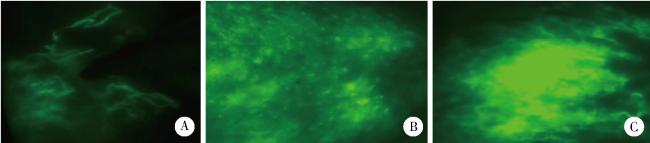
1.5 手术方法及术后处理
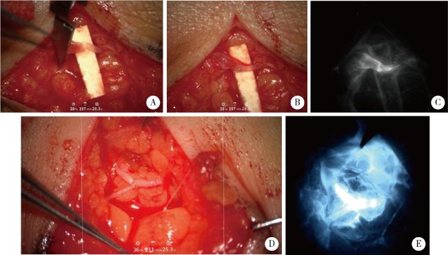
图2 淋巴管-静脉吻合术Figure 2 Lymphaticovenous anastomosis A, B, preoperative and postoperative images of end-to-end anastomosis between lymphatic vessels and veins under the microscope, respectively; C, fluorescence imaging after anastomosis; D, end-to-side anastomosis between lymphatic vessels and veins; E, fluorescence imaging after end-to-side anastomosis. |
1.6 观察指标
1.7 统计学分析
2 结果
2.1 不同ISL分期患者的基线特征
2.2 主要观察指标结果
2.2.1 不同ISL分期患者的%REV和术后周径减少情况
2.2.2 不同ICG分型患者的%REV比较
表1 不同ICG分型患者的%REV比较Table 1 Comparison of %REV in patients with different ICG patterns |
| Groups | n | %REV | 95%CI of %REV | Cohen’s d (vs. diffuse) |
| Splash | 6 | 63%± 6% | 59%- 67% | 3.5 (large effect) |
| Stardust | 14 | 56%± 7% | 52%- 60% | 2.4 (large effect) |
| Diffuse | 12 | 36%± 4% | 32%- 40% |
ICG, indocyanine green; %REV, percentage reduction of limb volume. |
2.2.3 不同ICG分型患者的术后周径减少情况
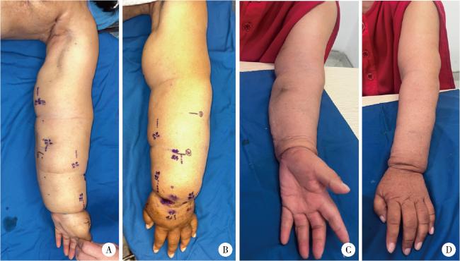


图3 乳腺癌术后上肢淋巴水肿患者LVA治疗前后对比Figure 3 Comparison of breast cancer-related upper extremity lymphedema patients before and after LVA treatment A, B, preoperative photographs showing obvious swelling of the right upper limb; C, D, photographs at 2 months postoperatively, showing resolution of limb swelling with good restoration of appearance and function. LVA, lymphaticovenous anastomosis |