Accuracy of dynamic navigation-assisted trephine method for bone harvesting

  • Jiayu LIU ,
  • Ning ZHU ,
  • Yuchen CHANG ,
  • Xianming GAO ,
  • Yu ZHANG , *
Expand
  • Department of Oral Implantology, Peking University School and Hospital of Stomatology & National Center for Stomatology & National Clinical Research Center for Oral Diseases & National Engineering Research Center of Oral Biomaterials and Digital Medical Devices & Beijing Key Laboratory of Digital Stomatology, Beijing 100081, China
ZHANG Yu, e-mail,

Received date: 2024-02-05

  Online published: 2026-02-25

Supported by

the Program for New Clinical Techniques and Therapies of Peking University School and Hospital of Stomatology(PKUSSNCT-22A01)

Copyright

All rights reserved. Unauthorized reproduction is prohibited.

Abstract

Objective: To evaluate the positional accuracy of dynamic navigation-assisted trephine bone harvesting in the symphysis and external oblique ridge. Methods: Ten standardized mandibular models were 3D-printed using polyetheretherketone (PEEK), mimicking natural mandibular mechanical properties. Pre-operative cone beam CT (CBCT) scans (70 kV, 70 mA, 0.25 mm×0.25 mm ×0.25 mm voxel) were acquired, and data were imported into dynamic navigation software (Dcarer, China). Two donor sites were designed in both the symphysis (≥15 mm from anterior teeth) and external oblique ridge (≥6 mm from molars), with 8 mm-diameter, 6 mm-deep cylindrical osteotomy tracts planned for each site.After calibrating the navigation system with 20 mm and 50 mm spherical burs, an 8 mm outer-diameter trephine prepared 40 tracts under real-time guidance. Post-operative CBCT scans were taken, and Mimics 20.0 software fitted actual tracts to standard cylinders. Superimposing actual and designed tracts via metal registration markers, we measured coronal/apical center point deviation, depth deviation, and axis angle deviation in order to compare site-specific accuracy. Results: Deviations of the dynamic navigation-assisted trephine method for bone harvesting was (1.91±0.69) mm at the coronal center point, (1.54±0.66) mm at the apical center point, (-0.83±0.77) mm at the depth of the apical center point and 3.02°±0.38° at the axis angle. The four deviations in symphysis and external oblique ridge were (1.32±0.36) mm and (2.50±0.35) mm at the coronal center point (P < 0.01), (1.06± 0.31) mm and (2.02±0.56) mm at the apical center point (P < 0.01), (-0.30±0.52) mm and (-1.38±0.57) mm at the depth of apical center point (P < 0.01), 3.03°± 0.38° and 3.00°± 0.39° at axis angle (P=0.80). Conclusion: Within the limitations of this study, dynamic navigation-assisted trephine harvesting shows good accuracy. The symphysis exhibits higher accuracy than the external oblique ridge, possibly due to surface morphology and operability differences. These findings support its clinical potential, but future clinical studies are needed to validate results.

Cite this article

Jiayu LIU , Ning ZHU , Yuchen CHANG , Xianming GAO , Yu ZHANG . Accuracy of dynamic navigation-assisted trephine method for bone harvesting[J]. Journal of Peking University(Health Sciences), 2026 , 58(2) : 365 -371 . DOI: 10.19723/j.issn.1671-167X.2026.02.022

在口腔种植学中,自体骨被认为是骨增量手术材料选择的金标准[1]。而口内供区来源的自体骨有着与牙槽骨相近的胚胎起源,在受骨区有着更快的血管化过程[2-3],有利于获得更稳定的骨增量治疗效果。环钻取骨是一种高效且省时的自体骨取骨方式并在临床中已经有着广泛的应用[4-6]。使用环钻获取的自体骨具有较低的感染风险和良好的生物活性[7-9]。1999年,Urban等[10]使用环钻于颏部获取后制备了大量自体碎骨,并结合使用骨组织再生技术(guided bone regeneration, GBR) 技术完成了萎缩牙槽骨无牙颌的骨重建病例。Streckbein等[11]则提出使用环钻制备自体柱状骨并预备对应尺寸的半柱状受植床。这种标准化的外置法植骨技术流程能够保证自体骨块在受骨区与天然基骨的紧密贴合,被证明有着良好的水平向骨增量效果。2015年,Giesenhagen[12]使用环钻和种植系列钻制备自体环状骨块以实现垂直向骨增量同期种植,这种骨环技术缩短了伴垂直向骨缺损缺牙患者的治疗周期,然而,环钻取骨手术准确性不足在一定程度上限制了其在临床的应用。环钻取骨时的偏差可能会对取骨区特定解剖结构,比如下牙槽神经和邻牙牙根造成损伤[13]。进一步提高环钻取骨的准确性不仅有助于减少取骨术中术后并发症的风险,也有助于实现种植术区原位取骨。同时,更加精准的环钻取骨也有利于实现可预期皮质/松质骨比的自体骨块获取,从而提高骨增量的治疗效果[14]
静态导板和动态导航等口腔数字化技术凭借其在手术准确性和可预期性方面的临床应用优势,在口腔种植学中得到了广泛应用[15]。现阶段数字化辅助取骨手术仍然主要依赖于静态导板技术[16]。动态导航系统作为一种光学定位的数字化辅助手术方式,在一定程度上克服了静态导板技术的局限性,提供了更好的手术灵活性和准确性[17-18], 其在辅助牙列缺损患者种植窝洞预备及种植体植入方面有着良好的准确性并得到了广泛应用[19]。然而,目前尚未见关于动态导航辅助在口内供区使用环钻取骨的相关研究报道。鉴于环钻和种植系列钻有着类似的驱动机制,动态导航辅助环钻取骨理论上具备可行性。但动态导航辅助环钻在常见口内供骨区(如颏部和外斜线区域)的工作准确性及差异仍不明确。
本研究旨在通过体外标准化下颌模型,评估动态导航辅助环钻取骨在颏部和外斜线区域的准确性表现及差异。

1 材料与方法

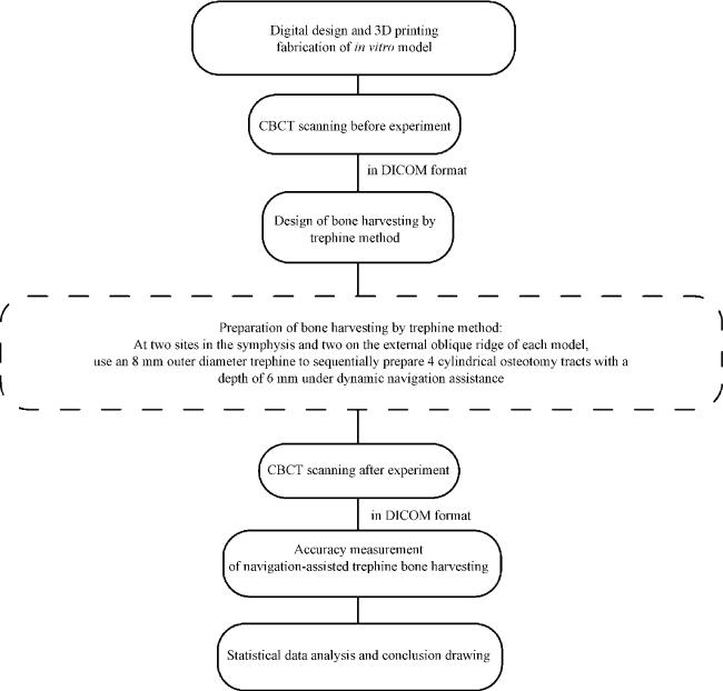
本研究设计并制作了10个下颌模型进行体外对照研究(图 1),研究于2022年11月至2023年2月进行,鉴于尚未见使用动态导航辅助环钻取骨的相关研究,本研究的样本量确定参考有关动态导航准确性的体外研究报道[20]。共计划在10个体外模型上使用动态导航辅助环钻完成40个圆柱形预备道,其中20个预备道在颏部完成,20个在外斜线区域完成。
图1 动态导航辅助环钻取骨准确性的体外研究流程图

Figure 1 Workflow chart of in vitro study on accuracy of dynamic navigation-assisted trephine bone harvesting

CBCT, cone beam computed tomography; DICOM, digital imaging and communications in medicine.

1.1 模型制作

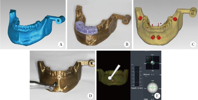
使用标准化下颌模型作为本研究的操作模型模板,拍摄锥型束CT(cone beam CT,CBCT)获取该模型的影像学数据,然后将其导入Mimics 20.0软件(Materialise公司,比利时)并进行三维重建。使用SolidWorks软件(达索公司,美国)设计了用于连接动态导航参考板的插槽结构,并以. stl格式导出。使用Geomagic Studio 2014软件(Geomagic公司,美国)将此插槽结构与重建的模型合并,获得本体外研究模型(图 2A)。最后,使用INTAMSUITE 3D打印软件以聚醚酮酮(polyetheretherketone, PEKK)材料制作10个相同下颌骨模型。
图2 导航辅助环钻取骨的方案设计及实施

Figure 2 Digital plan and conduction of the navigation-assisted trephine method for bone harvesting

A, digitally designed model of the in vitro study; B, registration device of dynamic navigation system was positioned on the lower anterior teeth; C, design digital trephine osteotomy tracts (red); D, use trephines to complete the osteotomy tracts under the guidance of dynamic navigation; E, real-time positioning and angular deviations of the trephine are displayed in the dynamic navigation system.

1.2 环钻取骨方案设计

使用聚醚橡胶在下颌模型的前牙区固定迪凯尔(DCARER)动态导航系统的注册装置(图 2B), 在检验注册装置的稳定性后拍摄70 kV管电压、70 mA管电流及0.25 μm扫描体积像素的锥形束CT,然后将影像数据导出到动态导航软件V2.5(迪凯尔公司,中国)中,在下颌骨颏部及外斜线各设计了2个直径8 mm和深度6 mm的圆柱形预备道,其中保证颏部的预备道至少距离下颌前牙15 mm,外斜线的预备道至少距离下颌磨牙6 mm,最后输出得到本研究的实验计划工程文件(图 2C)。

1.3 环钻取骨方案实施

实验前已协调设备厂家将环钻系统相关器械虚拟重构数据录入动态导航系统内,以实现对环钻器械的精准识别与实时空间定位,确保导航引导的有效性。实验中固定动态导航参考板并验证其具有良好稳定性,使用长度20 mm和50 mm的球钻完成动态导航标定步骤以标记种植手机相对于体外模型的空间位置,将动态导航配准装置在模型前牙区重新就位,将长度20 mm的球钻尖端依次置于配准装置的半球形凹槽中,完成了动态导航配准步骤,这样可以通过配准装置上的金属配准标记将实验前拍摄的模型CBCT与体外模型精确匹配。在完成导航标定和配准步骤后,就可以在实验前拍摄的CBCT上实时定位种植手机、工作车针与体外研究模型的相对空间位置,之后移除配准装置,在动态导航辅助下使用外径8 mm的环钻,依据计划的取骨方案依次于每个模型的外斜线及颏部区域完成直径8 mm和深度6 mm的取骨预备道制备(图 2DE),最终获得实验后的体外模型。

1.4 准确度测量与评价

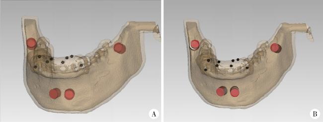
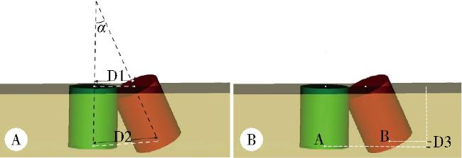
将迪凯尔动态导航配准装置在实验后的体外模型上重新就位,并拍摄具有相同参数的CBCT。将获得的影像学数据导入到Mimics 20.0软件(Materialise公司,比利时),首先设定阈值使得生成仅包含金属配准标记的掩模,并进行三维重建,之后将实验后体外模型上的环钻预备道拟合成实心圆柱形(图 3A)。考虑到环钻取骨操作过程可能导致实际预备孔径误差,本研究在准确性分析时根据环钻实际外径(8 mm)及预备深度(6 mm)进行实际预备道最佳拟合,确保分析基于统一尺寸标准,减少系统偏差后将这些代表实际预备道的圆柱形与三维重建获得的金属配准标记整合。同时,也将术前计划预备道及配准装置中的金属配准标记整合并以. stl文件格式导入,基于注册装置上的金属配准标记,在每个操作位点的环钻取骨实际预备道被叠加到对应的计划预备道上(图 3B)。最后测量动态导航辅助环钻取骨实际和计划预备道之间在进入中心点、结束中心点、预备深度及轴向角度上的误差(图 4AB)。本研究中进入中心点、结束中心点、轴向角度的误差为绝对误差,计算方法为实际位点到计划位点的距离(D1、D2)或中心轴角度偏差值的绝对值(|α|)。预备深度误差为相对误差,测量方法为预备深度误差D3=|实际预备结束中心点到颌骨平面距离| - |计划预备结束中心点到颌骨平面距离|。
图3 动态导航辅助环钻取骨准确性的数据分析

Figure 3 Statistical measurement for dynamic navigation-assisted trephine bone harvesting accuracy

A, actual osteotomy tracts were adapted in the post-lab CBCT (red); B, superimpose the actual osteotomy tracts (red) to the planned ones (white) based on the mental fiducial markers on the registration device (black).

图4 动态导航辅助环钻取骨准确性的评价指标

Figure 4 Experimental accuracy evaluation index

A, coronal center point deviation D1, apical center point deviation D2 and angulation deviation α; B, deviations in depths of apical centre points D3.

本研究的精度测量由2名经过培训的研究者独立完成,采用盲法测量。若2名研究者测量结果差异超过0.2 mm(角度差异超过0.3°),则由第3名资深研究者复核确定最终值。通过组内相关系数(ICC)检验一致性,结果显示ICC=0.92(95%CI:0.87~0.95),表明测量者间一致性良好。
所有准确度评价指标均被视为连续变量,使用Shapiro-Wilk检验、正态P-P图和箱线图来评估数据的正态性。使用SPSS 26.0软件(IBM公司,美国)进行统计分析,若在颏部及外斜线区域使用动态导航辅助环钻取骨的误差差异呈正态分布,则使用独立样本t检验评估精确度差异性; 如果不符合正态分布,则使用Mann-Whitney U检验研究动态导航辅助环钻在外斜线和颏部的准确性差异,检验水准为双侧α=0.05。

2 结果

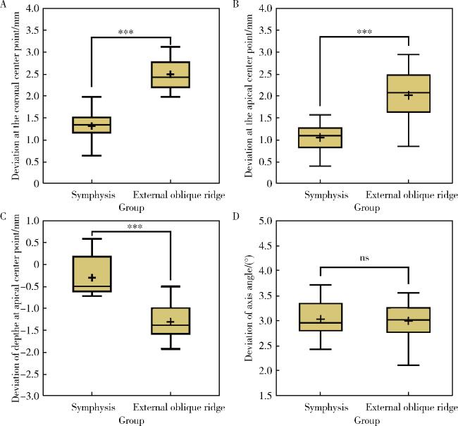
在本体外研究中,通过动态导航辅助在10个模型上共完成了40个位点的环钻取骨, 经Shapiro-Wilk检验、正态P-P图及箱线图分析,所有误差指标均符合正态分布(P>0.05,表 1),动态导航辅助环钻预备的进入中心点误差为(1.91±0.69) mm,结束中心点的误差为(1.54±0.66) mm,预备深度误差为(-0.83±0.77) mm,预备道中心轴角度差为3.02°±0.38°。其中, 在颏部与外斜线的进入中心点误差分别为(1.32±0.36) mm和(2.50±0.35) mm,结束中心点误差分别为(1.06±0.31) mm和(2.02±0.56) mm,深度误差分别为(-0.30±0.52) mm和(-1.38 ±0.57) mm,中心轴角度差分别为3.03°±0.38°和3.00°±0.39°(表 2)。使用动态导航辅助环钻在颏部及外斜线区域取骨的定位准确性,包括进入中心点(图 5A)、结束中心点(图 5B)及预备深度(图 5C)等差异有统计学意义,而在中心轴角度上则未见明显差异(图 5D)。
表1 动态导航辅助环钻取骨误差

Table 1 Descriptive values of deviations in dynamic navigation-assisted trephine methods for bone harvesting

Deviations n ${\bar x}$±s Minimum Maximum
Coronal center point/mm 40 1.91±0.69 0.64 3.12
Apical center point/mm 40 1.54±0.66 0.41 2.94
Depth of apical center point/mm 40 -0.84±0.77 -2.22 0.63
Axis angle/(°) 40 3.02±0.38 2.11 3.72
表2 动态导航辅助环钻取骨在颏部及外斜线区域的误差

Table 2 Descriptive values of deviations in dynamic navigation-assisted trephine methods for bone harvesting in the symphysis and external oblique line area

Deviations Donor sites n ${\bar x}$±s Minimum Maximum t P
Coronal center point/mm Symphysis 20 1.32±0.36 0.64 1.98 -10.42 <0.01
External oblique line 20 2.50±0.35 1.98 3.12
Apical center point/mm Symphysis 20 1.06±0.31 0.41 1.57 -6.70 <0.01
External oblique line 20 2.02±0.56 0.86 2.94
Depth of apical center point/mm Symphysis 20 -0.30±0.52 -1.13 0.63 4.81 <0.01
External oblique line 20 -1.38±0.57 -2.22 -0.55
Axis angle/(°) Symphysis 20 3.03±0.38 2.43 3.72 0.26 0.80
External oblique line 20 3.00±0.39 2.11 3.56

The independent sample t test was used to analyse the difference between the groups, the significant level was P<0.05.

图5 在颏部外斜线区使用动态导航辅助环钻取骨准确性的差异分析

Figure 5 Analysis of differences in bone harvesting accuracy using dynamic navigation-assisted trephine in extraoral oblique line and symphysis

A, deviations in coronal center points; B, deviations in apical center points; C, deviations in depth of apical center points; D, deviations in the axial angles. ns, correlation is not significant at the 0.05 level (2-tailed); * * * correlation is significant at the 0.001 level (2-tailed).

3 讨论

尽管动态导航技术被证明能够辅助种植系列钻实现精准可控的种植窝洞预备,但动态导航辅助环钻取骨的应用准确性仍有待进一步探究。本研究在体外模型中,动态导航辅助环钻能够获得良好的准确性表现,而动态导航技术在不同的取骨区辅助环钻取骨的定位准确性存在差异。
本研究使用的动态导航系统是一种国内常见的主动式红外光动态种植导航系统[21], 在临床应用过程中,其红外相机能够接受标定后参考板及种植手机主动发出的红外光,可在对种植手机空间位置及角度实现数字化呈现,辅助术者不断调整手术器械以精准完成手术操作[22]。然而不同研究报道的动态导航系统准确性存在差异,这可能是因为在实际临床应用中,导航系统误差、成像误差、配准误差及操作误差等在内的多种因素均会对导航准确性产生影响[23-24]。尽管一些影响因素,如影像学拍摄类型及拍摄参数是否对动态导航手术准确性存在影响依旧存在争议[25],但手术操作因素,包括术者操作经验及手术过程中相关因素均会对导航手术准确性产生影响。陶宝鑫等[26]研究发现,在前牙区及后牙区使用动态导航辅助种植窝洞预备的角度误差存在差异。王磊等[27]则发现斜坡型骨表面会造成更大的种植系列钻导航误差。这提示在口内不同术区,由于其骨表面形态、骨密度、操作空间等方面的差异,会造成不完全相同的动态导航手术准确性表现。刘思民等[20]则进一步探究了钻针预备深度对导航手术准确性的影响,发现随着工作深度的增加,导航手术偏差也会增大。本实验中动态导航辅助环钻取骨的定位偏差为(1.91±0.69) mm,误差值略高于相关研究报道的动态导航手术平均定位偏差(1.21 mm)[28],这可能是由于取骨环钻作为一种大直径,以约1 mm厚度圆环形切割刃作为工作端的中空取骨器械,尽管其与种植系列钻及根尖外开窗钻针等有着相同的驱动模式,但其往往存在着工作时稳定性不足、容易滑脱且难以控制的临床应用问题,同时,本研究是由同一名在50例体外模型上完成环钻取骨的口腔种植医师完成,然而相较于种植窝洞预备手术操作而言,环钻取骨操作经验尚缺乏。此外,口内常见供骨区,如颏部及外斜线区的骨表面形态及骨密度也与常规口腔临床动态导航手术的工作区域,如牙槽嵴顶等存在差异。然而,Aydemir等[29]的研究中动态导航辅助种植窝洞预备的实际应用定位误差也达到了1.83 mm,提示动态导航辅助环钻取骨准确性在可接受的范围内。此外,本研究中在颏部及外斜线区域导航辅助取骨的定位准确性存在差异,说明和动态导航辅助种植窝洞预备相似,骨表面形态、骨密度等可能也是影响导航下环钻取骨准确性的影响因素,然而不同取骨区的导航角度偏差无差异,这也和既往的研究结论一致[30],这在一定程度上说明了动态导航技术在工作器械中心轴角度的控制上的优越性。
本研究的局限性主要在于体外模型和操作空间与实际临床应用时的差异。使用不同材料和形态的体外研究会获得不同的动态导航手术误差结果,本研究通过3D打印技术,使用PEKK材料以不同的材料打印孔隙率模拟皮质和松质骨成分制作了用于操作的体外模型[31],尽可能使其具有与下颌骨相近的力学性能和表面形态,并使用常见取骨环钻以常规取骨深度完成操作,获得了可能更接近临床实际应用的误差值。这能够为临床初期应用动态导航辅助环钻取骨时的安全距离设定提供参考,然而,其仍无法充分模拟临床口内操作环境、医患体位、导航手机参考板可操作角度,以及医患主观配合度等导航取骨准确性的影响因素,因此,动态导航辅助环钻取骨的临床应用准确性还有待进一步研究。
综上所述,基于特定导航设备及体外模型的本研究结果显示动态导航辅助环钻取骨具有良好的准确性,但仍需要更多的临床试验来验证其应用表现。

利益冲突  所有作者均声明不存在利益冲突。

作者贡献声明  刘嘉昱:课题具体设计与实施,数据采集与整理,统计设计和结果分析,文章撰写;祝宁:课题设计,结果收集和数据整理,文章修改;张育祯、高贤明:结果收集和数据整理,文章修改;张宇:课题设计和实施,数据采集、归纳和整理指导,文章撰写与修改,总体把关和审定论文。所有作者均参与论文修改,并对最终文稿进行审读和确认。

1
Lichte P , Pape HC , Pufe T , et al. Scaffolds for bone healing: Concepts, materials and evidence[J]. Injury, 2011, 42 (6): 569- 573.

DOI

2
Omara M , Abdelwahed N , Ahmed M , et al. Simultaneous implant placement with ridge augmentation using an autogenous bone ring transplant[J]. Int J Oral Maxillofac Surg, 2016, 45 (4): 535- 544.

DOI

3
Khoury F , Hanser T . Mandibular bone block harvesting from the retromolar region: A 10-year prospective clinical study[J]. Int J Oral Maxillofac Implants, 2015, 30 (3): 688- 697.

DOI

4
Senos R , Hankenson KD . Calvaria critical-size defects in rats using piezoelectric equipment: A comparison with the classic trephine[J]. Injury, 2020, 51 (7): 1509- 1514.

DOI

5
Abdulrazaq SS , Issa SA , Abdulrazzak NJ . Evaluation of the trephine method in harvesting bone graft from the anterior iliac crest for oral and maxillofacial reconstructive surgery[J]. J Craniofacial Surg, 2015, 26 (8): e744- e746.

DOI

6
Chandra R , Suvvari N , Reddy A . Trephine core procedure versus bone-added osteotome sinus floor elevation in the augmentation of the sinus floor: A comparative clinical and radiographic study[J]. Int J Oral Maxillofac Implants, 2018, 33 (2): 425- 432.

DOI

7
Takamoto M , Takechi M , Ohta K , et al. Risk of bacterial contamination of bone harvesting devices used for autogenous bone graft in implant surgery[J]. Head Face Med, 2013, 9 (1): 3.

DOI

8
Maridati P , Dellavia C , Pellegrini G , et al. Histologic and radiographic comparison of bone scraper and trephine bur for autologous bone harvesting in maxillary sinus augmentation[J]. Int J Oral Maxillofac Implants, 2015, 30 (5): 1128.

9
Miron RJ , Gruber R , Hedbom E , et al. Bigger size and defatting of bone chips will increase cup stability[J]. Clin Implant Dent Rel Res, 2013, 15 (4): 481- 489.

DOI

10
Urban IA , Monje A , Lozada JL , et al. Long-term evaluation of peri-implant bone level after reconstruction of severely atrophic edentulous maxilla via vertical and horizontal guided bone regeneration in combination with sinus augmentation: A case series with 1 to 15 years of loading[J]. Clin Implant Dent Rel Res, 2017, 19 (1): 46- 55.

DOI

11
Streckbein P , Kähling C , Wilbrand JF , et al. Horizontal alveolar ridge augmentation using autologous press fit bone cylinders and micro-lag-screw fixation: Technical note and initial experience[J]. J Cranio Maxillofac Surg, 2014, 42 (5): 387- 391.

DOI

12
Giesenhagen B . Klinische fünfjahresergebnisse nach einzeitiger augmentation mit autologen knochenringen und ankylos-implantaten im atrophierten unterkiefer[J]. Zeitschrift Für Zahnärztliche Implantologie, 2015, 31 (1): 52- 62.

13
Streckbein P , Meier M , Kähling C , et al. Donor-site morbidity after retromolar bone harvesting using a standardised press fit cylinder protocol[J]. Materials, 2019, 12 (22): 3802.

DOI

14
Yeung R . Simultaneous placement of implant and bone graft in the anterior maxilla: A case report[J]. Int J Oral Maxillofac Implants, 2004, 19 (6): 892- 895.

15
Lin WS , Yang CC , Polido WD , et al. CAD-CAM cobalt-chromium surgical template for static computer-aided implant surgery: A dental technique[J]. J Prosthet Dent, 2020, 123 (1): 42- 44.

DOI

16
Zhu N , Liu J , Ma T , et al. A fully digital workflow for prosthetically driven alveolar augmentation with intraoral bone block and implant rehabilitation in an atrophic anterior maxilla[J]. J Prosthet Dent, 2023, 130 (5): 668- 673.

DOI

17
Block MS , Emery RW . Static or dynamic navigation for implant placement-choosing the method of guidance[J]. J Oral Maxillofac Surg, 2016, 74 (2): 269- 277.

DOI

18
Vasak C , Watzak G , Gahleitner A , et al. Computed tomography-based evaluation of template (NobelGuideTM)-guided implant positions: A prospective radiological study[J]. Clin Oral Implants Res, 2011, 22 (10): 1157- 1163.

DOI

19
Panchal N , Mahmood L , Retana A , et al. Dynamic navigation for dental implant surgery[J]. Oral Maxillofac Surg Clin N Am, 2019, 31 (4): 539- 547.

DOI

20
刘思民, 赵一姣, 王晓燕, 等. 动态导航下不同深度环钻定位精确度的体外评价[J]. 北京大学学报(医学版), 2022, 54 (2): 146- 152.

DOI

21
Wu D , Zhou L , Yang J , et al. Accuracy of dynamic navigation compared to static surgical guide for dental implant placement[J]. Int J Implant Dent, 2020, 6 (1): 78.

DOI

22
Cecchetti F , Di Girolamo M , Ippolito DG , et al. Computer-guided implant surgery: Analysis of dynamic navigation systems and digital accuracy[J]. J Bio Regulators Homeostatic Agents, 2020, 34 (3 Suppl 1): 9- 17.

23
Ma L , Jiang W , Zhang B , et al. Augmented reality surgical navigation with accurate CBCT-patient registration for dental implant placement[J]. Med Biol Eng Comput, 2019, 57 (1): 47- 57.

DOI

24
满毅, 周楠, 杨醒眉. 动态实时导航在口腔种植领域中的临床应用及新进展[J]. 口腔疾病防治, 2020, 28 (6): 341- 348.

25
Tao B , Shen Y , Sun Y , et al. Comparative accuracy of cone-beam CT and conventional multislice computed tomography for real-time navigation in zygomatic implant surgery[J]. Clin Implant Dent Rel Res, 2020, 22 (6): 747- 755.

DOI

26
陶宝鑫, 蓝耕良, 黄伟, 等. 动态导航技术辅助无牙颌种植精度分析[J]. 上海交通大学学报(医学版), 2022, 42 (9): 1353- 1360.

27
王磊, 伍颖颖, 满毅. 动态导航技术中植入位点骨质量对种植精度的影响[J]. 口腔颌面外科杂志, 2022, 32 (3): 174- 181.

28
Wei SM , Zhu Y , Wei JX , et al. Accuracy of dynamic navigation in implant surgery: A systematic review and meta-analysis[J]. Clin Oral Implants Res, 2021, 32 (4): 383- 393.

DOI

29
Aydemir CA , Arısan V . Accuracy of dental implant placement via dynamic navigation or the freehand method: A split-mouth randomized controlled clinical trial[J]. Clin Oral Implants Res, 2020, 31 (3): 255- 263.

DOI

30
Mediavilla Guzmán A , Riad Deglow E , Zubizarreta-Macho á , et al. Accuracy of computer-aided dynamic navigation compared to computer-aided static navigation for dental implant placement: An in vitro study[J]. J Clin Med, 2019, 8 (12): 2123.

DOI

31
林后学, 郭松松, 何佳宜, 等. 下颌骨颏部骨皮质增龄性变化的锥形束CT研究[J]. 中国CT和MRI杂志, 2016, 14 (9): 15- 18.

Outlines

/