Establishment of rabbit model of benign circumferential esophageal stricture

  • Lingyu MENG ,
  • Yonghui HUANG , * ,
  • Xiu'e YAN ,
  • Yingchun WANG
Expand
  • Department of Gastroenterology, Peking University Third Hospital, Beijing 100191, China
HUANG Yonghui, e-mail,

Received date: 2023-11-12

  Online published: 2025-11-10

Supported by

the National Natural Science Foundation of China(82070653)

Beijing Natural Science Foundation(7222207)

Capital's Funds for Health Improvement and Research(2022-2Z-40914)

Copyright

All rights reserved. Unauthorized reproduction is prohibited.

Abstract

Objective: To explore the method of establishing benign esophageal stricture in rabbits by using argon plasma coagulation under endoscopy, and to provide a convenient and stable animal model for subsequent research on the prevention of esophageal stricture. Methods: Twenty-two male New Zealand rabbits were randomly divided into three groups after completing esophageal radiography under X-ray. The blank control group (n=4) only received endoscopic examination. In the experimental group Ⅰ (n=9) and experimental group Ⅱ (n=9), argon knife was performed on the esophagus at 30 W and 50 W power, respectively, under endoscopy. Endoscopy was performed 1, 2, and 4 weeks after the operation to observe the changes in the esophagus, and the body weight and mental state were recorded. Four weeks after the operation, esophageal radiography under X-ray was performed to measure the inner diameter of the esophageal stricture and calculate the stricture index. All the experimental animals were sacrificed and esophageal specimens were obtained for histopathological examination and detection of hydroxyproline content in esophageal tissue. Results: In the blank control group, the body weight significantly increased 4 weeks after the operation compared with that before the operation [(4.13±0.25) kg vs. (3.10±0.39) kg, P < 0.05], and there was no significant change in the esophageal inner diameter [(12.89±0.83) mm vs. (12.83±1.07) mm, P>0.05]. In the experimental group Ⅰ, there was no significant change in the body weight and esophageal inner diameter 4 weeks after the operation compared with that before the operation [(2.91±0.28) kg vs. (2.91±0.54) kg; (11.19±0.97) mm vs. (12.06±0.32) mm; P>0.05]. In the experimental group Ⅱ, the body weight and esophageal inner diameter significantly decreased 4 weeks after the operation compared with that before the operation [(2.02±0.31) kg vs. (3.51±0.37) kg; (10.49±1.76) mm vs. (12.58±1.11) mm; P < 0.05]. The esophageal stricture index 4 weeks after the operation was significantly higher in the experimental group Ⅱ than in the experimental group Ⅰ (1.242±0.148 vs. 1.083±0.104, P < 0.05). The histopathological score and hydroxyproline content in the experimental group Ⅰ [2.55±0.52, (182.90±72.75) μg/g] and experimental group Ⅱ [4.55±0.52, (210.81±54.28) μg/g] were significantly higher than those in the blank control group [0, (91.37±29.74) μg/g] (P < 0.05). Conclusion: The induction of esophageal stricture in male New Zealand rabbits using 50 W argon knife under endoscopic guidance is a feasible, depth-controllable, and reproducible method. This animal model provides a reliable platform for the development and evaluation of novel therapeutic strategies for esophageal stricture.

Cite this article

Lingyu MENG , Yonghui HUANG , Xiu'e YAN , Yingchun WANG . Establishment of rabbit model of benign circumferential esophageal stricture[J]. Journal of Peking University(Health Sciences), 2026 , 58(2) : 380 -387 . DOI: 10.19723/j.issn.1671-167X.2026.02.024

食管癌是全球癌症相关死亡的第六大常见原因[1]。晚期食管癌患者的总体生存期较差,但早期发现则预后良好[2-3]。内镜黏膜下剥离术(endoscopic submucosal dissection,ESD)作为一种微创治疗方法,已被广泛应用于食管浅表癌、Barrett食管等多种食管病变的治疗中[4]。狭窄是ESD的主要并发症,尤其是黏膜大面积剥离时,有研究表明,当黏膜缺损周长>75%时,狭窄发生率高达70%~90%[5-7]。食管狭窄大大降低了生活质量,尽管其可通过内镜下球囊扩张来治疗[8],但治疗后会导致严重并发症,如穿孔和瘘管等[9]。为预防术后食管狭窄的发生,目前临床上有较多措施,包括局部类固醇注射、口服类固醇,以及放置可生物降解支架、聚乙醇酸片和组织工程细胞片等[10-15],但迄今为止,尚未见任何一种方法可达到完全预防的效果。
实验动物模型对于探索有效的治疗手段至关重要,本研究建立了一种便捷、稳定、可操控性强、更贴近临床实际的良性食管狭窄动物模型,为ESD术后食管狭窄的预防提供了研究基础。

1 材料与方法

1.1 实验动物及实验前准备

雄性新西兰兔22只,年龄4~6个月,体重2.4~3.5 kg,饲养于无特定病原体(specific pathogen free,SPF)级动物实验室,可自由进食和饮水;实验室光照时间6:00~18:00,温度18~22 ℃。本研究已获得北京大学生物医学伦理委员会实验动物福利伦理分会批准(批准号:LA2022342),所有操作均严格遵守动物福利伦理要求。
将动物随机分为实验Ⅰ组(n=9)、实验Ⅱ组(n=9)和空白对照组(n=4)。禁食12 h后肌肉注射盐酸塞拉嗪10 mg/kg+舒泰(左拉西泮和替来他明1 ∶ 1混合配制) 5 mg/kg麻醉,同时密切观察呼吸频率和节律,避免麻醉过度。麻醉成功标准:肌肉松弛,失去知觉,呼吸深慢且平稳,角膜反射消失。

1.2 模型建立方法

1.2.1 实验Ⅰ组

麻醉成功后将兔在试验台上摆成左侧卧位,右侧腹部备皮后用湿纱布湿润局部皮肤,覆以负极板。开口器打开口腔后,使用超细内镜(GIF-XP260N,日本奥林巴斯公司)行内镜检查,观察食管有无解剖异常及病变。经活检孔道置入氩气刀,在内镜直视下对食管中下段黏膜(据门齿约15~18 cm)进行点射式、环周逐点烧灼,选择氩离子凝固术(argon plasma coagulation,APC)模式,功率30 W,流量0.5 L/min,烧灼长度2 cm,同时需注意送气量,将内镜主机送气泵调整至LOW档,避免胃肠道注气过多。操作完成后观察食管有无明显出血、穿孔,然后吸气退镜。术后禁食1 d后给予正常饮食,每天观察进食状态并测量体重。

1.2.2 实验Ⅱ组

将氩气刀功率调整至50 W,其他参数不变,实验方法同Ⅰ组。

1.2.3 空白对照组

仅给予内镜检查,不进行氩气烧灼。

1.3 内镜下检查

各实验组和空白对照组分别在术后第1、2、4周进行内镜下检查,观察食管在内镜下的表现,并于第4周评估食管狭窄情况,评估标准参照人体食管内镜下狭窄评估标准:无狭窄,内镜下未见狭窄表现;轻度狭窄,内镜下见轻度管腔变窄,镜身顺畅通过;中度狭窄,内镜下见管腔显著狭窄,镜身有阻力通过;重度狭窄,内镜下见管腔严重狭窄,镜身无法通过。

1.4 食管造影检查及狭窄指数

各实验组和空白对照组在术前、术后第4周行食管造影检查,观察各组食管狭窄情况。麻醉状态下通过开口器经口留置胃管,胃管长度为距门齿10 cm,向胃管内推注泛影葡胺造影剂,同时在数字血管造影机检查床上进行曝光摄片。食管造影期间测量食管狭窄处内径,并使用胃管的外径作为参考进行放大校正。进一步计算实验后第4周的食管狭窄指数,即术前食管内径与术后第4周食管狭窄处内径的比值。实验过程中死亡的动物不进行食管造影检查。

1.5 组织病理学检查及羟脯氨酸测定

术后第4周食管造影后经耳缘静脉推注空气对所有实验动物实施安乐死,解剖食管下段后获取样本,并纵行分割。标本的一半置于10%(质量分数)甲醛溶液固定48 h,每个病灶按4 mm间隔连续取材,石蜡包埋后切片,进行苏木精-伊红染色,并进行Masson染色以评估胶原蛋白的表达。参考既往文献[16-17]对食管组织病理学改变进行半定量分析,具体评分标准见表 1。标本的另一半保存于-80 ℃低温冰箱,以进行羟脯氨酸含量测定。
表1 病变损伤评分标准

Table 1 Lesion damage scoring criteria

Damage category Lesion degree Score
Submucosal collagen deposition No submucosal collagen deposition 0
Mild (collagen deposition in submucosa < twice the thickness of muscularis mucosae) 1
Severe (collagen deposition in submucosa>twice the thickness of muscularis mucosae) 2
Mucosal muscularis injury No 0
Yes 1
Muscularis propria injury and collagen deposition No 0
Mild (collagen deposition around smooth muscle fibers) 1
Severe (in addition to the above changes, some smooth muscle fibers are replaced by collagen) 2
及时解剖实验过程中死亡的动物,观察食管及纵隔周围情况,了解死因,并将所获食管标本按照上述方法进行染色和羟脯氨酸含量测定。

1.6 统计学方法

采用SPSS 17.0统计软件分析数据。计量资料用均数±标准差表示,两组间比较使用独立样本t检验;计数资料用例数(百分数)表示,两组间比较应用Fisher确切检验。P<0.05为差异有统计学意义。

2 结果

2.1 一般情况

空白对照组实验前后的食欲和活动度无明显差异;实验Ⅰ组和Ⅱ组在术后1周左右均出现明显食欲下降、进食减少、精神萎靡和活动度下降。实验Ⅰ组1~2周后逐步好转,其中1只术后11 d死亡。实验Ⅱ组上述表现持续至第4周,其中2只分别于术后第13、21天死亡。所有死亡动物及时解剖后均未发现食管穿孔、纵隔感染等表现。观察实验前和实验后第1、2、4周各组体重,发现空白对照组体重在实验第4周较实验前显著增加,实验Ⅱ组体重在实验第4周较实验前显著降低,差异均有统计学意义(P<0.05,表 2)。
表2 实验前后各组体重

Table 2 Weight in each group before and after the operation

Group Before the operation 1 week after the operation 2 weeks after the operation 4 weeks after the operation
n Weight/kg n Weight/kg n Weight/kg n Weight/kg
Blank control group 4 3.10±0.39 4 3.25±0.34 4 3.48±0.33 4 4.13±0.25*
Experimental group Ⅰ 9 2.91±0.54 9 2.52±0.59 8 2.72±0.54 8 2.91±0.28
Experimental group Ⅱ 9 3.51±0.37 9 2.90±0.35 8 2.62±0.37 7 2.02±0.31*

Data are expressed as ${\bar x}$±s. *P<0.05, vs. before the operation.

2.2 食管在内镜下的表现

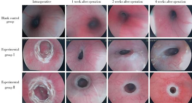
图 1为各组术中及术后食管内镜影像,可见空白对照组实验前后食管黏膜均光滑,管腔内径正常且无狭窄。实验Ⅰ组氩气烧灼后环周食管黏膜连续破坏,表现为黏膜发白、水肿;实验Ⅱ组局部还可见焦痂样改变。术后1周,实验Ⅰ组可见烧灼处环周溃疡形成,局部水肿,管腔不同程度变窄,实验Ⅱ组可见烧灼处溃疡伴黄白苔,局部可见坏死物质附着。术后2周,实验Ⅰ组食管环周溃疡仍有少许存在,局部可见白色瘢痕样黏膜,水肿及狭窄情况较术后第1周有所缓解;实验Ⅱ组可见烧灼处溃疡表面苔变薄、水肿消失,同时可见白色瘢痕形成,管腔不同程度变窄。造模后第4周,各实验组食管均可见白色瘢痕形成,管腔成不同程度狭窄;内镜下狭窄程度分级提示:实验Ⅰ组全部为轻、中度狭窄,5只(62.5%) 为轻度狭窄,3只(37.5%)为中度狭窄;实验Ⅱ组全部为中、重度狭窄,3只(42.9%)为中度狭窄,4只(57.1%)为重度狭窄;两组狭窄程度分布差异有统计学意义(P<0.05)。
图1 各组术中及术后食管内镜影像对比

Figure 1 Comparison of intraoperative and postoperative esophageal endoscopic images in each group

2.3 造影下食管内径及狭窄指数

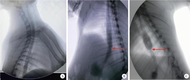
术前空白对照组及各实验组的食管造影均提示食管走行自然,无狭窄,术后4周各组食管造影情况见图 2,可见空白对照组食管内径与术前相比变化不大,实验Ⅰ组食管下段仅出现轻微管腔变窄,且狭窄上方食管伴有轻度扩张;实验Ⅱ组食管下段较前出现明显狭窄,且狭窄上方食管出现显著扩张。食管内径测量结果也提示,实验Ⅱ组较实验前明显变小[(10.49±1.76) mm vs. (12.58±1.11) mm,P < 0.05,表 3]。
图2 各组术后4周食管造影

Figure 2 Esophagography at 4 weeks after operation in each group

A, blank control group; B, experimental group Ⅰ; C, experimental group Ⅱ. The arrows show the sites of esophageal stenosis.

表3 造影下测得的食管内径及狭窄指数

Table 3 Esophageal inner diameter by angiography and the stricture index

Group Esophageal inner diameter/mm Stricture index
Preoperative 4 weeks after operation
Blank control group 12.83±1.07 12.89±0.83 0.995±0.028
Experimental group Ⅰ 12.06±0.32 11.19±0.97 1.083±0.104
Experimental group Ⅱ 12.58±1.11 10.49±1.76* 1.242±0.148#△

Data are expressed as ${\bar x}$±s. *P < 0.05,vs. preoperative; # P<0.05, vs. blank control group; △P<0.05, vs. experimental group Ⅰ.

进一步计算各组的食管狭窄指数,结果显示空白对照组、实验Ⅰ组、实验Ⅱ组狭窄指数分别为0.995±0.028、1.083±0.104和1.242±0.148,实验Ⅱ组与空白对照组和实验Ⅰ组相比差异均有统计学意义(P < 0.05,表 3),表明环周食管烧灼可导致不同程度的食管狭窄,但实验Ⅱ组的食管狭窄程度较实验Ⅰ组更明显。

2.4 组织病理学检查结果

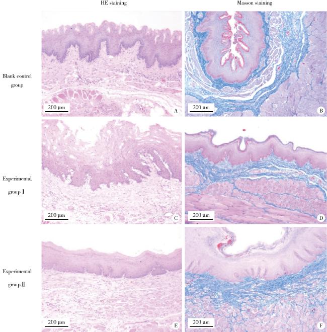
空白对照组在苏木精-伊红(hematoxylin eosin, HE)染色下可见正常的食管组织层次,包括黏膜上皮层、固有层、黏膜肌层、黏膜下层、肌层和外膜(图 3A);Masson染色可见正常食管组织胶原纤维有序存在于黏膜固有层及黏膜下层,在黏膜肌层及肌层间质可见少量胶原纤维(图 3B)。实验Ⅰ组在HE染色下可见食管黏膜上皮层结构存在,黏膜肌层大部分肌束缺失,固有层、黏膜肌层、黏膜下层纤维组织增生(图 3C);Masson染色提示食管黏膜固有层、黏膜肌层、黏膜下层及部分肌层胶原纤维轻微增生,分布紊乱(图 3D)。实验Ⅱ组在HE染色下可见上皮层结构存在,黏膜肌层大部分或全部肌束缺失,固有层、黏膜肌层、黏膜下层、部分固有肌层不同程度成纤维细胞或纤维组织增生,固有肌层完整(图 3E);Masson染色下可见食管黏膜固有层、黏膜肌层、黏膜下胶原纤维,其分布紊乱且呈不同程度增生(图 3F)。
图3 各组病理HE染色和Masson染色结果

Figure 3 Pathological HE staining and Masson staining results of each group

A, C and E are the hematoxylin eosin (HE) staining results of blank control group, experimental group Ⅰ and experimental group Ⅱ, repecitively; B, D and F are the Masson staining results of blank control group, experimental group Ⅰ and experimental group Ⅱ, repecitively.

各组组织病理学评分结果见表 4,可见实验Ⅱ组明显高于空白对照组和实验Ⅰ组(P < 0.05)。
表4 各组食管组织病理学评分和羟脯氨酸含量

Table 4 Esophageal histopathological score and hydroxyproline content in each group

Group Pathology score Hydroxyproline/(μg/g)
Blank control group (n=4) 0 91.37±29.74
Experimental group Ⅰ (n=9) 2.55±0.52* 182.90±72.75*
Experimental group Ⅱ (n=9) 4.55±0.52*# 210.81±54.28*

Data are expressed as ${\bar x}$±s. *P<0.05, vs. blank control group; # P<0.05, vs. experimental group Ⅰ.

2.5 羟脯氨酸含量测定

各实验组的羟脯氨酸含量均高于空白对照组,且差异有统计学意义(P<0.05)。实验Ⅱ组羟脯氨酸含量高于实验Ⅰ组,但差异无统计学意义(P>0.05),见表 4

3 讨论

ESD治疗早期食管癌的整体成功率达到95%,但大于3/4环周ESD后的狭窄会显著降低患者的生活质量,通常需要定期反复行内镜下球囊扩张。局部类固醇注射和口服类固醇可有效预防ESD后狭窄,但长期使用口服类固醇会导致脑脓肿等致命并发症的发生。有研究应用喷洒管对食管ESD术后创面进行局部喷洒,并利用喷洒时的压力将药物注射到残余的黏膜下层组织,以达到药物附着的效果[18]。我们前期的研究曾使用一种新型的预混凝胶(氢化可的松琥珀酸钠联合磷酸铝凝胶)预防ESD后的食管狭窄[19],研究纳入的均为接受食管3/4以上环周ESD的患者,其中14例接受预混凝胶口服,13例接受局部激素注射联合口服类固醇,术后2个月两组狭窄率分别为7.1%(1/14)和53.8%(7/13),提示口服预混凝胶可以有效预防食管狭窄,但这种预混凝胶的效果还需要更多的动物实验来印证。还有研究通过对比食管狭窄患者及食管正常者的食管黏膜,发现β-榄香烯可以抑制食管成纤维细胞的增殖和纤维化相关因子的水平,可能成为预防食管狭窄的新靶点[20]。所以制造一个稳定、便捷、可大量复制且又尽可能贴合临床的狭窄模型显得格外重要。
目前,有多种方法被用于诱发动物食管狭窄的形成,如ESD、电烧灼损伤、化学性腐蚀损伤、光动力等[21-25];用于制造模型的动物也有猪、狗、兔等多种选择。目前已有研究应用高频治疗钳对兔食管进行环周持续烧灼[26]、应用氢氧化钠对兔食管进行化学性腐蚀损伤[27],以及利用猪作为模型通过ESD来开展食管良性狭窄机制的研究[28]。狗和猪作为大型实验动物,可以行ESD,且更接近人体食管ESD后的损伤机制,但因其饲养成本、手术成本均过高,相对不易获得,且饲养周期较长,不适用于大样本量食管良性狭窄预防的研究。近期的研究也有以小鼠为实验对象,在腹腔制造切口后对胃进行造瘘,再以电刀经瘘口进入食管下段对食管黏膜进行非直视下烧灼后制造狭窄模型[9],但因其需要对瘘口及创面等进行缝合,对外科手术操作有较高要求。相比之下,兔体型及食管内径则允许用超细内镜进行多次内镜下检查及操作,且与猪等其他大型动物相比,其手术干预和维护成本更低,具有良好的经济性、可操作性及可重复性。
本研究在预实验中也曾考虑参照孟科[26]的方法,应用高频治疗钳对食管黏膜进行持续环周混合电凝制作狭窄模型,但在启动电凝过程时,发现治疗钳无法同时接触食管环周黏膜,而是部分黏膜先接触后造成损伤。考虑到高频电流对组织的烧灼程度与电流输出功率有关,也与电极与组织的接触面积和接触时间有关,故在这种情况下,部分食管黏膜接触时间相对延长,导致损伤不均匀,无法达到理想的食管损伤效果,而且也增加了局部食管穿孔的可能性,因此,我们尝试探索制作食管狭窄模型的更好方法。应用氩气刀对食管黏膜进行逐点环周、点射式烧灼,可以精准把控烧灼时间、烧灼位置和烧灼面积,能确保食管损伤程度一致;在高频高压电的作用下氩气刀能通过氩离子体电极和靶组织之间传递高频电流,对组织有干燥、凝固和灭活作用;此外,还可以通过调节氩气刀功率控制凝固范围和深度[29]。据此,本研究采用氩气刀制作动物食管狭窄模型,并通过控制烧灼时间,研究在同样烧灼时间内不同功率的氩气刀对食管黏膜造成的损伤程度,以探索更适宜的功率。
本研究应用氩气刀成功诱发了食管狭窄,术后4周内镜下可见实验Ⅰ组和Ⅱ组食管分别出现不同程度的狭窄,实验Ⅰ组均为轻、中度狭窄(轻度5只、中度3只),实验Ⅱ组均为中、重度狭窄(中度3只、重度4只),两组间差异有统计学意义,说明实验Ⅱ组的狭窄程度更严重,狭窄效果更为理想。食管造影结果提示,术后4周空白对照组和实验Ⅰ组食管内径与实验前相比均无明显变化(P=0.937,P=0.230),而实验Ⅱ组食管内径较实验前明显变小(P=0.012);且实验Ⅰ组和Ⅱ组间狭窄指数差异也有统计学意义(P < 0.05)。上述内镜和造影结果均提示,尽管两实验组都出现不同程度的食管狭窄,但实验Ⅱ组的狭窄程度更明显。
实验过程中两实验组共有3只动物中途死亡,解剖后发现其食管烧灼处均未发生穿孔及纵隔感染,死亡均出现在实验后2~3周,同时伴有显著体重下降;结合实验过程中每周进行的内镜下检查发现,食管烧灼后的第1周和第2周局部溃疡、炎症水肿的表现最为明显,且实验Ⅱ组因烧灼功率较大,上述表现更为明显,考虑死亡可能与实验2周左右出现的食管狭窄持续影响进食进而导致营养不良有关。但组织病理学结果显示,实验Ⅱ组的兔食管固有肌层完整,未发生破坏,故可以达到模拟食管环周ESD狭窄的效果。
对比孟科[26]应用30 W电凝功率建立的兔食管良性狭窄模型的实验方案,本研究发现应用50 W氩气刀可以达到较理想的效果,原因可能为两方案应用的是不同的器械对食管黏膜进行烧灼,且持续时间不同,本方案对黏膜的烧灼时间较短,所以需要更高功率以达到对黏膜层及黏膜下层足够的烧灼破坏。本实验仍存在以下不足:在食管烧灼过程中,内镜下可见食管黏膜由于受到电凝烧灼出现水泡、发白、坏死等表现,但由于是逐点烧灼,使得这些被逐点破坏的黏膜间残存正常黏膜组织。此前有研究指出食管狭窄的形成与黏膜下层缺失而不是损伤有关[28],本研究尽管对食管黏膜进行了环周的烧灼破坏,但肉眼下仍可见被烧灼的黏膜附着在食管壁,残存的正常黏膜可能会阻碍食管狭窄的形成。因此,下一步我们考虑将被烧灼的黏膜从食管壁表面移除,以改进该模型的质量。
综上,本研究通过应用氩气刀对兔食管进行环周电烧灼建立了一种兔食管狭窄模型,并引入食管狭窄指数(术前食管造影内径与术后4周食管狭窄处内径的比值)作为评估食管狭窄的新指标。本研究发现50 W氩气刀环周多点烧灼兔食管黏膜可引起黏膜下层甚至固有肌层的纤维化,导致食管良性狭窄且不增加穿孔风险,该方案可为预防食管良性狭窄的研究提供便捷、稳定的动物模型。

利益冲突  所有作者均声明不存在利益冲突。

作者贡献声明  黄永辉、闫秀娥:提出研究思路;孟伶宇、王迎春:设计研究方案:孟伶宇:收集、分析、整理数据,撰写论文:黄永辉:总体把关和审定论文。所有作者均参与论文修改,并对最终文稿进行审读和确认。

1
Ferlay J , Soerjomataram I , Dikshit R , et al. Cancer incidence and mortality worldwide: Sources, methods and major patterns in GLOBOCAN 2012[J]. Int J Cancer, 2015, 136 (5): E359- E386.

2
Yamashina T , Ishihara R , Nagai K , et al. Long-term outcome and metastatic risk after endoscopic resection of superficial esophageal squamous cell carcinoma[J]. Am J Gastroenterol, 2013, 108 (4): 544- 551.

DOI

3
Motoori M , Yano M , Ishihara R , et al. Comparison between radical esophagectomy and definitive chemoradiotherapy in patients with clinical T1bN0M0 esophageal cancer[J]. Ann Surg Oncol, 2012, 19 (7): 2135- 2141.

DOI

4
Oyama T , Tomori A , Hotta K , et al. Endoscopic submucosal dissection of early esophageal cancer[J]. Clin Gastroenterol Hepatol, 2005, 3 (7 Suppl 1): S67- S70.

5
Katada C , Muto M , Manabe T , et al. Esophageal stenosis after endoscopic mucosal resection of superficial esophageal lesions[J]. Gastrointest Endosc, 2003, 57 (2): 165- 169.

DOI

6
Hashimoto S , Kobayashi M , Takeuchi M , et al. The efficacy of endoscopic triamcinolone injection for the prevention of esophageal stricture after endoscopic submucosal dissection[J]. Gastrointest Endosc, 2011, 74 (6): 1389- 1393.

DOI

7
Ezoe Y , Muto M , Horimatsu T , et al. Efficacy of preventive endoscopic balloon dilation for esophageal stricture after endoscopic resection[J]. J Clin Gastroenterol, 2011, 45 (3): 222- 227.

DOI

8
Hanaoka N , Ishihara R , Takeuchi Y , et al. Intralesional steroid injection to prevent stricture after endoscopic submucosal dissection for esophageal cancer: A controlled prospective study[J]. Endoscopy, 2012, 44 (11): 1007- 1011.

DOI

9
Wu R , Fu M , Tao HM , et al. Benign esophageal stricture model construction and mechanism exploration[J]. Sci Rep, 2023, 13, 11769.

DOI

10
Grooteman KV , Wong Kee Song LM , Vleggaar FP , et al. Non-adherence to the rule of 3 does not increase the risk of adverse events in esophageal dilation[J]. Gastrointest Endosc, 2017, 85 (2): 332- 337.

DOI

11
Yamaguchi N , Isomoto H , Nakayama T , et al. Usefulness of oral prednisolone in the treatment of esophageal stricture after endoscopic submucosal dissection for superficial esophageal squamous cell carcinoma[J]. Gastrointest Endosc, 2011, 73 (6): 1115- 1121.

DOI

12
Iizuka T , Kikuchi D , Yamada A , et al. Polyglycolic acid sheet application to prevent esophageal stricture after endoscopic sub-mucosal dissection for esophageal squamous cell carcinoma[J]. Endoscopy, 2015, 47 (4): 341- 344.

13
Ohki T , Yamato M , Ota M , et al. Prevention of esophageal stricture after endoscopic submucosal dissection using tissue-engineered cell sheets[J]. Gastroenterology, 2012, 143 (3): 582- 588.

DOI

14
Saito Y , Tanaka T , Andoh A , et al. Novel biodegradable stents for benign esophageal strictures following endoscopic submucosal dissection[J]. Dig Dis Sci, 2008, 53 (2): 330- 333.

DOI

15
Nonaka K , Miyazawa M , Ban S , et al. Different healing process of esophageal large mucosal defects by endoscopic mucosal dissection between with and without steroid injection in an animal model[J]. BMC Gastroenterol, 2013, 13, 72.

DOI

16
Guven A , Demirbag S , Uysal B , et al. Effect of 3-amino benzamide, a poly(adenosine diphosphate-ribose) polymerase inhibitor, in experimental caustic esophageal burn[J]. J Pediatr Surg, 2008, 43 (8): 1474- 1479.

DOI

17
Berthet B , di Costanzo J , Arnaud C , et al. Influence of epidermal growth factor and interferon gamma on healing of oesophageal corrosive burns in the rat[J]. Br J Surg, 1994, 81 (3): 395- 398.

DOI

18
Goto A , Okamoto T , Ogawa R , et al. Intralesional steroid infusion using a spray tube to prevent stenosis after endoscopic submucosal dissection of esophageal cancer[J]. Clin Endosc, 2022, 55 (4): 520- 524.

DOI

19
Nie D , Yan X , Huang Y . Efficacy of hydrocortisone sodium succinate and aluminum phosphate gel for stricture prevention after ≥3/4 circumferential endoscopic submucosal dissection[J]. J Int Med Res, 2020, 48 (4): 300060519894122.

DOI

20
Hong C , Zhuang H , Cai B , et al. β-elemene attenuates fibrosis after esophageal endoscopic submucosal dissection via modulating the HIF-1α/HK2/p38-MAPK signaling axis[J]. ACS Biomater Sci Eng, 2021, 7 (7): 3399- 3408.

DOI

21
Hwang JC , Jin B , Kim JH , et al. Esophageal stricture induced by an ultraslim upper endoscope in a novel rabbit model of corrosive injury[J]. Scand J Gastroenterol, 2014, 49 (1): 30- 34.

22
Perry Y , Epperly MW , Fernando HC , et al. Photodynamic therapy induced esophageal stricture—an animal model: From mouse to pig[J]. J Surg Res, 2005, 123 (1): 67- 74.

DOI

23
Türkyilmaz Z , Sönmez K , Karabulut R , et al. Mitomycin C decreases the rate of stricture formation in caustic esophageal burns in rats[J]. Surgery, 2009, 145 (2): 219- 225.

DOI

24
Honda M , Hori Y , Nakada A , et al. Use of adipose tissue-derived stromal cells for prevention of esophageal stricture after circum-ferential EMR in a canine model[J]. Gastrointest Endosc, 2011, 73 (4): 777- 784.

DOI

25
Takagi R , Yamato M , Kanai N , et al. Cell sheet technology for regeneration of esophageal mucosa[J]. World J Gastroenterol, 2012, 18 (37): 5145- 5150.

26
孟科. 内镜下注射A型肉毒毒素对兔食管良性狭窄影响的研究[D]. 北京: 军医进修学院, 2012.

27
Yang K , Li X , Zhou B , et al. Sodium hydroxide-induced esophageal stricture via an endoscopic injection needle: A novel rabbit model of corrosive injury[J]. J Interv Med, 2018, 1 (1): 5- 8.

28
Liu BR , Liu D , Yang W , et al. Mucosal loss as a critical factor in esophageal stricture formation after mucosal resection: A pilot experiment in a porcine model[J]. Surg Endosc, 2020, 34 (2): 551- 556.

DOI

29
国家消化系统疾病临床医学研究中心(上海), 中华医学会消化内镜学分会, 中国医师协会内镜医师分会消化内镜专业委员会, 等. 消化内镜高频电技术临床应用专家共识(2020, 上海)[J]. 中华消化内镜杂志, 2020, 37 (7): 457- 465.

Outlines

/