Effect of LncRNA DANCR on the immune microenvironment of glioma cells by regulating the miR-656/BMPR1A axis

  • Ouyang WANG ,
  • Penglei ZHU ,
  • Jie LIN ,
  • Hao WU , *
Expand
  • Department of Neurosurgery, Wenzhou People' s Hospital, Wenzhou 325006, Zhejiang, China
WU Hao, e-mail,

Received date: 2024-04-18

  Online published: 2026-03-13

Supported by

the Wenzhou Science and Technology Bureau in 2023(Y2023462)

Copyright

All rights reserved. Unauthorized reproduction is prohibited.

Abstract

Objective: To investigate the effect of long non-coding RNA (LncRNA) differentiation antagonizing non-protein coding RNA (DANCR) on the immune microenvironment of glioma cells by regulating the miR-656/bone morphogenetic protein receptor type 1A (BMPR1A) axis. Methods: The expression levels of DANCR, miR-656 and BMPR1A in glioma cells were detected by quantitative real-time polymerase chain reaction (qRT-PCR). The U87 cells were transfected or co-transfected to form the following groups: sh-DANCR (transfected with sh-DANCR), overexpression (pcDNA 3.1) DANCR (transfected with pcDNA 3.1 DANCR), NC sh (transfected with negative control sh), pcDNA 3.1 (transfected with pcDNA 3.1 vector), sh-DANCR + miR-656 inhibitor (co-transfected with sh-DANCR and miR-656 inhibitor), sh-DANCR + NC inhibitor (co-transfected with sh-DANCR and NC inhibitor), sh-DANCR + pcDNA 3.1 BMPR1A (co-transfected with sh-DANCR and pcDNA 3.1 BMPR1A), and sh-DANCR + pcDNA 3.1 (co-transfected with sh-DANCR and pcDNA 3.1 BMPR1A). The untreated U87 cells were used as the blank group. The proliferation of U87 cells was detected by CCK-8; invasion and migration were detected by Transwell assay; apoptosis was detected by flow cytometry; the expression of DANCR, miR-656, and BMPR1A mRNA in cells was detected by qRT-PCR; the targeting relationship between DANCR and miR-656 was verified by dual-luciferase; and BMPR1A and immune escape factors [programmed death receptor 1 (PD-1) and programmed death-ligand 1 (PD-L1)] were detected by Western blot. Results: The mRNA expressions of DANCR and BMPR1A in U87, A172, LN229 and U251 cells were significantly increased, while the expression of miR-656 was significantly decreased compared with those in NHA cells (P < 0.05). Compared with the blank group and sh-DANCR group, the proliferation rate, invasion, migration number, DANCR, BMPR1A mRNA, BMPR1A, PD-1, PD-L1 expression of U87 cells in the sh-DANCR group were obviously reduced, while the apoptosis rate and miR-656 expression were obviously increased (P < 0.05). Compared with the pcDNA 3.1 group, the proliferation rate, invasion, migration number, DANCR, BMPR1A mRNA, BMPR1A, PD-1, and PD-L1 expression of U87 cells in the pcDNA 3.1 DANCR group were obviously increased, while the apoptosis rate and miR-656 expression were obviously reduced (P < 0.05). Compared with the sh-DANCR +NC inhibitor group, the proliferation rate, invasion, migration number, BMPR1A mRNA, BMPR1A, PD-1, and PD-L1 expression of U87 cells in the sh-DANCR+miR-656 inhibitor group were obviously increased, and the apoptosis rate and miR-656 expression were obviously reduced (P < 0.05), while the expression of DANCR was not obvious (P>0.05). Compared with the sh-DANCR+pcDNA 3.1 group, the proliferation rate, invasion, migration number, BMPR1A mRNA, BMPR1A, PD-1, and PD-L1 expression of U87 cells in the sh-DANCR+pcDNA 3.1 BMPR1A group obviously increased, and the apoptosis rate obviously decreased (P < 0.05), while here was no statistically obvious difference in miR-656 expression and DANCR expression (P>0.05). DANCR and miR-656 had a targeted negative regulatory relationship. Conclusion: LncRNA DANCR improves the immune microenvironment of glioma cells and inhibits the malignant behavior development of cancer cells by regulating the miR-656/BMPR1A axis.

Cite this article

Ouyang WANG , Penglei ZHU , Jie LIN , Hao WU . Effect of LncRNA DANCR on the immune microenvironment of glioma cells by regulating the miR-656/BMPR1A axis[J]. Journal of Peking University(Health Sciences), 2026 , 58(3) : 616 -623 . DOI: 10.19723/j.issn.1671-167X.2026.03.023

神经胶质瘤源于中枢神经系统,是全世界最常诊断的颅内恶性肿瘤,占原发性颅内肿瘤的75%以上,发病率、恶性程度较高且预后不良[1-2]。手术切除、放射治疗和化学治疗是神经胶质瘤治疗方法,然而其临床结果仍不令人满意[3]。因此了解胶质瘤进展的分子机制对该肿瘤的治疗意义重大。长链非编码RNA(long non-coding RNA,LncRNA)可调节某些肿瘤的进展和转移,包括神经胶质瘤[4]。LncRNA分化拮抗非蛋白编码RNA(differentiation antagonizing non-protein coding RNA,DANCR)作为LncRNA家族成员,在多种癌症中高度表达,并与肿瘤进展呈正相关[5]。Yu等[6]研究表明, DANCR在胶质瘤细胞中高表达,且增殖水平与DANCR表达呈正相关,凋亡水平则呈负相关。微小RNA(miRNA)是一类约21~22 nt的非编码小RNA,可以靶向mRNA、识别mRNA 3′-非翻译区(untranslated region, UTR)的位点并调节其稳定性,与许多人类癌症有关,包括胶质瘤细胞[7]。有学者发现,miR-656表达在神经胶质瘤细胞系和组织中显著下调,且与靶点骨形态发生蛋白受体1A(bone morphogenetic protein receptor type 1A,BMPR1A)表达呈负相关[8]。但其miR-656/BMPR1A轴是否受DANCR调控进而影响神经胶质瘤发展尚未见报道,生物信息学预测LncRNA DANCR与miR-656存在靶向结合位点,故本研究旨在探究LncRNA DANCR调节miR-656/BMPR1A轴在脑胶质瘤细胞中的作用机制。

1 材料与方法

1.1 主要材料与仪器

人神经胶质瘤细胞系(U87、A172、LN229和U251)和正常人星形胶质细胞(normal human astrocyte, NHA)购自美国ATCC(American Typer Culture Collection)细胞库,采用Roswell Park Memorial研究所-1640培养基[10%(体积分数)胎牛血清,双抗生素]在37 ℃和5%(体积分数)CO2的培养箱中培养细胞。
cDNA合成试剂盒(批号:RR037A)购自日本Takara公司,沉默DANCR重组载体(sh-DANCR)、过表达DANCR重组载体(pcDNA 3.1-DANCR)、过表达BMPR1A重组载体(pcDNA 3.1-BMPR1A)、miR-656模拟物、miR-656抑制剂及其相应阴性对照[negative control(NC) sh、空载体pcDNA 3.1、NC抑制剂、NC模拟物, 均购自中国上海基因制药有限公司,CCK-8试剂(批号:C0038)购自北京索莱宝科技有限公司,基质胶(批号:354234)购自美国BD Biosciences公司,结晶紫染色液(批号:C3886)及Annexin V-FITC/PI凋亡检测试剂盒(批号:K2350)购自美国Sigma-Aldrich公司,BMPR1A抗体(批号:ab219403)、程序性死亡受体1(programmed death-1, PD-1)抗体(批号:ab137116)及程序性死亡配体1(programmed death-ligand 1, PD-L1)抗体(批号:ab237726)均购自英国Abcam公司。
Synergy H1全功能酶标仪购自美国伯腾仪器有限公司,倒置显微镜购自日本Olympus公司,流式细胞仪购自美国Thermo Fisher Scientific公司。
本研究开始前已经温州市人民医院伦理委员会审查批准(伦理KY-2025 01-022号)。

1.2 实时荧光定量聚合酶链式反应(quantitative real-time polymerase chain reaction,qRT-PCR)检测细胞中DANCR、miR-656、BMPR1A表达水平

使用TRIzol试剂从神经胶质瘤细胞系中提取总RNA,对BMPR1A mRNA和DANCR进行检测,以cDNA合成SuperMix试剂盒将mRNA转录为cDNA,通过使用qPCR Master试剂盒进行实时PCR反应;以miRNA qPCR试剂盒检测miR-656表达,其中U6用作miRNA检测的内参照,β-actin用作mRNA和LncRNA检测的内部对照,采用2-ΔΔCt分析DANCR、miR-656、BMPR1A表达水平(表 1)。
表1 qRT-PCR引物序列

Table 1 Sequence of qRT-PCR primers

Primer name Orientation Sequence(5′ to 3′)
DANCR Forward GCGCCACTATGTAGCGGGTT
Reverse TCAATGGCTTGTGCCTGTAGTT
miR-656 Forward GTCAGAAAATGGAGTAACCTTA
Reverse GTCAGAAAATGGAGTAACCTTA
BMPR1A Forward TAGTTCGCTGAACCAATAAAGG
Reverse GTCAGAAAATGGAGTAACCTTA
β-actin Forward GCTAATATCTATAATC
Reverse GAGGCTATCTTCATAGAT
U6 Forward CTCGCTTCGGCAGCACA
Reverse AACGCTTCACGAATTTGCGT

qRT-PCR, quantitative real-time polymerase chain reaction; LncRNA, long non-coding RNA; DANCR, differentiation antagonizing non-protein coding RNA; BMPR1A, bone morphogenetic protein receptor type 1A.

1.3 细胞分组及处理

以U87细胞进行转染或共转染分组:利用Lipofectamine 3000试剂将sh-DANCR、pcDNA 3.1 DANCR、NC sh、pcDNA 3.1质粒分别转染细胞,并依次标记为sh-DANCR组、pcDNA 3.1 DANCR组、NC sh组、pcDNA 3.1组;Lipofectamine 3000试剂将sh-DANCR分别与miR-656抑制剂、NC抑制剂、pcDNA 3.1、pcDNA 3.1 BMPR1A共转染至细胞,并依次标记为sh-DANCR+miR-656抑制剂组、sh-DANCR+NC抑制剂组、sh-DANCR+pcDNA 3.1 BMPR1A组、sh-DANCR+pcDNA 3.1组,以未处理的U87细胞为空白组,48 h后分析各组指标变化。

1.4 CCK-8法检测细胞增殖

以每孔2 000个细胞接种在96孔板中并培养细胞,在处理后的各组细胞中加入CCK-8试剂,Synergy H1酶标仪检测450 nm波长下的光密度值并用于计算细胞增殖。

1.5 Transwell实验检测细胞迁移、侵袭

使用涂有基质胶的Transwell系统进行侵入试验,上室预先涂有基质胶并加入无血清培养基中(5×10⁵个细胞),含有10%胎牛血清(fetal bovine serum, FBS)的培养基加入底部,以诱导细胞侵袭,24 h后弃去培养基,用于固定侵袭的细胞,并用0.5%(质量分数)结晶紫染色。选择5个视野,将侵袭的细胞在倒置显微镜下拍照计数。迁移实验除不涂基质胶,其余同上。

1.6 流式细胞术检测细胞凋亡

转染后48 h收集细胞,并用磷酸盐缓冲液重悬细胞,避光条件下,将约10 μL染色溶液(包括膜联蛋白V-异硫氰酸荧光素、碘化丙锭)滴入样品中并孵育,流式细胞仪分析细胞凋亡率。

1.7 qRT-PCR检测U87细胞中DANCR、miR-656、BMPR1A表达水平

检测方法同1.3小节。

1.8 验证DANCR、miR-656的靶向关系

将DANCR的片段(包含与miR-656的相互作用位点)克隆到PmirGLO载体中,命名为DANCR-野生型(wild type,WT)。随后利用Mut Express Ⅱ快速诱变试剂盒使结合位点发生突变,并被插入到质粒中,命名为DANCR-突变型(mutant type,MUT)。用DANCR-MUT、DANCR-WT以及等量的NC mimics和miR-656 mimics共转染细胞,随后检测相对荧光素酶活性。

1.9 Western blot检测BMPR1A、PD-1、PD-L1蛋白表达水平

使用放射免疫沉淀法(radio-immunoprecipitation assay,RIPA)裂解缓冲液分离总蛋白,在4 ℃下以12 000 r/min离心20 min,收集上清液,使用二喹啉甲酸(bicinchoninic acid,BCA)方法估算总蛋白。蛋白质通过电泳分离后,转移到特定的膜上,膜中的蛋白质与BMPR1A、PD-1、PD-L1一抗在4 ℃孵育过夜,将膜洗涤三次后,与辣根过氧化物酶(horse radish peroxidase,HRP)标记的二抗在室温下孵育。使用增强型化学发光(enhanced chemiluminescence, ECL)显示蛋白质条带,并使用ImageJ软件定量分析条带强度。

1.10 统计学分析

采用SPSS 26.0统计学软件,符合正态分布且方差齐的计量资料以$\bar x \pm s$表示,两组间比较采用t检验;多组间比较采用单因素方差分析,进一步两两比较采用SNK-q检验,P<0.05认为差异有统计学意义。

2 结果

2.1 细胞DANCR、miR-656、BMPR1A mRNA表达水平

U87、A172、LN229和U251细胞较NHA细胞中DANCR、BMPR1A mRNA表达显著增加,miR-656表达显著降低(P<0.05,表 2)。鉴于U87细胞中以上三个基因的表达差异大,故作为本研究对象。
表2 比较细胞中DANCR、miR-656、BMPR1A mRNA的表达

Table 2 Comparison of mRNA expression of DANCR, miR-656 and BMPR1A in cells

Group DANCR miR-656 BMPR1A mRNA
NHA 0.94±0.10 0.92±0.10 1.02±0.11
LN229 1.51±0.16* 0.66±0.07* 1.41±0.15*
U251 1.55±0.16* 0.62±0.07* 1.47±0.15*
A172 1.62±0.17* 0.58±0.06* 1.57±0.16*
U87 2.03±0.21* 0.42±0.05* 2.03±0.21*

Data are $\bar x \pm s$, n=6. *P<0.05, compared with NHA cells. DANCR, differentiation antagonizing non-protein coding RNA; BMPR1A, bone morphogenetic protein receptor type 1A; NHA, normal human astrocytes.

2.2 各组U87细胞中的细胞增殖率变化

与空白组、sh-DANCR组相比,sh-DANCR组U87细胞增殖率显著降低(P<0.05);与pcDNA 3.1组相比,pcDNA 3.1 DANCR组增殖率显著增加(P<0.05);与sh-DANCR+NC抑制剂组相比,sh-DANCR+miR-656抑制剂组增殖率显著增加(P<0.05);与sh-DANCR+pcDNA 3.1组相比,sh-DANCR+pcDNA 3.1 BMPR1A组增殖率显著增加(P<0.05,表 3)。
表3 各组U87细胞增殖率的变化

Table 3 Changes in proliferation rate of U87 cells in each group

Group Proliferation rate/%
Blank 94.22±4.51
NC sh 94.11±4.49
sh-DANCR 47.52±4.82a,b
pcDNA 3.1 94.24±4.49
pcDNA 3.1 DANCR 125.34±6.85c
sh-DANCR+NC inhibitor 47.67±4.79
sh-DANCR+miR-656 inhibitor 72.14±7.31d
sh-DANCR+pcDNA 3.1 47.72±4.81
sh-DANCR+pcDNA 3.1 BMPR1A 72.54±7.38e

Data are $\bar x \pm s$, n=6. a, P<0.05, compared with the blank group; b, P<0.05, compared with the NC sh group; c, P<0.05, compared with pcDNA 3.1 group; d, P<0.05, compared with the sh-DANCR + NC inhibitor group; e, P<0.05, compared with the sh-DANCR+pcDNA 3.1 group. NC, negative control; sh, short hairpin; DANCR, differentiation antagonizing non-protein coding RNA; BMPR1A, bone morphogenetic protein receptor type 1A.

2.3 各组U87细胞中侵袭、迁移变化

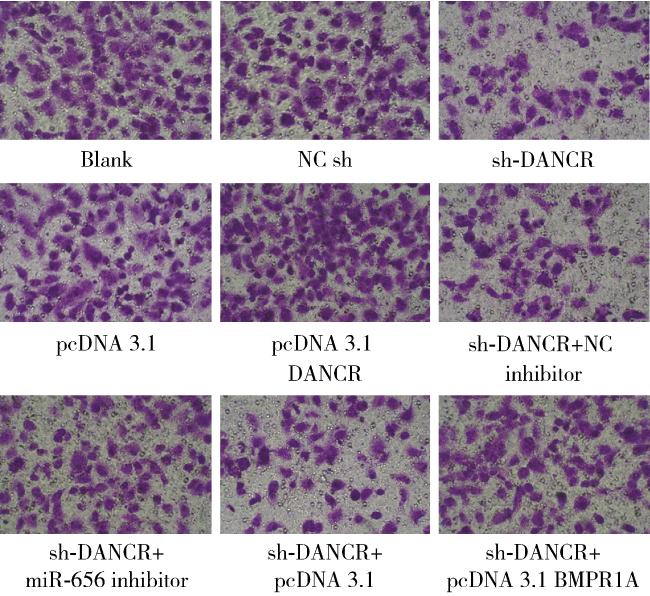
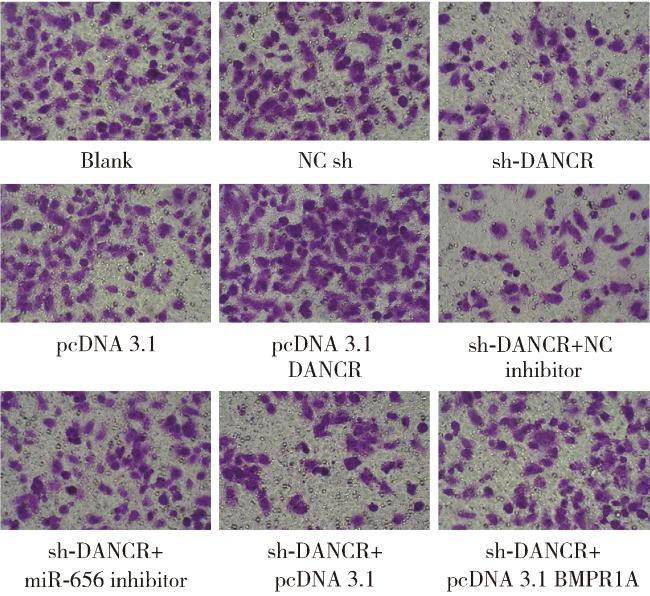
与空白组、sh-DANCR组相比,sh-DANCR组U87细胞侵袭、迁移数显著降低(P<0.05);与pcDNA 3.1组相比,pcDNA 3.1 DANCR组侵袭、迁移数显著增加(P<0.05);与sh-DANCR+NC抑制剂组相比,sh-DANCR+miR-656抑制剂组侵袭、迁移数显著增加(P<0.05);与sh-DANCR+pcDNA 3.1组相比,sh-DANCR+pcDNA 3.1 BMPR1A组侵袭、迁移数显著增加(P<0.05,图 12表 4)。
图1 观察U87细胞侵袭变化

Figure 1 Observation of invasion changes of U87 cells

NC, negative control; sh, short hairpin; DANCR, differentiation antagonizing non-protein coding RNA; BMPR1A, bone morphogenetic protein receptor type 1A.

图2 观察U87细胞迁移变化

Figure 2 Observation of migration changes of U87 cells

NC, negative control; sh, short hairpin; DANCR, differentiation antagonizing non-protein coding RNA; BMPR1A, bone morphogenetic protein receptor type 1A.

表4 U87细胞侵袭、迁移数变化

Table 4 Changes in invasion and migration numbers of U87 cells

Groups Invasion count Migration count
Blank 112.46±11.34 102.61±10.47
NC sh 108.94±11.27 105.81±10.62
sh-DANCR 70.88±7.18a, b 51.46±5.24a, b
pcDNA 3.1 111.74±11.20 104.55±10.53
pcDNA 3.1 DANCR 150.54±15.21c 145.84±14.75c
sh-DANCR+NC inhibitor 71.44±7.21 52.37±5.31
sh-DANCR+miR-656 inhibitor 108.72±11.04d 91.48±9.25d
sh-DANCR+pcDNA 3.1 71.67±7.20 52.53±5.29
sh-DANCR+pcDNA 3.1 BMPR1A 109.34±11.07e 91.66±9.28e

Data are $\bar x \pm s$, n=6. a, P<0.05, compared with the blank group; b, P<0.05, compared with the NC sh group; c, P<0.05, compared with pcDNA 3.1 group; d, P<0.05, compared with the sh-DANCR + NC inhibitor group; e, P<0.05, compared with the sh-DANCR+pcDNA 3.1 group. NC, negative control; sh, short hairpin; DANCR, differentiation antagonizing non-protein coding RNA; BMPR1A, bone morphogenetic protein receptor type 1A.

2.4 各组U87细胞中凋亡率变化

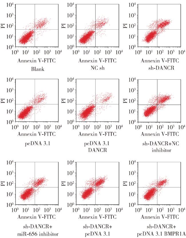
与空白组、sh-DANCR组相比,sh-DANCR组U87细胞凋亡率显著增加(P<0.05);与pcDNA 3.1组相比,pcDNA 3.1 DANCR组凋亡率显著降低(P<0.05);与sh-DANCR+NC抑制剂组相比,sh-DANCR+miR-656抑制剂组凋亡率显著降低(P<0.05);与sh-DANCR+pcDNA 3.1组相比,sh-DANCR+pcDNA 3.1 BMPR1A组凋亡率显著降低(P<0.05,图 3表 5)。
图3 观察U87细胞凋亡变化

Figure 3 Observation of apoptosis changes of U87 cells

NC, negative control; sh, short hairpin; DANCR, differentiation antagonizing non-protein coding RNA; BMPR1A, bone morphogenetic protein receptor type 1A.

表5 U87细胞各组细胞的凋亡率

Table 5 The apoptosis rates of U87 cells in each group

Group Apoptosis rate/%
Blank 12.51±1.28
NC sh 12.63±1.29
sh-DANCR 37.82±3.86a, b
pcDNA 3.1 12.58±1.28
pcDNA 3.1 DANCR 7.67±0.79c
sh-DANCR+NC inhibitor 37.64±3.79
sh-DANCR+miR-656 inhibitor 21.04±2.18d
sh-DANCR+pcDNA 3.1 37.74±3.82
sh-DANCR+pcDNA 3.1 BMPR1A 21.17±2.24e

Data are $\bar x \pm s$, n=6. a, P<0.05, compared with the blank group; b, P<0.05, compared with the NC sh group; c, P<0.05, compared with pcDNA 3.1 group; d, P<0.05, compared with the sh-DANCR + NC inhibitor group; e, P<0.05, compared with the sh-DANCR+pcDNA 3.1 group. NC, negative control; sh, short hairpin; DANCR, differentiation antagonizing non-protein coding RNA; BMPR1A, bone morphogenetic protein receptor type 1A.

2.5 各组U87细胞中DANCR、miR-656、BMPR1A mRNA表达

与空白组、sh-DANCR组相比,sh-DANCR组U87细胞DANCR、BMPR1A mRNA表达显著降低,miR-656表达显著增加(P<0.05);与pcDNA 3.1组相比,pcDNA 3.1 DANCR组DANCR、BMPR1A mRNA表达显著增加,miR-656表达显著降低(P<0.05);与sh-DANCR+NC抑制剂组相比,sh-DANCR+miR-656抑制剂组BMPR1A mRNA表达显著增加,miR-656表达显著降低(P<0.05),DANCR表达差异无统计学意义(P>0.05);与sh-DANCR+pcDNA 3.1组相比,sh-DANCR+pcDN A 3.1 BMPR1A组BMPR1A mRNA表达显著增加,miR-656表达、DANCR表达差异无统计学意义(P>0.05,表 6)。
表6 U87细胞DANCR、miR-656、BMPR1A mRNA表达变化

Table 6 Changes in mRNA expression of DANCR, miR-656, and BMPR1A in U87 cells

Group DANCR miR-656 BMPR1A mRNA
Blank 1.05±0.11 0.98±0.10 0.94±0.10
NC sh 1.06±0.11 1.02±0.11 0.98±0.10
sh-DANCR 0.45±0.05a, b 1.67±0.18a, b 0.55±0.06a, b
pcDNA 3.1 0.97±0.10 0.92±0.10 1.05±0.11
pcDNA 3.1 DANCR 1.55±0.16c 0.57±0.06c 1.72±0.18c
sh-DANCR+NC inhibitor 0.48±0.05 1.64±0.18 0.58±0.06
sh-DANCR+miR-656 inhibitor 0.46±0.05 1.08±0.11d 0.88±0.09d
sh-DANCR+pcDNA 3.1 0.47±0.05 1.62±0.18 0.56±0.06
sh-DANCR+pcDNA 3.1 BMPR1A 0.42±0.05 1.59±0.11 0.83±0.09e

Data are $\bar x \pm s$, n=6. a, P<0.05, compared with the blank group; b, P<0.05, compared with the NC sh group; c, P<0.05, compared with pcDNA 3.1 group; d, P<0.05, compared with the sh-DANCR + NC inhibitor group; e, P<0.05, compared with the sh-DANCR+pcDNA 3.1 group. NC, negative control; sh, short hairpin; DANCR, differentiation antagonizing non-protein coding RNA; BMPR1A, bone morphogenetic protein receptor type 1A.

2.6 验证DANCR、miR-656的靶向关系

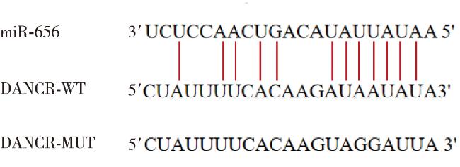
DANCR、miR-656预测结合位点见图 4。共转染miR-656模拟物与DANCR-WT的荧光素酶活性较NC模拟物与DANCR-WT降低(P<0.05,表 7)。
图4 DANCR、miR-656结合位点

Figure 4 Binding sites of DANCR and miR-656

DANCR, differentiation antagonizing non-protein coding RNA; WT, wild-type; MUT, mutant.

表7 DANCR、miR-656靶向关系

Table 7 Verification of targeting relationship between DANCR and miR-656

Group relative luciferase activity
NC mimics+DANCR-WT 1.07±0.11
miR-656 mimics+DANCR-WT 0.62±0.07a
NC mimics+DANCR-MUT 1.03±0.11
miR-656 mimics+DANCR-MUT 1.06±0.11

Data are $\bar x \pm s$. n=6. a, P <0.05, compared with the NC mimics+ DANCR-WT. NC, negative control; DANCR, differentiation antagonizing non-protein coding RNA; WT, wild-type; MUT, mutant.

2.7 各组U87细胞中BMPR1A、PD-1、PD-L1蛋白表达水平

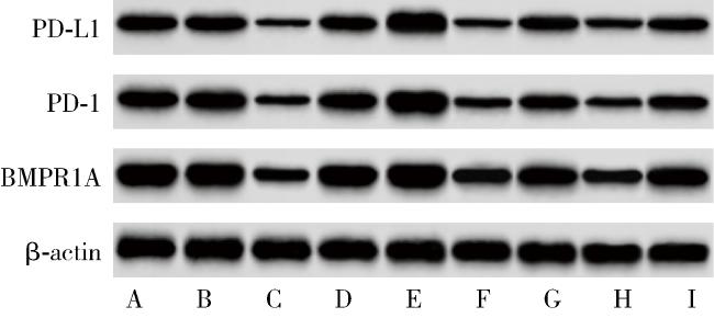
与空白组、sh-DANCR组相比,sh-DANCR组U87细胞BMPR1A、PD-1、PD-L1蛋白表达显著降低(P<0.05);与pcDNA 3.1组相比,pcDNA 3.1 DANCR组BMPR1A、PD-1、PD-L1蛋白表达显著增加(P<0.05);与sh-DANCR+NC抑制剂组相比,sh-DANCR+miR-656抑制剂组BMPR1A、PD-1、PD-L1蛋白表达显著增加(P<0.05);与sh-DANCR+pcDNA 3.1组相比,sh-DANCR+pcDNA 3.1 BMPR1A组BMPR1A、PD-1、PD-L1蛋白表达显著增加(P<0.05,图 5表 8)。
图5 细胞中BMPR1A、PD-1、PD-L1蛋白表达

Figure 5 Expression of BMPR1A, PD-1, and PD-L1 proteins in cells

A, blank group; B, NC sh group; C, sh-DANCR group; D, pcDNA 3.1 group; E, pcDNA 3.1 DANCR group; F, sh-DANCR + NC inhibitor group; G, sh-DANCR + miR-656 inhibitor group; H, sh-DANCR + pcDNA 3.1 group; I, sh-DANCR + pcDNA 3.1 BMPR1A group. NC, negative control; sh, short hairpin; DANCR, differentiation antagonizing non-protein coding RNA; BMPR1A, bone morphogenetic protein receptor type 1A.

表8 U87细胞BMPR1A、PD-1、PD-L1表达变化

Table 8 Changes in the expression of BMPR1A, PD-1, and PD-L1 in U87 cells

Groups BMPR1A/β-actin PD-1/β-actin PD-L1/β-actin
Blank 1.24±0.13 0.83±0.09 0.77±0.08
NC sh 1.27±0.13 0.87±0.09 0.71±0.08
sh-DANCR 0.65±0.07a,b 0.41±0.05a,b 0.34±0.04a,b
pcDNA 3.1 1.25±0.13 0.85±0.09 0.75±0.08
pcDNA 3.1 DANCR 1.68±0.17c 1.37±0.14c 1.14±0.12c
sh-DANCR+NC inhibitor 0.68±0.07 0.48±0.05 0.36±0.04
sh-DANCR+miR-656 inhibitor 0.92±0.10d 0.78±0.08d 0.64±0.07d
sh-DANCR+pcDNA 3.1 0.65±0.07 0.46±0.05 0.37±0.04
sh-DANCR+pcDNA 3.1 BMPR1A 0.97±0.10e 0.72±0.08e 0.66±0.07e

Data are $\bar x \pm s$, n=6. a, P<0.05, compared with the blank group; b, P<0.05, compared with the NC sh group; c, P<0.05, compared with pcDNA 3.1 group; d, P<0.05, compared with the sh-DANCR + NC inhibitor group; e, P<0.05, compared with the sh-DANCR+pcDNA 3.1 group. NC, negative control; sh, short hairpin; DANCR, differentiation antagonizing non-protein coding RNA; BMPR1A, bone morphogenetic protein receptor type 1A.

3 讨论

神经胶质瘤是最常见的中枢神经系统原发性恶性肿瘤,侵袭性较强,导致患者死亡率增加[9],神经胶质瘤的发生受遗传和环境因素以及不当的生活方式等因素的影响[10],识别新的治疗靶点在神经胶质瘤的发展、侵袭和转移过程中,有助于治疗效果和总生存期的改善[11],因此,迫切需要寻找新的治疗靶点。
LncRNA虽不能编码蛋白质,但长度超过200个核苷酸,参与转录和转录后调控,与各种癌症类型的发展有关,因此可作为癌症诊断和预后的生物标志物[12]。越来越多的研究表明,作为一种新发现的LncRNA,DANCR在多种人类癌症中呈现异常表达,并在癌症进展中发挥重要作用[13],如DANCR通过靶向miR-185-5p促进前列腺癌的恶性行为发展[14]。PD-1/PD-L1信号通路是免疫治疗的重要手段,阻断其发展有助于肿瘤微环境的改善,抑制肿瘤免疫逃逸[15]。本研究发现DANCR在神经胶质瘤细胞中高表达,与先前研究相吻合[16-17],沉默DANCR表达可明显抑制PD-1、PD-L1表达,阻止U87细胞的恶性行为发展,但过表达DANCR则提高PD-1、PD-L1表达,促进U87细胞恶性行为发展,提示DANCR可能通过改善肿瘤微环境来参与神经胶质瘤的发展,但其机制尚不明确。
LncRNA发挥作用的调控机制很复杂,研究者尚未完全了解,但其可通过表观遗传重塑、转录激活和转录后修饰等机制参与影响疾病发展[18]。据报道,LncRNAs通过直接结合miRNA中的互补序列并竞争性阻断miRNA,与其靶mRNA的相互作用而发挥其竞争性内源性RNA的作用[19]。如Jia等[20]研究表明,DANCR可通过靶向miR-5194/VEGFB信号治疗黑色素瘤。为了确定DANCR在神经胶质瘤中的调控模式,本研究通过数据库和荧光素酶报告分析确定miR-656是DANCR的潜在靶点,qRT-PCR结果显示miR-656在胶质瘤细胞系表达显著降低,且与Guo等[8]提出的miR-656通过抑制BMPR1A从而抑制胶质瘤发生的结论相一致。本研究进行相关实验验证,结果同样证实了抑制miR-656表达或过表达BMPR1A逆转了沉默DANCR对U87细胞恶性行为的抑制作用,表明DANCR参与神经胶质瘤的发展,可能与调节miR-656/BMPR1A轴有关。
综上所述,沉默LncRNA DANCR可改善肿瘤微环境,抑制U87细胞恶性生物学行为,可能与miR-656/BMPR1A轴有关,但由于涉及靶点较多,机制复杂,本课题组后续会继续开展相关机制研究。

利益冲突  所有作者均声明不存在利益冲突。

作者贡献声明  王欧洋:提出研究思路,设计研究方案,收集、整理、分析数据,撰写论文;朱鹏磊:协助细胞试验,收集、整理数据;林杰:协助收集、整理数据;吴昊:总体把关和审定论文。所有作者均参与论文修改,并对最终文稿进行审读和确认。

1
Chen Q , Wang W , Wu Z , et al. Over-expression of lncRNA TMEM161B-AS1 promotes the malignant biological behavior of glioma cells and the resistance to temozolomide via up-regulating the expression of multiple ferroptosis-related genes by sponging hsa-miR-27a-3p[J]. Cell Death Discov, 2021, 7, 311.

DOI

2
Qi ZY , Wang LL , Qu XL . lncRNA LINC00355 acts as a novel biomarker and promotes glioma biological activities via the regulation of miR-1225/FNDC3B[J]. Dis Markers, 2021, 2021, 1683129.

3
Liu R , Wu M , Xu G , et al. Ropivacaine inhibits proliferation, migration, and invasion while inducing apoptosis of glioma cells by regulating the SNHG16/miR-424-5p axis[J]. Open Life Sci, 2020, 15 (1): 988- 999.

DOI

4
Cai Y , Wang M , Cui Y , et al. Differential expression profile of lncRNA in glioma cells and the effect of lncRNA NKX3-1 on glioma cells through Fem1b/SPDEF pathway[J]. Front Oncol, 2021, 11, 706863.

DOI

5
Wang YY , Chen C . lncRNA-DANCR promotes taxol resistance of prostate cancer cells through modulating the miR-33b-5p-LDHA axis[J]. Dis Markers, 2022, 2022, 9516774.

6
Yu W , Ma L , Li X . DANCR promotes glioma cell autophagy and proliferation via the miR-33b/DLX6/ATG7 axis[J]. Oncol Rep, 2023, 49 (2): 39- 51.

DOI

7
Jin Z , Li H , Long Y , et al. microRNA-1269 is downregulated in glioblastoma and its maturation is regulated by long non-coding RNA SLC16A1 antisense RNA 1[J]. Bioengineered, 2022, 13 (5): 12749- 12759.

DOI

8
Guo M , Jiang Z , Zhang X , et al. miR-656 inhibits glioma tumorigenesis through repression of BMPR1A[J]. Carcinogenesis, 2014, 35 (8): 1698- 1706.

DOI

9
Shree B , Tripathi S , Sharma V . Transforming growth factor-beta-regulated LncRNA-MUF promotes invasion by modulating the miR-34a Snail1 axis in glioblastoma multiforme[J]. Front Oncol, 2022, 11, 788755.

DOI

10
Li Z , Li M , Xia P , et al. Targeting long non-coding RNA PVT1/TGF-β/Smad by p53 prevents glioma progression[J]. Cancer Biol Ther, 2022, 23 (1): 225- 233.

DOI

11
Zhong X , Cai Y . Long non-coding RNA (lncRNA) HOXD-AS2 promotes glioblastoma cell proliferation, migration and invasion by regulating the miR-3681-5p/MALT1 signaling pathway[J]. Bioengineered, 2021, 12 (2): 9113- 9127.

DOI

12
Jia H , Yan X . lncRNA MEG3 inhibits the proliferation and growth of glioma cells by downregulating bcl-xL in the PI3K/Akt/NF-κB signal pathway[J]. BioMed Res Int, 2022, 2022, 3729069.

DOI

13
Ping Q , Shi Y , Yang M , et al. LncRNA DANCR regulates lymphatic metastasis of bladder cancer via the miR-335/VEGF-C axis[J]. Transl Androl Urol, 2021, 10 (4): 1743- 1753.

DOI

14
Sun W , Zu S , Shao G , et al. Long non-coding DANCR targets miR-185-5p to upregulate LIM and SH3 protein 1 promoting prostate cancer via the FAK/PI3K/AKT/GSK3β/snail pathway[J]. J Gene Med, 2021, 23 (7): e3344.

DOI

15
陈兰玉, 胡凯文. 基于PD-1/PD-L1信号通路中医药改善肿瘤免疫抑制微环境研究进展[J]. 中国中医药信息杂志, 2020, 27 (2): 135- 137.

16
Feng L , Lin T , Che H , et al. Long noncoding RNA DANCR knockdown inhibits proliferation, migration and invasion of glioma by regulating miR-135a-5p/BMI1[J]. Cancer Cell Int, 2020, 20 (1): 53.

DOI

17
Han J , Yu X , Wang S , et al. IGF2BP2 induces U251 glioblastoma cell chemoresistance by inhibiting FOXO1-mediated PID1 expression through stabilizing lncRNA DANCR[J]. Front Cell Dev Biol, 2022, 9, 659228.

DOI

18
Zhao H , Li J , Yan X , et al. LncRNA MAFG-AS1 suppresses the maturation of miR-34a to promote glioblastoma cell proliferation[J]. Cancer Manag Res, 2021, 13, 3493- 3501.

DOI

19
Zheng D , Chen D , Lin F , et al. LncRNA NNT-AS1 promote glioma cell proliferation and metastases through miR-494-3p/PRMT1 axis[J]. Cell Cycle, 2020, 19 (13): 1621- 1631.

DOI

20
Jia J , Zhu X , Xue K , et al. LncRNA DANCR enhances angiogenesis to promote melanoma progression via sponging miR-5194[J]. J Cancer, 2023, 14 (7): 1161- 1173.

DOI

Outlines

/