Clinical and imaging characteristics and etiology of 544 cases with chronic sialadenitis

  • Jing YANG ,
  • Xiaoyun XU ,
  • Danni ZHENG ,
  • Xiaotong LING ,
  • Liuyang QU ,
  • Denggao LIU , *
Expand
  • Department of Oral and Maxillofacial Radiology, Peking University School and Hospital of Stomatology & National Center for Stomatology & National Clinical Research Center for Oral Diseases & National Engineering Research Center of Oral Biomaterials and Digital Medical Devices & Beijing Key Laboratory of Digital Stomatology & NHC Research Center of Engineering and Technology for Computerized Dentistry & NMPA Key Laboratory for Dental Materials, Beijing 100081, China
LIU Denggao, e-mail,

Received date: 2024-04-18

  Online published: 2025-03-07

Copyright

All rights reserved. Unauthorized reproduction is prohibited.

Abstract

Objective: To investigate the etiology of chronic sialadenitis, and to analyze their clinical and imaging characteristics. Methods: This retrospective analysis reviewed the clinical and imaging data of patients with chronic sialadenitis who underwent sialendoscopy at the Peking University Hospital of Stomatology between January 2021 and August 2023. Inclusion criteria: (1) with a history of recurrent swelling of major salivary glands; (2) complete medical records with detailed information about potential causes; (3) sialography images were available; and (4) patients had undergone interventional endoscopy. Exclusion criteria: (1) salivary stones; (2) juvenile recurrent parotitis; (3) IgG4-related sialadenitis; (4) Sjögren syndrome; and (5) neoplastic diseases. Based on the latest research results and clinical data, chronic sialadenitis was classified into radioactive iodine-induced sialadenitis (RAIS), allergy-related sialodochitis (ARS), adult chronic recurrent parotitis (ACRP), sialadenosis with sialodochitis, and idiopathic sialadenitis. Idiopathic sialadenitis was defined as a type of chronic sialadenitis with duct stenosis of unknown etiology (allergic causes, autoimmune disorders, radioactive iodine exposure, history of "parotitis" in childhood, etc.). The proportions of five types of sialadenitis were calculated, and their relationships with age, gender, type of affected glands, number of affected glands, duration of symptoms, and imaging characteristics were statistically analyzed. Results: A total of 544 consecutive patients diagnosed with chronic sialadenitis were enrolled in this study. They consisted of 192 males and 352 females, and their ages ranged from 14 to 83 years [mean age: (47.44±13.52) years]. Idiopathic sialadenitis accounted for the largest proportion (71.7%, 390 cases), followed by ARS (12.5%, 68 cases), RAIS (6.4%, 35 cases), ACRP (4.8%, 26 cases), and sialadenosis with sialodochitis (4.6%, 25 cases). Among the 2 176 available glands of the 544 patients, 1 120 (51.5%) glands were affected, including 880 (78.6%) parotid glands and 240 (21.4%) submandibular glands. These five types of sialadenitis exhibited significant differences in gender, age, type and number of affected glands, and duration of symptoms (P < 0.05). Among them, RAIS patients showed the lowest male to female ratio (male ∶ female=1 ∶ 4.83), ARCP patients were the youngest [(32.50±8.60) years], and RAIS and ARS patients had relatively higher number of affected glands. Sialography showed ductal atresia in 23.2% of affected glands with ARIS, "snowflake" pattern in 46.5% of affected glands with ARS, "punctate and globular" ectasia of terminal ducts in 80.4% of affected glands with ARCP, and clustered branch ducts in 71.4% of affected glands with sialadenosis with sialodochitis. Moreover, stenosis and/or dilatation of the main and branch ducts represented the most typical sialography appearance of idiopathic sialadenitis. Conclusion: Idiopathic sialadenitis, RAIS, ARS, ACRP, and sialadenosis with sialodochitis are the five common types of chronic sialadenitis. Among these, idiopathic sialadenitis is the most common type of chronic sialadenitis. Clarification of the etiology, clinical manifestations and imaging characteristics of chronic sialadenitis is important for clinicians to develop personalized treatment plans and improve treatment outcomes.

Cite this article

Jing YANG , Xiaoyun XU , Danni ZHENG , Xiaotong LING , Liuyang QU , Denggao LIU . Clinical and imaging characteristics and etiology of 544 cases with chronic sialadenitis[J]. Journal of Peking University(Health Sciences), 2026 , 58(3) : 650 -657 . DOI: 10.19723/j.issn.1671-167X.2026.03.027

慢性唾液腺炎是大唾液腺最常见的良性疾病,主要表现为受累腺体反复肿胀,多与进食相关,通常发生于单侧,也可双侧发生。目前已知的能够引起慢性唾液腺炎的病因较多,包括唾液腺阴性结石、导管狭窄、黏液栓、异物或导管系统的解剖变异等,也可能与放射性碘治疗、自身免疫性因素、过敏性因素、代谢性因素或特发性因素有关[1-4]。传统类型的慢性唾液腺炎还包括慢性复发性腮腺炎、唾液腺结石伴发的唾液腺炎等。近年来,一些新的唾液腺炎类型逐渐被发现,主要包括嗜酸性唾液腺导管炎、131I相关唾液腺炎和IgG4相关唾液腺炎[5]
嗜酸性唾液腺导管炎近年来备受关注,许多报道认为过敏是该病的主要病因,并强调富含嗜酸性粒细胞的黏液栓、外周血嗜酸性粒细胞增多和血清IgE升高是该病的重要特征。此外,组织病理学研究显示, 该病大唾液腺导管周围有嗜酸性粒细胞浸润。多年来,不同的学者对该疾病有不同的命名,本研究将其统称为过敏相关唾液腺炎[6-11]131I相关唾液腺炎是分化型甲状腺癌131I辅助治疗后出现的并发症[12],它的分子病理学基础在于唾液腺和甲状腺都表达钠-碘共转运蛋白(sodium iodide symporter,NIS),NIS在唾液腺中主要分布于导管上皮的基底膜上,负责摄取和分泌碘离子[12-13]。服用的131I约24%通过唾液排泄,唾液中的131I浓度可达血浆的20~100倍[14-16]131I在衰变过程中释放β射线,使暴露于其中的细胞经历急、慢性炎症反应,受累细胞发生炎性肿胀,部分细胞脱落后形成黏液栓,导致导管狭窄和分泌功能障碍[5, 17-18]。IgG4相关性疾病是由免疫介导的纤维炎症性疾病,可累及多个器官及组织,尤以胰腺、唾液腺、胆管、肾和肺为常见,累及唾液腺者称为IgG4相关唾液腺炎[19-22]
目前,尚未见有研究分析慢性唾液腺炎的不同病因,也未见有研究对不同病因导致的慢性唾液腺炎的临床及影像学特点进行对比。因此,本研究回顾了北京大学口腔医院收治的慢性唾液腺炎患者,按不同病因进行分类后分析其临床和影像学特点,以期指导临床诊断和治疗。

1 资料与方法

1.1 研究对象及资料来源

选择2021年1月至2023年8月就诊于北京大学口腔医院并行唾液腺内镜检查的各类慢性唾液腺炎患者。纳入标准:(1)有腺体反复肿胀等明显临床症状;(2)有完整可用的病历资料;(3)病历中有关于病因的详细记录;(4)有造影图像;(5)有唾液腺内镜诊疗史。排除标准:(1)唾液腺结石病;(2)儿童复发性腮腺炎;(3)IgG4相关唾液腺炎;(4)干燥综合征;(5)唾液腺肿瘤等其他病变。本研究为回顾性病例对照研究,获得北京大学口腔医院生物医学伦理委员会批准(批准号:PKUSSIRB-202162018),所有患者均知情同意。
研究对象的临床及影像学资料来源于北京大学口腔医院电子病历资料、医学影像科Carestream Health 11.0 PACS系统及Mindray Resona 7超声工作站。收集患者的年龄、性别、受累腺体类别和数目、病程等信息,其中,受累腺体类别包括仅累及腮腺、仅累及下颌下腺、同时累及腮腺及下颌下腺,受累腺体数目分为1、2、3或4,病程为首次发病至首次就诊的时间间隔(成人复发性腮腺炎的病程为幼年初次发病至来院就诊时间间隔)。

1.2 分类标准

本研究基于临床经验和国内外最新研究成果[5-11, 16, 19],依据以下标准对慢性唾液腺炎进行初步分类。
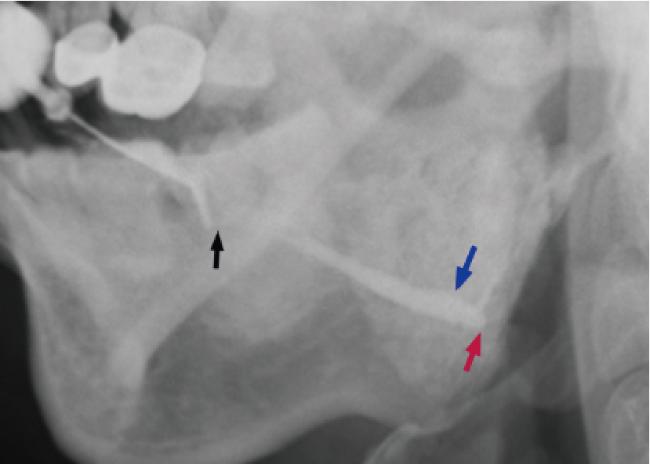
131I相关唾液腺炎:(1)有131I治疗史;(2)131I治疗前无慢性阻塞性唾液腺炎病史;(3)131I治疗后出现唾液腺区肿痛症状;(4)造影表现为主导管狭窄、扩张或中断闭锁,分支导管大致正常或显影少,腺体可有萎缩,排空迟缓(图 1)。
图1 131I相关唾液腺炎的造影图像

Figure 1 Sialogram of radioactive iodine-induced sialadenitis

A 48-year-old female patient with recurrent swelling of bilateral parotid and submandibular glands for a period of 8 months. These symptoms occurred one year after surgery for thyroid cancer and radioactive iodine therapy. Sialogram of the right parotid gland showed stenosis of distal duct (black arrow), ectasia of hilum duct (blue arrow) and atresia of intraglandular duct (red arrow).

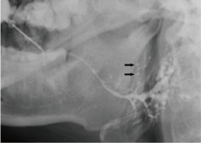
过敏相关唾液腺炎:(1)有过敏性鼻炎、哮喘、荨麻疹等过敏性疾病史;(2)多个腺体反复肿胀,可伴痒;(3)常分泌黏液栓子;(4)实验室检查末梢血象嗜酸性粒细胞增高或IgE水平增高;(5)造影表现为主导管有狭窄及扩张表现,分支导管可呈“雪花样”扩张,排空迟缓(图 2);(6)可除外IgG4相关唾液腺炎表现。
图2 过敏相关唾液腺炎的造影图像

Figure 2 Sialogram of allergy-related sialodochitis

A 29-year-old female with comorbid allergic rhinitis complained of recurrent swelling and mucus plug exudation of four major salivary glands for 3 years. Laboratory tests showed an elevated level of immunoglobulin E (128.9 IU/mL; Normal range: ≤100 IU/mL). Sialogram of the right parotid gland showed ectasia of the main duct and "snowflake" appea-rance of the branch ducts (black arrow).

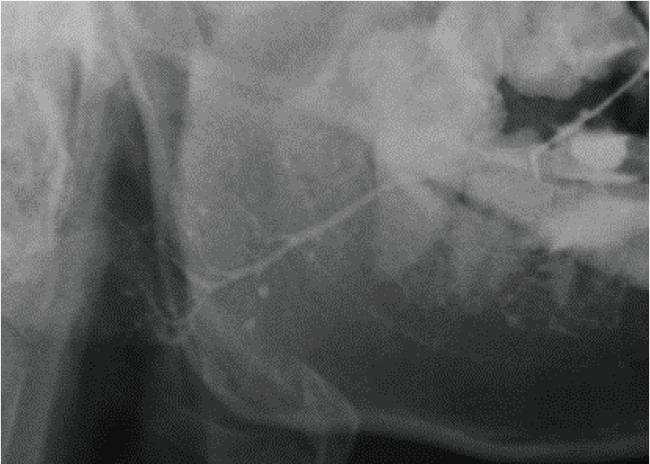
成人复发性腮腺炎:(1)有幼儿腮腺炎病史;(2)挤压腺体可见导管口有脓液或胶冻状液体溢出;(3)造影表现为主导管无明显异常或稍扩张不整,末梢导管可见“点球状”扩张,排空迟缓(图 3);(4)可除外干燥综合征。
图3 成人复发性腮腺炎的造影图像

Figure 3 Sialogram of adult chronic recurrent parotitis

A 24-year-old male patient with recurrent swelling and pain of the left parotid gland for half a year. He complained of a history of "parotitis" 10 years ago. Sialography showed "punctate and globular" ectasia of the terminal ducts of the left parotid gland.

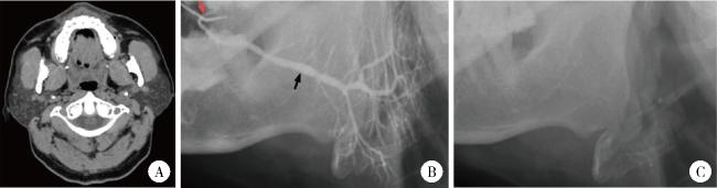
腮腺良性肥大伴管炎:(1)有腮腺反复肿胀史;(2)腮腺弥漫性肿大,柔软无肿块,常为双侧性,导管口无红肿,分泌物较清亮;(3)有肝病、糖尿病或嗜酒等病史;(4)螺旋CT或超声检查显示腺体弥漫性增大,无异常占位征象(图 4A);(5)腮腺造影显示腺体体积明显增大,分支呈丛簇状,主导管存在狭窄与扩张,排空迟缓(图 4BC)。
图4 腮腺良性肥大伴管炎的造影及CT图像

Figure 4 Sialogram and CT images of sialadenosis with sialodochitis

A 45-year-old male with long-term drinking and smoking habits complained of persistent swelling of bilateral parotid glands for 7-8 years. A, enlarged bilateral parotid glands on the axial CT image; B, stenosis (red arrow) and ectasia (black arrow) of the main duct, and clustered branch ducts on the sialogram of the right parotid gland; C, persistent contrast media on the functional film.

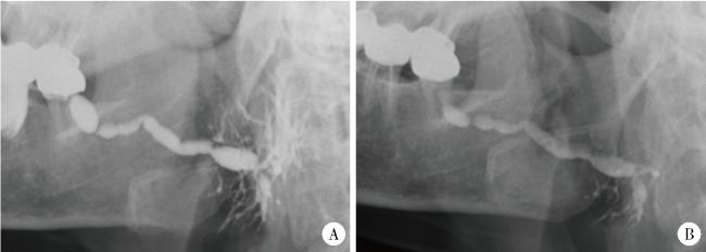
原发性导管狭窄性唾液腺炎:(1)未发现可能导致导管狭窄的病因(包括过敏因素、幼儿期腮腺炎病史、自身免疫性疾病、放射治疗或 131I治疗等);(2)有反复进食肿胀史,挤压腺体,导管口流出混浊液体;(3)造影表现为主导管、叶间、小叶间导管部分狭窄、部分扩张,可呈腊肠样改变,排空迟缓(图 5)。
图5 原发性导管狭窄性唾液腺炎的造影图像

Figure 5 Sialogram of idiopathic sialadenitis

A 72-year-old female with a 3-month history of recurrent swelling of the right parotid gland. A, sausage-like appearance of the main duct of the right parotid gland; B, persistent contrast media on the functional film.

1.3 统计学方法

采用SPSS 24.0对数据进行统计分析。计量资料用均值和标准差描述,采用Welch检验和Tamhane T2检验进行组间比较;计数资料用频数及百分比描述,采用卡方检验进行组间比较。显著性水平α为0.05。

2 结果

共纳入544例慢性唾液腺炎患者,包括男性192例、女性352例,男女比例约为1 ∶ 1.8,平均就诊年龄(47.44±13.52)岁(14~83岁)。其中,原发性导管狭窄性唾液腺炎最多,占71.7%(390例);过敏相关唾液腺炎次之,占12.5%(68例);131I相关唾液腺炎占6.4%(35例),成人复发性腮腺炎占4.8%(26例),腮腺良性肥大伴管炎占4.6%(25例)。慢性唾液腺炎主要发生于腮腺,417例(76.7%)仅累及腮腺,73例(13.4%)仅累及下颌下腺,54例(9.9%)同时累及腮腺和下颌下腺(表 1)。
表1 五类慢性唾液腺炎的临床特征

Table 1 Clinical features of five types of chronic sialadenitis

Item Total (n=544) Idiopathic sialadenitis (n=390) ARS (n=68) RAIS (n=35) ACRP (n=26) Sialadenosis (n=25) P value
Gender, n(%) 0.025
  Male 192 (35.3) 137 (35.1) 23 (33.8) 6 (17.1) 12 (46.2) 14 (56.0)
  Female 352 (64.7) 253 (64.9) 45 (66.2) 29 (82.9) 14 (53.8) 11 (44.0)
Age/years, $\bar x \pm s$ 47.44±13.52 49.15±13.47 46.16±11.82 39.71±10.56 32.50±8.60 50.72±13.36 < 0.001
Type of affected glands, n(%) < 0.001
  PG only 417 (76.7) 319 (81.8) 28 (41.2) 19 (54.3) 26 (100.0) 25 (100.0)
  SMG only 73 (13.4) 53 (13.6) 20 (29.4) 0 (0.0) 0 (0.0) 0 (0.0)
  PG & SMG 54 (9.9) 18 (4.6) 20 (29.4) 16 (45.7) 0 (0.0) 0 (0.0)
Number of affected glands, n(%) < 0.001
  1 68 (12.5) 61 (15.6) 3 (4.4) 2 (5.7) 1 (3.8) 1 (4.0)
  2 424 (77.9) 313 (80.3) 45 (66.2) 17 (48.6) 25 (96.2) 24 (96.0)
  3 or 4 52 (9.6) 16 (4.1) 20 (29.4) 16 (45.7) 0 (0.0) 0 (0.0)
Duration of symptoms/years, n(%) < 0.001
  ≤1 282 (51.8) 215 (55.1) 21 (30.9) 30 (85.7) 0 (0.0) 16 (64.0)
  >1 & ≤10 205 (37.7) 148 (37.9) 42 (61.8) 5 (14.3) 2 (7.7) 8 (32.0)
  >10 57 (10.5) 27 (6.9) 5 (7.4) 0 (0.0) 24 (92.3) 1 (4.0)

ARS, allergy-related sialodochitis; RAIS, radioactive iodine-induced sialadenitis; ACRP, adult chronic recurrent parotitis; PG, parotid gland; SMG, submandibular gland.

卡方检验显示,五类唾液腺炎在性别构成上差异有统计学意义(P=0.025),除腮腺良性肥大伴管炎患者男性比女性略多外(男∶女=1 ∶ 0.79),其余四类唾液腺炎均为女性多发,其中131I相关唾液腺炎女性占比最高(男∶女=1 ∶ 4.83)。Welch检验显示五类唾液腺炎在就诊年龄上差异亦存在统计学意义(P < 0.05);Tamhane T2检验多重比较提示,131I相关唾液腺炎患者的年龄与过敏相关唾液腺炎之间差异无统计学意义,但明显小于腮腺良性肥大伴管炎及原发性导管狭窄性唾液腺炎;成人复发性腮腺炎患者的平均年龄最小,与其他四类唾液腺炎之间差异均存在统计学意义(P < 0.05,表 1)。
卡方检验显示,五类唾液腺炎的受累腺体类别差异存在统计学意义(P<0.05)。成人复发性腮腺炎和腮腺良性肥大仅累及腮腺;390例原发性导管狭窄性唾液腺炎患者中,81.8%仅累及腮腺,13.6%仅累及下颌下腺,4.6%同时累及腮腺和下颌下腺;68例过敏相关唾液腺炎患者中,41.2%仅累及腮腺,29.4%仅累及下颌下腺,29.4%同时累及腮腺和下颌下腺;131I相关唾液腺炎的患者腮腺均受累,54.3%仅累及腮腺,45.7%同时累及腮腺和下颌下腺(表 1)。
本研究纳入的544例患者中,4例于初次就诊前因腺体反复肿胀已行腺体摘除术,故共存在2 171侧腺体(1 086侧腮腺和1 085侧下颌下腺),其中1 120侧(51.6%)腺体受累,包括腮腺880侧(78.6%)和下颌下腺240侧(21.4%)。卡方检验显示五类唾液腺炎在受累腺体数目上差异存在统计学意义(P < 0.05)。成人复发性腮腺炎和腮腺良性肥大多累及双侧腮腺,少数累及单侧腮腺;131I相关唾液腺炎和过敏相关唾液腺炎多累及双侧腮腺和/或双侧下颌下腺,受累腺体数目较多,多数为2或4个;原发性导管狭窄性唾液腺炎多数(68.5%)累及双侧腮腺,少数可累及下颌下腺,或同时累及腮腺和下颌下腺,受累腺体数目为1~4个(表 1)。
卡方检验显示五类唾液腺炎在病程分布上差异存在统计学意义(P < 0.05)。原发性导管狭窄性唾液腺炎患者中病程≤1年者占55.1%,>1年且≤10年者占37.9%,>10年者占6.9%;腮腺良性肥大患者中病程≤1年者占64.0%,>1年且≤10年者占32.0%,>10年者占4.0%;131I相关唾液腺炎患者的病程均≤10年,且绝大多数(85.7%)病程≤1年;过敏相关唾液腺炎患者中大多数(61.8%)病程>1年且≤10年;成人复发性腮腺炎患者的病程均>1年,且绝大多数(92.3%)病程>10年(表 1)。
131I相关唾液腺炎的造影特点是主导管狭窄及扩张或中断闭锁,99侧受累腺体中22侧(23.2%)造影表现为导管闭锁,内镜检查显示14侧(14.1%)导管闭锁。过敏相关唾液腺炎的造影也有主导管狭窄及扩张表现,分支导管可呈雪花样扩张(46.5%,80/172)。成人复发性腮腺炎的造影检查显示主导管多有狭窄及扩张表现,51侧受累腺体中41侧(80.4%)的分支导管有末梢“点球状”扩张表现。腮腺良性肥大伴管炎的CT检查显示腺体体积明显增大,造影见主导管粗细不均改变,多数(71.4%)受累腺体分支导管呈丛簇状表现。原发性导管狭窄性唾液腺炎的造影表现以主导管(主导管和分支导管)狭窄和(或)扩张为主。

3 讨论

对于慢性唾液腺炎病因的探索一直是研究者关注的问题,了解其发病机制对疾病的治疗和预防至关重要。临床上常见的唾液腺导管狭窄、阴性结石、黏液栓等都可能导致唾液排泌受阻,进而造成唾液淤滞和腺体炎症,临床上可通过超声、造影、CT和唾液腺内镜等检查进一步明确[1-4]。这些现象背后的病因可能包括过敏性因素、131I治疗、自身免疫性因素(包括干燥综合征、IgG4相关唾液腺炎、嗜酸性粒细胞淋巴肉芽肿等)、唾液腺退行性肿大、发育因素、创伤性瘢痕等[1-4, 19, 23-25]。目前,唾液腺内镜已成为各类唾液腺炎性疾病的重要诊疗手段,但各类慢性唾液腺炎性疾病的概念较为混乱,缺乏统一的诊断标准[5]。本研究基于临床经验和国内外最新的研究成果[5-11, 16, 19],对慢性唾液腺炎进行了初步的分类,可帮助临床医师厘清不同类型的慢性唾液腺炎在发病原因和临床特点上的差异,进而指导疾病的诊断及治疗。此外,本研究还统计分析了不同病因所致慢性唾液腺炎比例,这将有助于进一步研究慢性唾液腺炎的发病机制。
唾液腺良性阻塞性疾病主要包含唾液腺结石和唾液腺炎两大类,它们可互为彼此的致病因素,也可同时发生[26-28],但二者在治疗方法和预后方面存在较大差异[29-30],故本研究排除了唾液腺结石患者。IgG4相关唾液腺炎及干燥综合征这两类疾病属于自身免疫性疾病,其明确诊断有严格的临床、影像、实验室及组织学标准,主要依赖药物治疗[19],因此,本研究也未纳入这两类唾液腺炎患者。关于儿童复发性腮腺炎,一般认为与腮腺发育不全、免疫功能低下和细菌逆行感染有关[31-33];由于患者年龄较小,多不配合检查和治疗,且随着年龄增长,其症状会逐渐缓解或痊愈,因此绝大多数采取保守治疗[31];另外,此类患者的临床资料也多不完整,故本研究未纳入此部分患者。成人复发性腮腺炎为儿童复发性腮腺炎延期未愈导致[31-33],本研究中占比为4.8%,其诊断主要依据临床表现和腮腺造影[32]。目前尚无研究表明腮腺良性肥大是慢性唾液腺炎的明确病因,但在本研究中腮腺良性肥大伴管炎占据一定比例(4.6%),不排除腮腺良性肥大可能与慢性唾液腺炎的发生存在某种潜在关联。
过敏相关唾液腺炎和131I相关唾液腺炎是两种新型唾液腺炎,除原发性导管狭窄性唾液腺炎外,它们在本研究患者中的占比相对较大。过敏相关唾液腺炎的诊断标准目前仍有待明确[7],近年来该类患者数量增多,在本研究中占12.5%,可能与环境因素有关,也可能与近期的相关研究增加了医生对该疾病的认识有关[5]。甲状腺癌术后采用131I治疗的目的是消融甲状腺残余物,治疗残余或转移性肿瘤[34]。近年来随着甲状腺癌的发病率增高,131I治疗被广泛地应用,因此131I相关唾液腺炎患者也明显增多[5]131I相关唾液腺炎的病程最长为10年,但大多数(85.7%)不超过1年,可能与131I相关唾液腺炎发展到后期会发生腺体萎缩有关。唾液腺萎缩后,唾液排泌功能下降,腺体肿痛症状缓解,可能会降低患者的就诊欲望[15, 35]
尽管上述几类唾液腺炎的病因相对明确,但目前临床上仍有相当比例的患者病因不明。本研究回顾了所有研究对象的病史、临床表现及辅助检查结果,对于不能被归为131I相关唾液腺炎、过敏相关唾液腺炎、成人复发性腮腺炎或腮腺良性肥大伴管炎的慢性唾液腺炎,结合临床和影像学表现,将其命名为原发性导管狭窄性唾液腺炎,也可称为单纯导管狭窄性唾液腺炎,这部分患者在本研究中占大多数(71.7%)。分析其原因,可能主要由局部因素(如磨牙咀嚼时反复刺激颊黏膜使导管口形成瘢痕,或修复体微电流刺激、医疗操作等引起导管口肿胀或瘢痕)导致导管口狭窄,唾液排出不畅,从而诱发炎症反应;导管腔内的慢性炎症逐渐进展,最终使导管的其他部位也发生狭窄,进一步加重唾液腺炎症[27, 36-37]。该类唾液腺炎导管狭窄的数目、位置、范围、程度等特征尚有待深入探索。
本组慢性唾液腺炎患者多为中年人[平均年龄(47.44±13.52)岁],且女性较男性多(男女比例约为1 ∶ 1.8),各类唾液腺炎的性别构成差异也存在统计学意义。成人复发性腮腺炎患者的年龄在各类唾液腺炎中最小,可能原因是成人复发性腮腺炎为儿童复发性腮腺炎迁延不愈而来[31-33],较其他疾病发病早,因此就诊时间也较早。慢性唾液腺炎主要累及腮腺,其次是下颌下腺,累及舌下腺者罕见。各类慢性唾液腺炎的受累腺体类别和数目差异存在统计学意义:成人复发性腮腺炎和腮腺良性肥大只累及腮腺,多为双侧发生;原发性导管狭窄性唾液腺炎多数双侧发生,绝大多数(>80%)仅累及腮腺,少数累及下颌下腺;过敏相关唾液腺炎和131I相关唾液腺炎亦多数有腮腺受累,而58.8%的过敏相关唾液腺炎和45.7%的131I相关唾液腺炎有下颌下腺受累,因此这两类唾液腺炎的受累腺体数目相对较多,多为2~4个,这可能与这两种新型唾液腺炎为全身性疾病所致有关[19]
慢性唾液腺炎的治疗方法主要包括保守治疗和手术治疗。保守治疗方法仅对轻症有效。对于症状顽固、迁延不愈者,传统的手术治疗方法是腺体摘除术,但会导致腺体功能丧失、面部外形塌陷、神经损伤等严重并发症[1]。过敏相关因素和131I治疗是导致慢性唾液腺炎的重要原因[5],这在过去经常被忽视,从而未能及时预防和给予更有效的治疗。过敏相关唾液腺炎患者常分泌黏液栓,黏液栓由脱落的上皮细胞、嗜酸性粒细胞和浓缩的黏蛋白聚集而成,是导致导管阻塞的重要原因,临床上可采用内镜冲洗治疗和网篮套索来清除。此外,避免过敏源刺激并进行抗过敏治疗,也是防治该疾病的必要手段。131I相关唾液腺炎的治疗以唾液腺自身维护疗法为基础,药物治疗(硝酸毛果芸香碱等催唾剂)疗效不确切,有条件时可选择唾液腺内镜治疗[5]。对于腮腺良性肥大伴管炎患者,除常规保守治疗和内镜治疗外,还应重视去除致病原因,指导患者控制全身疾病、戒酒、均衡饮食等[38]。儿童或成人复发性腮腺炎有一定的自愈性,采用保守治疗和药物灌注治疗可获得明显疗效,对症状较重者可进行内镜治疗[32]。虽然这几类慢性唾液腺炎均可采用内镜治疗,但在治疗细节和预后上有所差别,因此,确定慢性唾液腺炎的病因将有利于临床医师优化治疗方案并提高疗效。
本研究的不足之处:(1)临床上对唾液腺炎患者通常不采用活体组织检查,分类时主要依据临床和影像特点,部分结合了实验室检查结果,因此,本研究提出的分类结果可能不够完善和精准;(2)本研究为回顾性研究,病因记录的完整性和准确性受患者的回忆和医师的主观判断影响,部分成人复发性腮腺炎患者本人和亲属可能无法确认是否存在幼儿期发病史而未被纳入研究;(3)个别可疑为过敏相关唾液腺炎的患者因未能完善免疫学相关实验室检查而被归类为原发性导管狭窄性唾液腺炎,可能低估了过敏相关唾液腺炎的占比;(4)研究样本来自在我院行唾液腺内镜诊疗的病例资料,可能难以反映自然人群中不同类型唾液腺炎的构成比。
综上,目前临床上较为常见的慢性唾液腺炎类型分别为原发性导管狭窄性唾液腺炎、过敏相关唾液腺炎、131I相关唾液腺炎、成人复发性腮腺炎及腮腺良性肥大伴管炎,其在性别、年龄、受累腺体的类别和数目以及病程分布上均存在明显差异,并具有不同的临床及影像学特征。本研究对慢性唾液腺炎的分类将有助于临床医师针对不同病因的患者进行个性化治疗,以获得更好的治疗效果,但由于其确切发病机制尚不明确,有关慢性唾液腺炎的分类和概念仍有待未来进一步补充和完善。

利益冲突   所有作者均声明不存在利益冲突。

作者贡献声明  杨静:设计研究方案,整理文献,收集、分析、整理数据,撰写论文;许晓韵、郑丹妮、凌晓彤、屈留洋:收集、分析数据;柳登高:提出研究思路,总体把关和审定论文。所有作者均参与论文修改,并对最终文稿进行审读和确认。

1
Vashishta R , Gillespie MB . Salivary endoscopy for idiopathic chronic sialadenitis[J]. Laryngoscope, 2013, 123 (12): 3016- 3020.

DOI

2
Erdoǧan O , Özcan C , İsmi O , et al. Effectiveness of sialendoscopy on the symptoms of chronic obstructive sialadenitis and patient satisfaction degree[J]. J Stomatol Oral Maxillofac Surg, 2022, 123 (3): 314- 319.

DOI

3
Pace CG , Hwang KG , Papadaki M , et al. Interventional sialoendoscopy for treatment of obstructive sialadenitis[J]. J Oral Maxillofac Surg, 2014, 72 (11): 2157- 2166.

DOI

4
Sánchez Barrueco á , Santillán Coello JM , González Galán F , et al. Correction to: Epidemiologic, radiologic, and sialendoscopic aspects in chronic obstructive sialadenitis[J]. Eur Arch Otorhinolaryngol, 2023, 280 (4): 2061- 2062.

DOI

5
俞光岩, 柳登高, 李巍, 等. 3类新型慢性唾液腺炎的诊断和治疗[J]. 北京大学学报(医学版), 2022, 54 (1): 13- 17.

DOI

6
Zhu WX , Chen Y , Liu DG , et al. Eosinophilic sialodochitis: A type of chronic obstructive sialadenitis related to allergy[J]. Laryngoscope, 2021, 131 (3): E800- E806.

7
Carey B , O' Neill N , Brown J , et al. Eosinophilic sialodochitis: An emerging atopic condition[J]. Oral Dis, 2022, 28 (3): 648- 656.

DOI

8
Baer AN , Okuhama A , Eisele DW , et al. Eosinophilic sialodochitis: Redefinition of 'allergic parotitis' and 'sialodochitis fibrinosa'[J]. Oral Dis, 2017, 23 (7): 840- 848.

DOI

9
Flores Robles BJ , Brea álvarez B , Sanabria Sanchinel AA , et al. Sialodochitis fibrinosa (kussmaul disease) report of 3 cases and literature review[J]. Medicine (Baltimore), 2016, 95 (42): e5132.

DOI

10
Zhao YN , Zhang LQ , Zhang YQ , et al. Allergy-related sialodochitis: A preliminary cohort study[J]. Laryngoscope, 2021, 131 (9): 2030- 2035.

DOI

11
Kawamura Y , Ikeda R , Hori T , et al. Sialodochitis fibrinosa: Salivary duct obstruction by eosinophil extracellular traps?[J]. Oral Dis, 2020, 26 (7): 1459- 1463.

DOI

12
Wu JQ , Feng HJ , Ouyang W , et al. Systematic evaluation of salivary gland damage following 131I therapy in differentiated thyroid cancer patients by quantitative scintigraphy and clinical follow-up[J]. Nucl Med Commun, 2015, 36 (8): 819- 826.

DOI

13
Spitzweg C , Joba W , Schriever K , et al. Analysis of human sodium iodide symporter immunoreactivity in human exocrine glands[J]. J Clin Endocrinol Metab, 1999, 84 (11): 4178- 4184.

14
Mandel SJ , Mandel L . Radioactive iodine and the salivary glands[J]. Thyroid, 2003, 13 (3): 265- 271.

DOI

15
Choi JS , Park IS , Kim SK , et al. Morphometric and functional changes of salivary gland dysfunction after radioactive iodine ablation in a murine model[J]. Thyroid, 2013, 23 (11): 1445- 1451.

DOI

16
Singer MC , Marchal F , Angelos P , et al. Salivary and lacrimal dysfunction after radioactive iodine for differentiated thyroid cancer: American Head and Neck Society Endocrine Surgery Section and Salivary Gland Section joint multidisciplinary clinical consensus statement of otolaryngology, ophthalmology, nuclear medicine and endocrinology[J]. Head Neck, 2020, 42 (11): 3446- 3459.

DOI

17
Lee SM , Lee JW , Kim SY , et al. Prediction of risk for symptomatic sialadenitis by post-therapeutic dual 131I scintigraphy in patients with differentiated thyroid cancer[J]. Ann Nucl Med, 2013, 27 (8): 700- 709.

DOI

18
Abedi SM , Yarmand F , Motallebnejad M , et al. Vitamin E protects salivary glands dysfunction induced by ionizing radiation in rats[J]. Arch Oral Biol, 2015, 60 (9): 1403- 1409.

DOI

19
俞光岩, 宿骞, 张艳, 等. 唾液腺疾病与全身系统性疾病的相关性[J]. 北京大学学报(医学版), 2023, 55 (1): 1- 7.

DOI

20
Fragoulis GE , Zampeli E , Moutsopoulos HM . IgG4-related sialadenitis and Sjögren's syndrome[J]. Oral Dis, 2017, 23 (2): 152- 156.

DOI

21
俞光岩, 洪霞, 李巍, 等. IgG4相关唾液腺炎的临床病理特点及诊断[J]. 北京大学学报(医学版), 2019, 51 (1): 1- 3.

DOI

22
Witte T , Schulze-Koops H . IgG4-related sialadenitis: IgG4 is helpful, but biopsies are still crucial[J]. Arthritis Res Ther, 2015, 17, 368.

DOI

23
De Luca R , Trodella M , Vicidomini A , et al. Endoscopic management of salivary gland obstructive diseases in patients with Sjögren's syndrome[J]. J Craniomaxillofac Surg, 2015, 43 (8): 1643- 1649.

DOI

24
Gallo A , Martellucci S , Fusconi M , et al. Sialendoscopic management of autoimmune sialadenitis: A review of literature[J]. Acta Otorhinolaryngol Ital, 2017, 37 (2): 148- 154.

DOI

25
Davis AB , Hoffman HT . Management options for sialadenosis[J]. Otolaryngol Clin North Am, 2021, 54 (3): 605- 611.

DOI

26
Moorthy A , Bachalli PS , Krishna S , et al. Sialendoscopic management of obstructive salivary gland pathology: A retrospective analysis of 236 cases[J]. J Oral Maxillofac Surg, 2021, 79 (7): 1474- 1481.

DOI

27
Yu CQ , Yang C , Zheng LY . Sialendoscopic findings in patients with obstructive sialadenitis: Long-term experience[J]. Br J Oral Maxillofac Surg, 2013, 51 (4): 337- 341.

DOI

28
Perez-Tanoira R , Aarnisalo A , Haapaniemi A , et al. Bacterial biofilm in salivary stones[J]. Eur Arch Otorhinolaryngol, 2019, 276 (6): 1815- 1822.

DOI

29
Capaccio P , Torretta S , Pignataro L , et al. Salivary lithotripsy in the era of sialendoscopy[J]. Acta Otorhinolaryngol Ital, 2017, 37 (2): 113- 121.

DOI

30
Plonowska KA , Ochoa E , Ryan WR , et al. Sialendoscopy in chronic obstructive sialadenitis without sialolithiasis: A prospective cohort study[J]. Otolaryngol Head Neck Surg, 2021, 164 (3): 595- 601.

DOI

31
Wood J , Toll EC , Hall F , et al. Juvenile recurrent parotitis: Review and proposed management algorithm[J]. Int J Pediatr Otorhinolaryngol, 2021, 142, 110617.

DOI

32
王松灵, 丁刚. 慢性腮腺炎性疾病的诊断与治疗[J]. 中国实用口腔科杂志, 2008, 1 (3): 132- 135.

33
Baurmash HD . Chronic recurrent parotitis: A closer look at its origin, diagnosis, and management[J]. J Oral Maxillofac Surg, 2004, 62 (8): 1010- 1018.

DOI

34
Corrêa NL , de Sá LV , de Mello RC . Estimation of second primary cancer risk after treatment with radioactive iodine for differentiated thyroid carcinoma[J]. Thyroid, 2017, 27 (2): 261- 270.

DOI

35
Horvath E , Skoknic V , Majlis S , et al. Radioiodine-induced salivary gland damage detected by ultrasonography in patients treated for papillary thyroid cancer: Radioactive iodine activity and risk[J]. Thyroid, 2020, 30 (11): 1646- 1655.

DOI

36
Sánchez Barrueco Á , Santillán Coello JM , González Galán F , et al. Epidemiologic, radiologic, and sialendoscopic aspects in chronic obstructive sialadenitis[J]. Eur Arch Otorhinolaryngol, 2022, 279 (12): 5813- 5820.

DOI

37
Koch M , Zenk J , Iro H . Stenosis and stenosis-like lesions in the submandibular duct: Detailed clinical and sialendoscopy-based analysis and proposal for a classification[J]. Oral Surg Oral Med Oral Pathol Oral Radiol, 2020, 130 (5): 486- 495.

DOI

38
俞光岩. 涎腺疾病[M]. 北京: 北京医科大学、中国协和医科大学联合出版社, 1994: 77- 78.

Outlines

/