
根据GLOBOCAN 2022的数据[1],前列腺癌(prostate cancer, PCa)的发病率在全球男性恶性肿瘤中居第二位,死亡率位列第五。近年来,随着我国人均寿命的延长和肿瘤检测技术的发展,前列腺癌的发病率在我国呈逐年上升趋势,严重威胁我国中老年男性健康[2]。在中国, 虽然前列腺癌的发病率仍较欧美国家低,但是初诊患者中不良预后的比例远超欧美国家[3-4]。根据中国前列腺癌联盟的报道,在初诊的前列腺癌患者中,中低分化腺癌的比例高达77%,显著高于欧洲(21%)和美国(29%)的初诊患者比例[5]。除了肿瘤分化程度,中国初诊前列腺癌患者的高分期肿瘤比例同样远超欧美国家[6], 这一现状主要归因于我国前列腺癌的早期筛查尚未得到有效普及。因此,对高危人群开展前列腺癌的早期筛查与诊断,是改善我国前列腺癌患者预后的关键。血清总前列腺特异性抗原(total prostate specific antigen, tPSA)检测是前列腺癌筛查的基石,然而其特异性较低,存在潜在的过度穿刺与过度诊断风险,因此,传统前列腺特异性抗原筛查模式的利弊长期备受争议。过去十年间,磁共振成像(magnetic resonance imaging, MRI)技术的长足进步及其临床推广,显著提升了临床显著前列腺癌(clinically significant prostate cancer, csPCa)的诊断水平[7-8]。瑞典STHLM3-MRI研究证实[9],以“tPSA联合MRI”为核心的精准筛查模式,不仅能提升穿刺活检的依从性与准确性,更对提高csPCa的检出率具有重要价值。在此背景下,该联合模式在中国人群中能否有效减少过度诊断和切实提高csPCa的检出率,仍有待进一步探索。目前,我国南京等地已开展了基于tPSA与多参数MRI(multiparametric MRI, mpMRI)的筛查模式探索,并取得了初步成果[10]。然而,现有研究缺乏与传统的tPSA筛查模式的直接比较,导致该模式在我国人群中的有效性尚未得到充分论证。另外,mpMRI作为影像学评估的重要工具,其标准流程包含T2加权成像、弥散加权成像和动态对比增强三个核心序列,但因扫描时间较长(约30~40 min)且需使用含钆对比剂,在推广中面临一定限制。近年来,一种省略动态对比增强序列的双参数MRI(biparametric MRI, bpMRI)方案逐渐受到关注,该方案将扫描时间缩短到15~20 min,并避免了对比剂注射相关风险与监护需求,在提升检查效率的同时也提高了安全性[11]。已有研究证实,bpMRI与mpMRI在检测csPCa方面具有相近的诊断效能[12-13]。尽管bpMRI在前列腺癌筛查中展现出良好的应用前景,但目前仍缺乏针对其在筛查场景中实际效果的系统研究。因此,本研究基于tPSA + bpMRI筛查模式,对北京地区2 251例社区居民进行前列腺癌筛查实践,系统评估该筛查模式在中国人群中的适用性与效能,为未来制定精准筛查策略提供依据。
1 资料与方法
1.1 研究对象
共纳入北京市三家社区卫生服务中心2024年5月至2025年5月的2 251例男性居民作为研究对象。入组标准:(1)年龄≥50岁,身体状况良好,且预期寿命在10年以上的健康男性;(2)在所在社区连续居住时间长于3年,且未被诊断过前列腺癌;(3)自愿参加本项目。排除标准:(1)既往被诊断为前列腺癌者;(2)既往5年内患有任何恶性肿瘤者;(3)既往3个月内曾服用可影响PSA的药物者(包括但不限于非那雄胺);(4)近6周内患有泌尿系统感染、泌尿系结石、急性尿潴留,或近4周内曾接受经尿道操作(如留置尿管、膀胱镜等)者;(5)患其他严重疾病或生活不能自理者。本研究开始前已经北京肿瘤医院医学伦理委员会审查批准(批件号:2022KT16-GZ02),所有研究对象均签署了知情同意书。
1.2 筛查流程
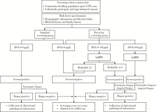
本研究采用方便抽样方法抽取3个北京市社区卫生服务中心,再采用随机数字表法将社区按2 ∶ 1比例随机分为精准筛查组与标准筛查组。每例居民均接受血清tPSA检测,并记录年龄、职业危险因素、生活习惯、饮食情况、疾病史、前列腺癌家族史等信息。根据预设分组及tPSA结果对受试者进行不同的随访管理推荐:对于精准筛查组,tPSA≥4 μg/L的受试者建议行bpMRI检查,若前列腺影像报告和数据系统(prostate imaging reporting and data system, PI-RADS)评分≥3分,建议行前列腺系统穿刺联合靶向穿刺;对于tPSA≥10 μg/L的受试者,若PI-RADS评分 < 3分,建议行前列腺系统穿刺;tPSA < 4 μg/L或者tPSA < 10 μg/L且PI-RADS评分 < 3分的受试者建议每2年进行一次前列腺特异性抗原筛查;对于标准筛查组,tPSA≥4 μg/L的受试者建议行前列腺系统穿刺,tPSA < 4 μg/L者则每2年进行一次前列腺特异性抗原筛查(图 1)。标准筛查组中,筛查阳性定义为tPSA≥4 μg/L;精准筛查组中,筛查阳性定义为满足以下任一条件:(1)tPSA≥4 μg/L且PI-RADS评分≥3分,(2)tPSA>10 μg/L。
1.3 血清tPSA检测
所有受试者在采集血样前均满足以下条件:抽血前48 h内无性生活或射精,未进行直肠指检、导尿、膀胱镜检查等操作,且1周内未接受过前列腺按摩。采集空腹肘静脉血3 mL,以3 000 r/min离心5 min,分离血清备用。血清tPSA检测采用全自动电化学发光免疫分析仪(Cobas e601,Roche Diagnostics GmbH, Mannheim, Germany)及配套的总前列腺特异性抗原化学发光免疫分析试剂盒进行,所有操作均由专业人员严格遵循标准化规程执行。为确保关键数据的准确性,对所有初次检测tPSA > 4.0 μg/L的样本进行复测,并以复测结果作为最终数据。
1.4 bpMRI
所有患者均采用3.0T磁共振扫描仪(GE Discovery MR750,通用电气医疗集团,Milwaukee, Wisconsin, USA)进行检查,使用32通道体部相控阵线圈作为接收线圈。扫描序列包括轴位、冠状位及矢状位T2加权成像(T2WI)快速自旋回波序列;弥散加权成像(DWI)采用自旋-平面回波成像序列,b值分别为0、800、1 400 s/mm2。ADC图于ADW 4.6工作站上通过functool软件,选取b值为0与800 s/mm2的弥散加权成像(diffusion-weighted imaging, DWI)序列进行重建获得。所有MRI图像均由两名具备五年以上前列腺MRI诊断经验的放射科医师独立评估。图像评估采用PI-RADS第2.1版,并将评分大于等于3分的病灶定义为MRI阳性[14]。
1.5 前列腺穿刺
前列腺穿刺即经直肠超声引导下的前列腺穿刺活检,对于PI-RADS评分≥ 3分的患者,先行靶向穿刺,对MRI阳性病灶穿刺2针,随后按标准模板行12针系统性穿刺;对于PI-RADS评分 < 3分的患者,则仅行12针系统性穿刺。超声仪器采用超声诊断仪(Logiq E9,通用电气医疗集团,Milwaukee, Wisconsin, USA),探头IC5-9。所有穿刺操作均由同一名资深超声科医师完成。穿刺组织标本由一名专职泌尿系统病理医师按照2014年国际泌尿病理学会共识标准进行制片与诊断,并报告Gleason评分和国际泌尿病理学会(International Society of Urological Pathology, ISUP)分级[15]。本研究将csPCa定义为穿刺病理为ISUP分级评分≥2的前列腺癌[16]。
1.6 统计学分析
采用SPSS 26.0软件,计量资料首先进行正态性检验(Shapiro-Wilk检验)及方差齐性检验(Levene检验),符合正态分布且方差齐者,以均数±标准差表示,两组间比较采用独立样本t检验;符合正态分布但方差不齐者,采用校正t检验(Welch’ s t检验);不符合正态分布的计量资料,以M(P25,P75)表示,两组间比较采用Mann-Whitney U检验。计数资料以n(%)表示,组间比较采用卡方检验或Fisher确切概率法。有序等级资料同样采用Mann-Whitney U检验进行组间比较。双侧检验,P < 0.05认为差异有统计学意义。
2 结果
2.1 筛查人群基线
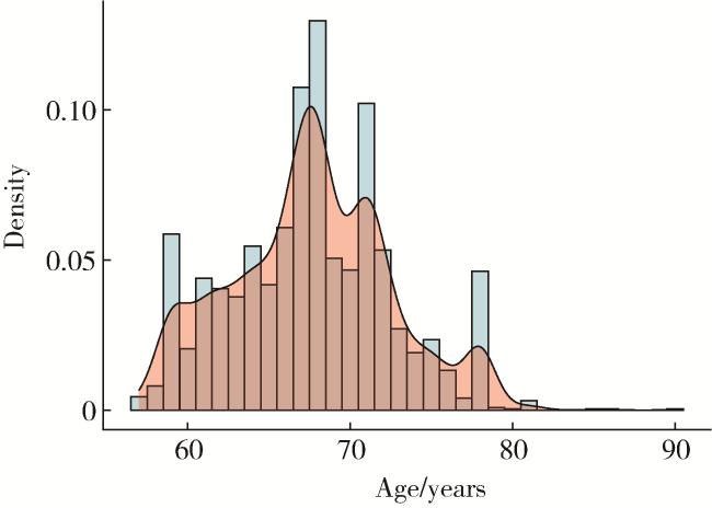


图2 筛查人群的年龄分布及密度曲线Figure 2 Age distribution and density curve of the screening population |
表1 筛查人群的基线数据Table 1 Baseline characteristics of the screening population |
| Variable | Total | Precision screening group | Standard screening group | P value |
| Number of participants | 2 251 | 985 | 1 266 | |
| Age/years,M (P25, P75) | 68 (64, 71) | 68 (64, 71) | 68 (64, 71) | 0.666 |
| tPSA/(μg/L), M (P25, P75) | 1.32 (0.78, 2.30) | 1.34 (0.77, 2.42) | 1.31 (0.79, 2.25) | 0.814 |
| Recommended for biopsy, n(%) | 140 (6.2) | 29 (2.9) | 111 (8.8) | |
| Actual biopsy performed | 46 | 20 | 26 | |
| PCa, n(%) | 0.141 | |||
| Yes | 29 (63.0) | 15 (75.0) | 14 (53.8) | |
| No | 17 (37.0) | 5 (25.0) | 12 (46.2) | |
| csPCa, n(%) | 0.004 | |||
| Yes | 21 (45.7) | 14 (70.0) | 7 (26.9) | |
| No | 25 (54.3) | 6 (30.0) | 19 (73.1) | |
| PCa detection rate/% | 1.29 | 1.52 | 1.11 | 0.384 |
| csPCa detection rate/% | 0.93 | 1.42 | 0.55 | 0.033 |
tPSA, total prostate specific antigen; PCa, prostate cancer; csPCa, clinically significant PCa. |
2.2 PCa筛查结果
在2 251例接受前列腺癌筛查的受试者中,tPSA≥4 μg/L者共有226例,其中tPSA≥10 μg/L者有33例。
在标准筛查组中,111例(8.8%)受试者tPSA≥ 4 μg/L,建议进行前列腺穿刺,其中26例接受了前列腺系统穿刺,共检出14例PCa,包括7例csPCa和7例临床无意义前列腺癌,总体穿刺阳性率为53.8%,csPCa穿刺阳性率为26.9%(表 1)。
在精准筛查组中,115例受试者tPSA≥4 μg/L,其中19例tPSA≥10 μg/L;共有30例接受了bpMRI检查,其中20例PI-RADS评分≥3分。精准筛查组中共29例受试者建议进行前列腺穿刺,其中20例接受了靶向穿刺联合系统穿刺,共检出前列腺癌15例,包括14例csPCa和1例临床无意义前列腺癌。精准筛查组总体穿刺阳性率为75.0%,与标准筛查组(58.3%)相比,差异无统计学意义(P=0.141);而精准筛查组csPCa穿刺阳性率(70.0%)显著高于标准筛查组(26.9%),差异有统计学意义(P=0.004)。
通过病历回顾、电话随访及社区记录,对未依从者进行原因追溯。标准筛查组拒绝穿刺的85例受试者中,患者个人主观拒绝占52.9%(45例),包括畏惧穿刺操作疼痛及潜在并发症32例和健康认知不足,认为无需进一步检查或受时间及就医成本限制13例;失访或转诊外院占35.3%(30例),原因不明或记录缺失占11.8%(10例)。精准筛查组未行bpMRI的85例受试者中,患者个人主观拒绝占60.0%(51例),包括健康认知不足,认为无需进一步检查或受时间及就医成本限制26例,畏惧穿刺操作疼痛及潜在并发症20例,存在幽闭恐惧5例;失访或转诊外院占29.4%(25例),原因不明或记录缺失占10.6%(9例)。精准筛查组未穿刺的9例受试者中,患者个人主观拒绝占55.6%(5例,其中3例畏惧穿刺疼痛及潜在并发症、2例健康认知不足,认为无需进一步检查),失访或转诊外院占44.4%(4例)。
2.3 前列腺癌治疗选择
在确诊前列腺癌的29例患者中,局灶期、局部进展期和转移性患者分别为17例(58.6%)、10例(34.5%)和2例(6.9%)。治疗方面,21例患者接受了前列腺癌根治性手术,4例患者接受主动监测,1例接受放射治疗,3例接受内分泌联合新型抗雄激素治疗(表 2)。对于接受了前列腺癌根治手术的21例患者,进一步比较了前列腺穿刺与手术标本的Gleason评分,结果发现精准筛查组中前列腺穿刺样本与手术样本的Gleason评分一致性为70.0%,高于标准筛查组(36.4%),但两组差异无统计学意义(P=0.198)。
表2 前列腺癌患者的临床、病理分期和病理分级Table 2 Clinical stage, pathological stage, and pathological grade of patients with prostate cancer |
| Variable | Total | Precision screening group | Standard screening group | P value |
| PCa, n | 29 | 15 | 14 | |
| ISUP grade | 0.012 | |||
| 1 | 8 (27.6) | 1 (6.7) | 7 (50.0) | |
| 2-3 | 10 (34.5) | 5 (33.3) | 5 (35.7) | |
| 4-5 | 11 (37.9) | 9 (60.0) | 2 (14.3) | |
| Clinical T stage | ||||
| T1-T2 | 17 (58.6) | 8 (53.3) | 9 (64.3) | 0.550 |
| T3-T4 | 12 (41.4) | 7 (46.7) | 5 (35.7) | |
| Clinical N stage | ||||
| N0 | 27 (93.1) | 13 (86.7) | 14 (100.0) | 0.483 |
| N1 | 2 (6.9) | 2 (13.3) | 0 (0) | |
| Clinical M stage | ||||
| M0-Mx | 27 (93.1) | 13 (86.7) | 14 (100.0) | 0.483 |
| M1 | 2 (6.9) | 2 (13.3) | 0 (0) | |
| Treatment selection, n (%) | 0.170 | |||
| Radical surgery | 21 (72.4) | 10 (66.7) | 11 (78.6) | |
| Radiotherapy | 1 (3.4) | 1 (6.7) | 0 (0) | |
| Active surveillance | 4 (13.8) | 1 (6.7) | 3 (21.4) | |
| Systemic therapy | 3 (10.3) | 3 (2.0) | 0 (0) |
Data are n (%). PCa, prostate cancer; ISUP, International Society of Urological Pathology. |
3 讨论
当前,前列腺特异性抗原筛查对于前列腺癌特异性死亡率的影响尚无定论,且其对过度诊断与过度治疗的贡献引发了日益增长的担忧。欧洲前列腺癌筛查随机研究(European randomized study of screening for prostate cancer, ERSPC)是一项欧洲多中心随机试验,旨在评估每2~4年一次PSA筛查对前列腺癌死亡率的影响。ERSPC研究的中位随访(23年)结果表明,筛查组前列腺癌死亡率较对照组降低13%(RR=0.87, 95%CI:0.80~0.95),绝对风险降低为0.22%(95%CI:0.10~0.34)[20]。英国前列腺癌PSA检测的集群随机试验(cluster randomized trial of PSA testing for prostate cancer, CAP)探索了单次前列腺特异性抗原检测对前列腺癌特异性死亡率的影响,15年随访结果显示,与对照组相比,单次前列腺特异性抗原筛查组受试者前列腺癌死亡风险降低了8%(0.69% vs. 0.78%;RR=0.92,95%CI:0.85~0.99;P=0.03)[21]。但是,前列腺、肺、结直肠和卵巢癌筛查试验(the prostate, lung, colorectal and ovarian cancer screening trial, PLCO)研究对76 693名55~74岁男性进行了近17年的随访,研究结果显示,其年度筛查方案(每年一次前列腺特异性抗原检测和直肠指诊)并未能降低前列腺癌特异性死亡率[22],而且筛查组中Gleason评分为2~6分的低级别前列腺癌发病率显著上升,提示了过度诊断风险[22]。尽管筛查能使前列腺癌死亡率的绝对风险降低0.22%,但为防止1例死亡,需要邀请456人进行筛查并随之诊断出12例前列腺癌[20]。本研究仅依赖tPSA异常行前列腺穿刺,前列腺穿刺阳性率为53.8%,而csPCa比例为50.0%,提示大量tPSA异常患者面临着不必要的前列腺穿刺,这表明前列腺特异性抗原筛查在降低死亡率方面获益有限,同时可能伴随着显著的过度诊断及过度治疗,前列腺癌筛查方案有待进一步优化。
MRI将解剖序列和功能序列相结合,提供了更为全面的图像信息,提高了对前列腺癌的检测灵敏度和特异度,在tPSA异常的基础上,联合MRI进行前列腺癌筛查,可以更精准地筛选出csPCa并降低临床无意义前列腺癌的检出率,为优化前列腺癌早期筛查策略提供了新的方向[23]。STHLM3-MRI研究是国际上首个将MRI应用于基于人群前列腺癌筛查的随机对照研究[9],研究结论显示“PSA初筛+ MRI精筛+靶向穿刺”筛查模式在诊断csPCa上不劣于传统PSA筛查组(21% vs. 18%,P < 0.001),并且可以显著减少穿刺人数和临床无意义前列腺癌的检出。以STHLM3-MRI研究为代表的“PSA+MRI”新模式,为超越传统筛查的局限提供了全新范式。在南京、宁夏等地的初步实践成功验证了该筛查策略在我国医疗环境下的可操作性及诊断价值[10, 19]。但目前缺乏“PSA+MRI”筛查模式与传统PSA筛查模式的直接比较。本研究利用大规模社区队列筛查,探索两种筛查模式对于前列腺癌的筛查效能。研究结果显示,与传统PSA筛查相比,“PSA+bpMRI”筛查模式能够显著提高csPCa的穿刺阳性率,减少三分之一不必要的前列腺穿刺。此外,该模式能够提高csPCa的总体检出率。这些结果表明,“PSA+bpMRI”筛查模式具有双重优势,即在有效规避过度诊断与过度治疗的同时,更能精准识别有临床意义的前列腺癌,从而实现筛查效益的最大化。
在治疗层面,本研究证实,“PSA+bpMRI”筛查模式相较于传统前列腺特异性抗原筛查,其术前穿刺与术后病理分级的一致性更高。前列腺癌的病理分级是风险分层与临床治疗决策的核心依据。然而,既往研究传统穿刺所获得的Gleason评分与术后病理的一致性较低,仅为50%左右;即使在穿刺仅单针阳性的患者中,术后Gleason评分升级的发生率仍高达32.3%[17]。这种普遍的评分升级现象,凸显了系统穿刺在术前低估肿瘤恶性程度的巨大风险。相比之下,MRI引导下的靶向穿刺联合系统穿刺,能够更全面地评估肿瘤的空间分布与异质性,从而实现对整体肿瘤负荷的精准判断,有力推动了前列腺癌的精准诊治。
本研究存在一定局限性:第一,本研究受试者的整体依从性仍有待提高,在标准筛查组中,115例建议穿刺者仅有23.4%接受了前列腺穿刺,精准筛查组中PSA异常患者接受bpMRI检查的比例也仅为26.1%,对未依从者的原因追溯显示,标准筛查组中主观拒绝占52.9%,失访/转诊外院占35.3%,原因不明占11.8%;精准筛查组未行bpMRI的85例中,主观拒绝占60.0%,失访/转诊外院占29.4%,原因不明占10.6%;精准筛查组未穿刺的9例中,主观拒绝占55.6%,失访/转诊外院占4 4.4%。本研究患者依从性与国内社区筛查报道(21.0%~35.4%) 相近[10, 18-19]。患者个人主观拒绝是未能完成穿刺或bpMRI的首要原因,其核心在于对前列腺癌缺乏足够认知,以及因担忧有创操作并发症而产生的心理回避;而低依从性可能导致选择性偏倚,从而影响对前列腺癌真实发病率的准确估计。因此,未来应加强健康教育、建立转诊绿色通道、探索医共体模式、主动随访并加强与患者沟通,以消除顾虑、提高复诊率。第二,当前筛查工作尚处于初始阶段,存在长期随访数据不足的局限,后续将持续积累远期数据,以全面评估该筛查模式的长期效果。第三,本研究采用方便抽样方法,样本仅来自北京3家社区卫生服务中心,样本量较小,存在地域及医疗资源可及性等方面的选择偏倚,未来需开展多中心、不同层级医疗机构的研究,以验证该筛查模式的普适性。
综上所述,PSA+MRI序贯筛查模式在中国人群中展现出良好的适用性与筛查效能,该策略不仅有望提升csPCa的早期诊断率,还能精准优化穿刺决策,从而为改善当前我国前列腺癌确诊晚、预后欠佳的局面提供有效路径。未来应在更大规模高危人群中推进该策略的应用,并积累长期随访数据,以进一步评估其成本效益与临床适用性。