Visceral leishmaniasis with suspected autoimmune disease: A report of 2 cases

  • Fan SUN 1 ,
  • Maomao CHEN 1 ,
  • Aixin HUO 1 ,
  • Wen ZHANG 2 ,
  • Zhuo LI 1 ,
  • Lan CHENG 1 ,
  • Yuhong LIU , 1, *
Expand
  • 1. Department of Rheumatology and Immunology, Yan'an University Affiliated Hospital, Yan'an 716000, Shaanxi, China
  • 2. Department of Hematology, Yan'an University Affiliated Hospital, Yan'an 716000, Shaanxi, China
LIU Yuhong,

Received date: 2024-07-25

  Online published: 2025-03-07

Copyright

All rights reserved. Unauthorized reproduction is prohibited.

Abstract

Visceral leishmaniasis (VL) is a rare parasitic infection characterized by distinctive features, including the overproduction of various autoantibodies by B cells, which may mimic manifestations and antibody profiles associated with autoimmune diseases. In this report, we presented two patients diagnosed with visceral leishmaniasis who were initially suspected of having an autoimmune disease. We discussed the similarities and differences between visceral leishmaniasis and autoimmune diseases based on clinical presentation, physical examination findings, laboratory tests, diagnosis, and treatment. Case 1 involved a 69-year-old female patient who was admitted to the hospital due to irregular fever. Physical examination revealed splenomegaly. Laboratory investigations indicated pancytopenia along with an abnormal autoantibody profile. The initial bone marrow aspirate demonstrated: Proliferative changes in the bone marrow, and abnormal lymphocytes in peripheral blood accounting for approximately 6%. Auto-immune disease was initially considered, and hormone therapy did not respond well. Further screening for immunological disorders, infections, tumors, and other related conditions was conducted. A second bone marrow aspirate was performed actively which revealed Leishman-Donovan body. Subsequent nuc-leic acid detection of pathogenic microorganisms confirmed the presence of Leishmania through real-time fluorescence PCR analysis; thus, confirming a diagnosis of visceral leishmaniasis. Case 2 involved a 41-year-old male patient who presented with fever and abdominal distension. Similar to Case 1, physical examination also showed splenomegaly alongside an abnormal autoantibody profile. During treatment, he tested positive for Leishmania infection and was ultimately diagnosed with visceral leishmaniasis complicated by hemophagocytic syndrome. The symptoms observed in the two patients included recurrent fever, anorexia, and splenomegaly. To further investigate the cause of pancytopenia and to rule out factors such as surgery, infection, tumors, and other related conditions, an abnormal antinuclear antibody profile was assessed. The initial misdiagnosis pointed towards autoimmune diseases; however, during treatment, visceral leishmaniasis infection was identified, leading to a final diagnosis of visceral leishmaniasis. The gold standard for diagnosing visceral leishmaniasis involves detecting Leishmania bodies through aspiration of bone marrow, lymph nodes, or spleen. The positive rate for bone marrow puncture is approximately 80% to 90%. However, clinical practice may encounter missed detections due to limitations associated with the puncture site. This underscores the necessity for secondary punctures when warranted. Addi-tionally, patients presenting with splenomegaly accompanied by trilineage cytopenia often seek care from Department of Hematology or Department of Rheumatology and Immunology rather than Department of Infection. This tendency can contribute to both misdiagnosis and missed diagnoses. It is hoped that this case report will offer valuable insights for clinicians in enhancing their diagnostic capabilities and improving the clinical detection rates of visceral leishmaniasis.

Cite this article

Fan SUN , Maomao CHEN , Aixin HUO , Wen ZHANG , Zhuo LI , Lan CHENG , Yuhong LIU . Visceral leishmaniasis with suspected autoimmune disease: A report of 2 cases[J]. Journal of Peking University(Health Sciences), 2026 , 58(4) : 889 -893 . DOI: 10.19723/j.issn.1671-167X.2026.04.031

系统性红斑狼疮(systemic lupus erythematosus,SLE)是一种病因不明的慢性自身免疫疾病。据文献记载,SLE患者中过度活跃的B细胞会产生多种类型的自身抗体。另外,这种多克隆B细胞活化也可能发生在慢性传染病中[1],如慢性病毒感染或寄生虫感染。而内脏利什曼病(visceral leishmaniasis,VL,又称黑热病)作为一种罕见的寄生虫感染,具有与自身免疫疾病相似的特征[2-3]。本文报告2例曾被怀疑为自身免疫疾病的VL患者,在诊疗过程中通过临床表现、体格检查、实验室检查等逐渐明确诊断,并对VL与自身免疫疾病的相似点与不同点进行了讨论。

1 病例资料

1.1 病例1

1.1.1 一般资料

患者,女,69岁,长期居住于延安市宝塔区,主因“不规则发热2年,加重伴乏力半月余”于2023年12月8日就诊于延安大学附属医院风湿免疫科。2年前患者无明显诱因出现不规则发热伴全身乏力,2023年11月20日患者症状加重,出现持续性腹痛,放射至腰背部,且食纳欠佳,伴有头晕、头痛、恶心,无呕吐,无皮疹、雷诺现象(Raynaud phenomenon)、关节疼痛症状,于外院A查白细胞(white blood cell,WBC)2.83×10⁹/L、红细胞(red blood cell,RBC)3.85×10¹²/L、血红蛋白(hemoglobin,HB)106 g/L、血小板(platelet,PLT)78×109/L,腹部超声及增强CT提示脾大。11月26日就诊于我院血液科,入院后查WBC 1.88×10⁹/L、RBC 3.32×10¹²/L、HB 91 g/L、PLT 82×109/L,全血细胞呈进行性下降;骨髓穿刺提示增生性骨髓象;外周血可见异型淋巴细胞(约占6.0%)。抗着丝点B蛋白(+++)、抗核小体抗体(+)、抗组蛋白抗体(+)、抗核抗体滴度1 ∶1 000,补体(-),免疫球蛋白G 35.5 g/L、免疫球蛋白E 537 IU/mL,考虑为:(1)免疫性全血细胞减少,(2)系统性红斑狼疮。住院第4天,患者突然出现高热,体温可达40 ℃,红细胞沉降率(erythrocyte sedimentation rate,ESR)129 mm/h、C-反应蛋白(C-reactive protein,CRP)89 mg/L、铁蛋白466 μg/L,经抗感染治疗, 体温恢复正常后出院。患者出院后症状反复,间断发热,夜间为著,伴畏寒、肌肉酸痛及全身乏力,收住我院风湿免疫科。既往史:高血压、脑梗死病史15年,否认外地旅居史。查体:心肺查体无异常,颈部浅表淋巴结可触及肿大,左上腹压痛阳性,肝脏肋下未触及肿大,脾脏肋下可触及,超出肋缘1 cm。

1.1.2 实验室检查

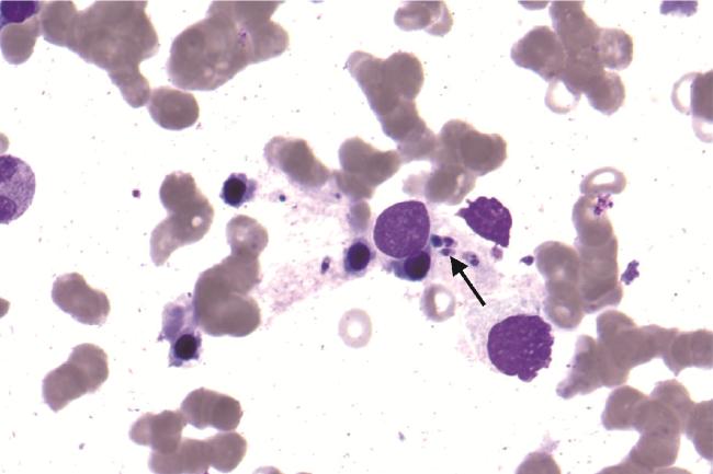
为明确患者反复发热伴全血细胞减少的原因,我科从免疫、炎症、感染、肿瘤等方面着手,进行了详细的化验检查。(1)免疫指标:抗核抗体谱:抗着丝点B蛋白(+++)、抗核小体抗体(+)、抗组蛋白抗体(+)、抗核抗体滴度>1 ∶3 200;免疫球蛋白G 34.8 g/L、免疫球蛋白E 516 IU/mL,补体、心磷脂系列正常。(2)炎症指标:降钙素原1.65 ng/L、CRP 179 mg/L、ESR 136 mm/h、铁蛋白652 μg/L。(3)感染指标:呼吸道病原体、巨细胞病毒、EB病毒、真菌检查及多次血培养结果均为阴性,易引起发热表现的疾病如布鲁氏菌病、感染性心内膜炎等相关检查均正常,胆道及泌尿系疾病无临床影像征象。(4)肿瘤指标:胸部CT提示结节灶,条索影;腹部CT提示脂肪肝、门静脉增宽、脾大,未见占位;针对消化道和肺的肿瘤标记物均无明显异常;骨髓穿刺、CD55、CD59等检查无明显异常,暂可排除血液系统恶性肿瘤。(5)其他:血常规:WBC 2.88×10⁹/L、RBC 3.36×10¹²/L、HB 92 g/L、PLT 46×109/L; 患者肝功能、肾功能、甲状腺功能检查及血糖等均未见明显异常,凝血酶原时间12.9 s,凝血酶原活动度63.6%,活化部分凝血活酶时间26.8 s。故暂不考虑内分泌系统疾病、急慢性肝病、慢性肾功能衰竭及药物因素影响。尽管入院相关检查未发现感染灶存在,但考虑到正值流感季节,遂给予三代抗生素对症治疗,治疗后患者仍然持续高热,故考虑可能存在免疫功能异常,SLE不除外。加用激素后体温缓解仍不明显,复查外周血常规呈全血细胞下降状态(图 1),凝血功能变化见表 1。于2023年12月18日行第二次骨髓穿刺,提示:增生性骨髓象,PLT少见;全片偶可见利-杜小体(Leishman-Donovani body,即杜氏利什曼原虫无鞭毛体形态,图 2)。病原微生物核酸检测明确为杜氏利什曼原虫(实时荧光聚合酶链反应法)。
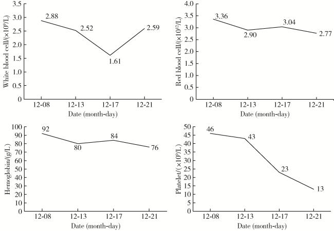
图1 患者的外周血变化

Figure 1 Changes in the patient's peripheral blood

表1 患者的凝血功能变化

Table 1 Changes in coagulation function in patients

Date PT/s PTA/% D-dimer/(mg/L) APTT/s FIB/(g/L) TT/s FDP/(mg/L)
2023-11-27 12.2 62.3 1.01 26.0 3.51 20.0 78
2023-12-09 12.9 63.6 14.25 26.8 4.02 21.1 108
2023-12-18 14.2 64.1 23.42 27.6 3.14 17.2 144

PT, prothrombin time; PTA, prothrombin activity; APTT, activated partial thromboplastin time; FIB, fibrinogen; TT, thrombin time; FDP, fibrin degradation products.

图2 患者的骨髓涂片(瑞氏-姬姆萨染色×1 000)

Figure 2 Patient's bone marrow smear(Wright-Giemsa staining ×1 000)

The black arrow indicates the presence of Leishmania.

1.1.3 诊断

患者最终诊断为VL,继发噬血细胞综合征待查。由于VL需要特殊药物治疗,遂将患者转诊至某上级医院就诊,未在本院再行自然杀伤(natural killer, NK)细胞活性及sCD25(可溶性白细胞介素-2受体)等检测,因此未能明确是否继发噬血综合征。

1.1.4 治疗及转归

患者于该上级医院予以两性霉素B脂质体治疗后突发房颤,改用葡萄糖酸锑钠肌肉注射4 d,后未再发热,病情好转出院。2024年1月随访时,患者复查病原微生物检测提示未检出杜氏利什曼原虫,同时期的抗核抗体谱也恢复正常,随访至2024年7月未见复发。

1.2 病例2

1.2.1 一般资料

患者,男,41岁,长期居住于延安市延川县。主因“发热伴腹胀半年,加重2月”于2021年8月就诊于延安大学附属医院血液科。2个月前患者无明显诱因出现消瘦、乏力、纳差, 曾于外院B诊断为“脾大,脾功能亢进”。本次入院期间查血常规:WBC 2.13×10⁹/L、HB 63 g/L、PLT 40×109/L;查体:脾肋下可触及,超出脐水平线及前正中线,Ⅰ线18 cm、Ⅱ线24 cm、Ⅲ线+6.5 cm。风湿免疫科会诊后考虑为脾功能亢进、结缔组织病,不除外自身免疫性肝炎,转入风湿免疫科进一步诊治。

1.2.2 实验室检查

血常规:WBC 0.59×10⁹/L、HB 59 g/L、PLT 12×109/L;血脂:血清甘油三酯5.04 mmol/L;抗核抗体谱:抗着丝点B蛋白(+++)、抗SSA/Ro 52(+)、抗组蛋白抗体(++)、抗核小体抗体(+)、抗核抗体滴度1 ∶1 000;自身免疫性肝炎抗体谱:抗肝细胞溶质抗原Ⅰ型抗体(++);骨髓穿刺结果提示:增生性骨髓象,PLT减少,浆细胞比例偏高,可见吞噬细胞;NK细胞活性降低(2.8%);杜氏利什曼原虫病原微生物核酸检测阳性。

1.2.3 诊断

诊断为VL继发噬血细胞综合征。

1.2.4 治疗及转归

因患者病情危重,且诊断明确后未积极治疗,在1个月内死亡。

2 讨论

VL是一种慢性地方性人畜共患寄生虫病,通过白蛉叮咬感染[4],陕西省是现阶段VL的主要疫区之一。VL的临床表现主要是不规则发热、纳差、腹痛、腹泻等,可能与许多免疫异常有关,其全血细胞减少主要是由脾肿大和脾功能亢进导致。据国内外文献报道,VL极易被误诊为结缔组织病,尤其与SLE的临床及实验室特征相似[5-6]。Liberopoulos等[7]追踪报道了16例入院时没有任何自身免疫疾病临床表现的VL患者,在检查时发现存在多种自身抗体,其中7例患者在治疗后抗体水平恢复正常。机制研究表明,杜氏利什曼原虫释放的物质能促使B细胞有丝分裂,使B细胞过度活跃,进而引起高丙种球蛋白血症并导致多种自身抗体(如抗核抗体)阳性,使患者出现自身免疫疾病表现[1]。此外,杜氏利什曼原虫抗原和核糖核蛋白之间的分子模拟也可能导致VL感染期间自身抗体的形成[7-8]。对于被诊断为SLE的患者,特别是在流行地区,必须与VL进行鉴别诊断。
本文报告了2例VL患者,均疑似自身免疫疾病。根据相关文献,可从以下5点辅助鉴别诊断:(1)巨脾在SLE中并不常见;(2)关节炎不是VL的常见临床特征;(3)除非存在感染,否则CRP升高在SLE患者中并不常见;(4)SLE患者中可见血清补体水平降低,但VL患者血清补体水平往往正常;(5)VL患者通常会出现抗杜氏利什曼原虫抗体阳性,而该抗体在SLE患者中表现为阴性[1, 6]。病例1随访中可见抗核抗体谱转为阴性,确定之前的异常是由于杜氏利什曼原虫感染模拟所致。综上所述,VL与自身免疫疾病存在相似性,在临床鉴别诊断中应该引起高度重视。
确诊VL的金标准是骨髓、淋巴结、脾穿刺检出利-杜小体[6],其中骨髓穿刺阳性率为80%~90%,脾穿刺阳性率高达90%~99%[9]。由于穿刺部位受限,有可能会出现漏检,提示有必要行二次穿刺。本文病例1二次骨髓穿刺结果对确诊起到关键作用。也有研究表明,直接凝集试验(direct agglutination test,DAT)和rk39抗体检测在VL的血清学诊断上也具有一定价值,或可成为VL特异抗体的快速检测方法[10-11]
噬血细胞综合征,又称噬血细胞性淋巴组织细胞增多症,是一种原发或继发的免疫过度活化综合征引起的全身性疾病,常伴有多脏器损伤,病情发展迅猛,且诊断困难,易被延误,病死率高,其未经治疗的中位生存时间不超过2个月[12]。临床上将噬血细胞综合征分为原发性(遗传性)和继发性(获得性)两类,后者更为常见,感染、肿瘤、免疫功能异常等通常是继发性噬血细胞综合征的因素。继发性噬血细胞综合征作为VL的严重并发症[13],其主要临床表现为持续性发热、全血细胞减少、肝肿大、脾肿大,噬血现象可在骨髓、肝、脾及淋巴结组织中发现[14]。国内外很多报道中,VL与噬血细胞综合征总是同时存在。依据《中国噬血细胞综合征诊断与治疗指南(2022年版)》[12]给出的诊断标准,病例1符合发热、脾大、血细胞减少、血清铁蛋白升高4条标准,但由于未检测NK细胞活性和sCD25等,不能确诊继发噬血细胞综合征。病例2符合VL继发噬血细胞综合征的诊断。
目前VL的首选药物仍然是5价锑制剂[15],常用药物为葡萄糖酸锑钠,该药物具有疗效快、治疗周期短、副反应少等优点,然而由于药物滥用,部分VL患者对葡萄糖酸锑钠具有耐药现象。对于锑剂治疗无效的患者也可选择使用两性霉素B脂质体治疗,国内外多项临床回顾性研究均报道该药最终治愈率在95%以上,且不良反应率较低[16],但由于其价格偏高,较难推广。临床证实,口服药物米替福新也可替代锑剂治疗VL,且使用安全,治愈率高,但具有一定复发风险[16-17]。本报告中病例1经两性霉素B脂质体、葡萄糖酸锑钠治疗后好转,随访至2024年7月未见复发。
本文中的两例患者症状相似,主要为反复发热、纳差,查体脾大,为进一步查找引起全血细胞减少的原因,我们先排除了手术、感染、肿瘤等相关因素,由于抗核抗体谱异常,初期将其误诊为自身免疫疾病,治疗过程中发现杜氏利什曼原虫感染,最终确诊为VL。临床上,这种疑似自身免疫疾病的VL并不少见,崔银风等[2]、郑迈等[5]、Garg等[18]均报道过疑似SLE的VL,这些患者具有与本文病例相似的临床特征,容易与自身免疫疾病相混淆。Tunccan等[19]也曾报道1例疑似自身免疫性肝炎、原发性胆汁性肝硬化和SLE重叠的VL。相对应的是,在VL疑似自身免疫疾病的同时,自身免疫疾病中发生的VL也容易被误诊为疾病复发,Ossandon等[20]报道了1例狼疮患者感染VL,提示若能及时识别出狼疮患者伴有杜氏利什曼原虫感染,避免误诊、误治,可有效改善患者的预后。事实上,无论是疑似自身免疫疾病而被误诊的VL患者,还是自身免疫疾病基础上合并杜氏利什曼原虫感染,都需要临床医生保持一定的警惕性。对于红细胞、白细胞、血小板均减少的患者,若首次就诊于血液科或风湿免疫科而非感染科时,应考虑到VL的可能性,以免造成误诊和漏诊。

利益冲突  所有作者均声明不存在利益冲突。

作者贡献声明  孙帆:选择病例,提出思路,撰写论文;陈毛毛、霍爱鑫:文献检索,参与病例讨论;张雯:提供相关病例资料;李卓、程兰:收集、分析、整理病历资料;刘宇宏:论文思路的整体设计和论文审定。所有作者均参与论文修改,并对最终文稿进行审读和确认。

1
Voulgari PV , Pappas GA , Liberopoulos EN , et al. Visceral leishmaniasis resembling systemic lupus erythematosus[J]. Ann Rheum Dis, 2004, 63 (10): 1348- 1349.

DOI

2
崔银风, 张莉芸, 许珂, 等. 误诊为系统性红斑狼疮的黑热病一例[J]. 中华风湿病学杂志, 2022, 26 (1): 39- 41.

3
Bueno GCL , Koerich ATS , Burg LB , et al. Visceral leishmaniasis mimicking systemic lupus erythematosus[J]. Rev Soc Bras Med Trop, 2019, 52, e20180208.

DOI

4
张路钱, 李欣欣, 年云鹏, 等. 2011-2022年陕西省人群黑热病流行特征分析[J]. 中华地方病学杂志, 2023, 42 (9): 727- 729.

5
郑迈, 刘爽, 崔若玫, 等. 误诊为系统性红斑狼疮的黑热病2例[DB/OL]. 中国临床案例成果数据库, 2022, 4(1): E07198. (2022-11-30)[2024-06-28]. https://rs.yiigle.com/cmaid/1435631.

6
Santana IU , Dias B , Nunes EA , et al. Visceral leishmaniasis mimicking systemic lupus erythematosus: Case series and a systematic literature review[J]. Semin Arthritis Rheum, 2015, 44 (6): 658- 665.

DOI

7
Liberopoulos E , Kei A , Apostolou F , et al. Autoimmune manifestations in patients with visceral leishmaniasis[J]. J Microbiol Immunol Infect, 2013, 46 (4): 302- 305.

DOI

8
Chen X , Zhou Q , Liu J , et al. Autoimmune manifestations of visceral leishmaniasis in Chinese patients[J]. Ann Palliat Med, 2021, 10 (12): 12699- 12705.

DOI

9
毕红霞, 刘焱斌. 《黑热病诊疗方案(2023年版)》解读[J]. 中国抗生素杂志, 2024, 49 (7): 729- 736.

10
Getnet M , Minaye Dejen A , Abebaw D , et al. Diagnostic accuracy of serological rk-39 test for visceral Leishmaniasis: Systematic review and meta-analysis[J]. PLoS Negl Trop Dis, 2024, 18 (3): e0011938.

DOI

11
Roberts T , Keddie SH , Rattanavong S , et al. Accuracy of the direct agglutination test for diagnosis of visceral leishmaniasis: A systematic review and meta-analysis[J]. BMC Infect Dis, 2023, 23 (1): 782.

DOI

12
中国医师协会血液科医师分会, 中华医学会儿科学分会血液学组, 噬血细胞综合征中国专家联盟. 中国噬血细胞综合征诊断与治疗指南(2022年版)[J]. 中华医学杂志, 2022, 102 (20): 1492- 1499.

13
陆丹倩, 吴为强, 周梅, 等. 黑热病继发噬血细胞综合征1例[J]. 中国感染与化疗杂志, 2023, 23 (5): 630- 632.

14
Bode SF , Lehmberg K , Maul-Pavicic A , et al. Recent advances in the diagnosis and treatment of hemophagocytic lymphohistiocytosis[J]. Arthritis Res Ther, 2012, 14 (3): 213.

DOI

15
李瑞娟, 康文, 连建奇, 等. 唐都医院2010-2020年收治的黑热病患者流行病学及临床特征分析[J]. 空军军医大学学报, 2022, 43 (8): 867- 870.

16
van Griensven J , Diro E . Visceral leishmaniasis: Recent advances in diagnostics and treatment regimens[J]. Infect Dis Clin North Am, 2019, 33 (1): 79- 99.

DOI

17
Melcon-Fernandez E , Galli G , García-Estrada C , et al. Miltefo-sine and nifuratel combination: A promising therapy for the treatment of leishmania donovani visceral leishmaniasis[J]. Int J Mol Sci, 2023, 24 (2): 1635.

DOI

18
Garg S , Kundu M , Dwivedi AN , et al. Visceral leishmaniasis or systemic lupus erythematosus flare?[J]. Case Reports Immunol, 2012, 2012, 523589.

19
Tunccan OG , Tufan A , Telli G , et al. Visceral leishmaniasis mimicking autoimmune hepatitis, primary biliary cirrhosis, and systemic lupus erythematosus overlap[J]. Korean J Parasitol, 2012, 50 (2): 133- 136.

DOI

20
Ossandon A , Bompane D , Alessandri C , et al. Leishmania in SLE mimicking an exacerbation[J]. Clin Exp Rheumatol, 2006, 24 (2): 186- 190.

Outlines

/