Journal of Peking University(Health Sciences) ›› 2019, Vol. 51 ›› Issue (5): 931-936. doi: 10.19723/j.issn.1671-167X.2019.05.023
Previous Articles Next Articles
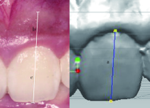
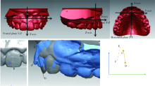
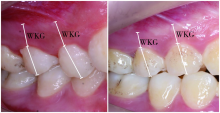
Effect of orthodontic tooth movement on keratinized gingival width
Gao-nan WANG1,Jian JIAO2,Yan-heng ZHOU1,Jie SHI1,△( )
)
- 1. Department of Orthodontics, Peking University School and Hospital of Stomatology & National Clinical Research Center for Oral Diseases & National Engineering Laboratory for Digital and Material Technology of Stomatology & Beijing Key Laboratory of Digital Stomatology, Beijing 100081, China
2. Department of Periodontology, Peking University School and Hospital of Stomatology & National Clinical Research Center for Oral Diseases & National Engineering Laboratory for Digital and Material Technology of Stomatology & Beijing Key Laboratory of Digital Stomatology, Beijing 100081, China
CLC Number:
- R783.5


| [1] | Closs LQ, Branco P, Rizzatto SD , et al. Gingival margin alterations and the pre-orthodontic treatment amount of keratinized gingiva[J]. Braz Oral Res, 2007,21(1):58-63. |
| [2] | Coatoam GW, Behrents RG, Bissada NF . The width of keratinized gingiva during orthodontic treatment: its significance and impact on periodontal status[J]. J Periodontol, 1981,52(6):307-313. |
| [3] | Trentini CM, Moriarty JD, Phillips C , et al. Evaluation of the use of orthodontic records to measure the width of keratinized tissue[J]. J Periodontol, 1995,66(6):438-442. |
| [4] | Melsen B, Allais D . Factors of importance for the development of dehiscences during labial movement of mandibular incisors: a retrospective study of adult orthodontic patients [J]. Am J Orthod Dentofacial Orthop, 2005, 127(5): 552-561; quiz 625. |
| [5] | Wyrębek B, Orzechowska A, Cudziło D , et al. Evaluation of changes in the width of gingival in children and youth[J]. Dev Period Med, 2015,19(2):212-216. |
| [6] | Cho MY, Choi JH, Lee SP , et al. Three-dimensional analysis of the tooth movement and arch dimension changes in Class I malocclusions treated with first premolar extractions: A guideline for virtual treatment planning[J]. Am J Orthod Dentofacial Orthop, 2010,138(6):747-757. |
| [7] | Artun J, Krogstad O . Periodontal status of mandibular incisors following excessive proclination. A study in adults with surgically treated mandibular prognathism[J]. Am J Orthod Dentofacial Orthop, 1987,91(3):225-232. |
| [8] | Dorfman HS . Mucogingival changes resulting from mandibular incisor tooth movement[J]. Am J Orthod, 1978,74(3):286-297. |
| [9] | Djeu G, Hayes C, Zawaideh S . Correlation between mandibular central incisor proclination and gingival recession during fixed appliance therapy[J]. Angle Orthod, 2002,72(3):238-245. |
| [10] | Dannan A, Darwish MA, Sawan MN . Keratinized gingiva width alteration during orthodontic alignment and leveling phase; a preliminary investigation [J/OL]. Int J Dent Sci, 1995, 7(2): 1-6[2018-01-31]. |
| [11] | King KO, Sadler JF, Higgason JD , et al. The effects of angulation upon pre- and postoperative photographs[J]. J Periodontol, 1963,34(2):139-141. |
| [12] | Erkan M, Pikdoken L, Usumez S . Gingival response to mandibular incisor intrusion[J]. Am J Orthod Dentofacial Orthop, 2007, 132(2): 143. e9-13. |
| [13] | Pikdoken L, Erkan M, Usumez S . Gingival response to mandibular incisor extrusion[J]. Am J Orthod Dentofacial Orthop, 2009, 135(4): 432. e1-6. |
| [14] | Zawawi KH, Al-Harthi SM, Al-Zahrani MS . Prevalence of gingival biotype and its relationship to dental malocclusion[J]. Saudi Med J, 2012,33(6):671-675. |
| [15] | Zawawi KH, Al-Zahrani MS . Gingival biotype in relation to incisors’ inclination and position[J]. Saudi Med J, 2014,35(11):1378-1383. |
| [1] | Bochun MAO,Yajing TIAN,Xuedong WANG,Jing LI,Yanheng ZHOU. Soft and hard tissue changes of hyperdivergent class Ⅱ patients before and after orthodontic extraction treatment [J]. Journal of Peking University (Health Sciences), 2024, 56(1): 111-119. |
| [2] | DAI Fan-fan, LIU Yi, XU Tian-min, CHEN Gui. Exploring a new method for superimposition of pre-treatment and post-treatment mandibular digital dental casts in adults [J]. Journal of Peking University(Health Sciences), 2018, 50(2): 271-278. |
| [3] | . Stress change of periodontal ligament of the anterior teeth at the stage of space closure in lingual appliances: a 3-dimensional finite element analysis [J]. Journal of Peking University(Health Sciences), 2018, 50(1): 141-147. |
| [4] | ZHANG Da, WANG Lin-chuan, ZHOU Yan-heng, LIU Xiao-mo, LI Jing. Precision of three-dimensional printed brackets#br# [J]. Journal of Peking University(Health Sciences), 2017, 49(4): 704-708. |
| [5] | SHEN Xiao, SHI Jie, XU Li, JIAO Jian, LU Rui-fang, MENG Huan-xin. Clinical evaluation of periodontal-orthodontic treatment in patients with aggressive periodontitis and malocclusion [J]. Journal of Peking University(Health Sciences), 2017, 49(1): 60-066. |
| [6] | WEN Fu-jia,CHEN Gui,LIU Yi. Morphological analysis of roots and alveolar bone changes after upper anterior #br# retraction with maximum anchorage based on cone-beam computed tomography [J]. Journal of Peking University(Health Sciences), 2016, 48(4): 702-708. |
| [7] | HUANG Jun-qiang, LIU Shi-yao, JIANG Jiu-hui. Therapeutic evaluation of the correction of the severe bi-maxillary protrusion cases by Tweed-Merrifield technique [J]. Journal of Peking University(Health Sciences), 2016, 48(3): 555-561. |
| [8] | SONG Guang-Ying, JIANG Ruo-Ping, ZHANG Xiao-Yun, LIU Si-Qi, YU Xiao-Nan, CHEN Qing, WENG Xuan-Rong, WU Wei-Zi, SU Hong, REN Chong, DAN Ru-Kai, GENG Zhi, XU Tian-Min, JIAN Li-Zhong-Guo-Zheng-Ji-Liao-Xiao-Ping-Jia-Biao-Zhun-Ke-Ti-Zu. Validation of subjective and objective evaluation methods for orthodontic treatment outcome [J]. Journal of Peking University(Health Sciences), 2015, 47(1): 90-97. |
| [9] | PAN Yi-Chun, ZHAO Jian-Hui. Clinical outcome evaluation of midpalatal mini-implant anchorage system in orthodontic treatment [J]. Journal of Peking University(Health Sciences), 2014, 46(6): 969-974. |
|
||